

 PDF
PDF ePub
ePub Citation
 Print
Print


INTRODUCTION
Paratesticular tumors are infrequent tumor of mesenchymal origin that can affect the testicular tunics, epididymis, and spermatic cord. The most common neoplasm arising in paratesticular area is adenomatoid tumor (1). Schwannoma of the scrotum and testis is extremely rare. We report the case of an intrascrotal and extratesticular tumor that was histopathologically shown to be schwannoma.
CASE REPORT
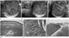
A 59-year-old man presented with a painless, slowly growing, palpable, scrotal tumor of ten years of duration. On physical examination, 2 cm sized round and movable mass was palpated in the lower extratesticular region within the left scrotum. It was located just below scrotal skin but was not attached to the skin or any other regional structure. In particular, the mass was separated from the left testis, and both testes appeared to be normal. Lymphadenopathy was not detected. Ultrasonography depicted a 2 × 2 cm sized smooth, round, well-circumscribed, heterogeneous, hyperechoic mass within the left scrotum (Fig. 1A). The tumor was separate from the left testis and epididymis (Fig. 1B). Doppler examination showed that the mass was well-vascularized (Fig. 1C). We initially considered this extratesticular mass a benign tumor such as adenomatoid tumor or leiomyoma. Therefore, curative surgical excision of tumor was performed. At the resection, the tumor was observed to be separated from the testis, based on ultrasound findings. Grossly, the resected mass appeared yellowish, round, and soft with focal hemorrhage. Microscopically, tumor showed to be a well demarcated solid tumor with multiple dilated hyalinized vessels (Fig. 1D). The tumor was comprised of spindle cells with elongated nuclei and fibrillary cytoplasm. Tumor cells showed hypercellular areas with nuclear palisading and alternating with hypocellular areas (Fig. 1E). Immunohistochemically, tumor cells were diffusely positive for S-100 protein (Fig. 1F). Histological and immunohistochemical findings were consistent with schwannoma.
DISCUSSION
Schwannoma is a benign encapsulated neoplasm that usually arises from cranial, spinal, and peripheral nerves. They mostly affect the head and neck region along a nerve sheath (2). However, although schwannoma is the most common peripheral nerve sheath tumor, it is extremely rare in the paratesticular area. To date, intrascrotal schwannomas have been reported in only seven cases in the medical literature written in English (3, 4, 5, 6, 7, 8, 9).
Histologically, schwannoma is an encapsulated tumor with biphasic architectural pattern composed of Antoni A and Antoni B areas. Antoni A areas correspond to compacted spindle cells that are often arranged in palisades or adopt an organoid arrangement (Verocay bodies), whereas Antoni B areas are characterized by loose-textured tissue in a myxomatous matrix that may appear microcystic. Immunohistochemistry studies show uniform positive staining for S-100 protein (2).
The diagnosis of schwannoma is challenging, and the clinical and radiographic findings of scrotal schwannoma are non-specific, which means the tumor can be easily misdiagnosed as another solid tumor. Patients usually present with an asymptomatic, slowly growing scrotal mass with duration of several months to a year (3, 4, 5, 6). Only one patient complained of local pain in the scrotum for 20 days before presentation (7). Chan et al. (3) reported ultrasonographic findings of a well-circumscribed ovoid heterogeneous mass separate from the testis, which measured 7 cm in greatest diameter. Bergeron et al. (4) reported a solid voluminous, heterogeneous, lobulated mass, with greatest diameter of 5.5 cm; and Shahid et al. (5) also described a well-circumscribed heterogeneous mass. On ultrasound, all tumors were depicted as a well-circumscribed heterogeneous mass, well-separated from testis and epididymis. There has been no case reporting of a cystic portion or calcification.
The most common extratesticular tumor is adenomatoid tumor, which occurs at a peak age of between 20 and 50 years. These patients usually present with an asymptomatic scrotal tumor. Adenomatoid tumors are smooth, round, well-defined, and varying in size. On ultrasound, they are typically homogeneous and hyperechogenic in nature (1). Leiomyoma is the second most common tumor of the epididymis. This tumor most commonly manifests as a slow-growing, nontender scrotal mass, which occurs at a peak age during the fifth decade of life. Leiomyomas usually present as well-demarcated tumor surrounded by a gray-white fibrous capsule and would range from 1 to 4 cm in size. This tumor has a variable sonographic appearance whether it is predominantly solid or cystic, and it may contain calcifications. Leiomyomas can be associated with a hydrocele in half of the cases (1, 10).
However, it is difficult to accurately diagnose paratesticular tumors, preoperatively. Despite its rarity in the scrotum, schwannoma should be included in the differential diagnosis of paratesticular tumor with other more common benign tumors.
 XML Download