

 PDF
PDF ePub
ePub Citation
 Print
Print


INTRODUCTION
A dorsal epidural migration of sequestered lumbar disc fragment, known as complete isolation of the disc fragment from the parent disc may occur in approximately one-third of symptomatic disc herniations (1). Low back pain and radiculopathy due to nerve root compression as a result of dorsal or dorsal-lateral disc rupture are the most common symptoms of the disease (2). Clinically, the differential diagnosis of a dorsal epidural migration of sequestered disc fragment is important, because it can influence both, the treatment and prognosis of the patient.
A dorsal epidural migration of a sequestered lumbar disc fragment is very rare and there are few case reports on the migration of lumbar disc fragment to the dorsal aspect of the epidural space only (2,3,4,5,6). Herein we present 3 pathologically confirmed cases of dorsal epidural migration of sequestered lumbar disc fragment and review the literature on the dorsal epidural migration of the sequestrated disc fragment. Also we discuss the occurrence tendency of such a migrated disc fragment in the more affected age group.
CASE REPORT
We retrospectively reviewed the imaging features of dorsal epidural migration of sequestered lumbar disc fragment in three male patients. Magnetic resonance (MR) imaging was performed in all three patients. All patients underwent surgical excision and a pathological diagnosis was achieved.
The first patient was a 95-year-old man who presented with motor weakness of both lower extremities for 2 weeks. The motor strength of the right hip flexion was 3/5 and the left hip flexion, both knee extension, both ankle dorsiflexion, both great toe dorsiflexion and both ankle plantarflexion were 0/5 on motor examination. However, the sensory examination was intact.
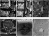
Sagittal (Fig. 1A-C) and axial (Fig. 1D) MR images of the patient showed an abnormal mass-like lesion located in the dorsal aspect of the epidural space at L3/4 level. The signal intensity of the lesion was low on T2-weighted images (T2WI) (Fig. 1A, D) and intermediate on T1-weighted images (T1WI) (Fig. 1B), similar to the signal intensity of the adjacent intervertebral discs on both, T1WI and T2WI. The lesion showed a rim contrast enhancement on fat saturated T1WI following the intravenous gadolinium enhancement (Fig. 1C).
The second man was a 78-year-old patient who presented with severe pain of the low back and both lower extremities for one month. Motor and sensory examinations were intact. MR images of the patient showed an abnormal mass-like lesion located in dorsal aspect to the epidural space at L4/5 level also. The signal intensity of the lesion was low and intermediate on T2WI and intermediate on T1WI, similar to the signal intensity of the lesion of the first patient. Also the lesion showed rim contrast enhancement on fat saturated T1WI following intravenous gadolinium enhancement.
These two patients underwent a midline circumferential decompression and surgical excision of the mass lesion and the pathology revealed a fresh and degenerated fibrocartilage, consistent with the disc material (Fig. 1E, F).
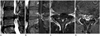
The third patient, a 62-year-old male had developed severe pain of the low back and both lower extremities for a week after lifting a heavy object. He underwent a microscopic decompression of L4/5 and discectomy of L3/4 for right herniated intervertebral disc disease five months ago. The motor strength of right ankle dorsiflexion was 4/5, great toe elevation was 3/5 but the other motor and sensory examinations were intact on motor examination. Sagittal (Fig. 2A, B) and axial (Fig. 2C, D) MR images of the patient showed an abnormal mass-like lesion located in the dorsal aspect of the epidural space at L4/5 level also. The signal intensity of the lesion was low on T2WI (Fig. 2A, C), similar to the signal intensity of the adjacent intervertebral disc. The lesion showed a rim contrast enhancement on fat saturated T1WI following the intravenous gadolinium enhancement (Fig. 2B, D). The patient underwent a surgical excision of the mass lesion also and the pathology revealed also fresh and degenerated fibrocartilage. After surgery, the pain and motor weakness gradually resolved in the low back and both lower extremities and the patient was discharged without further complications.
On MR images of all patients, there were degenerative lumbar spondylosis causing central spinal stenosis at L4/5 or L3/4 and the free disc fragment migrated into the dorsal epidural space.
DISCUSSION
MR imaging is the modality of choice to investigate a disc herniation. The MR imaging features of dorsally sequestrated disc fragments have been reported to be usually hypointense on T1WI, and 80% are hyperintense on T2WI (1, 4).
Those fragments often appear heterogeneous, whereas tumors usually appear homogeneous on T2WI (1, 4). A peripheral enhancement around the sequestrated disc fragment is commonly found on contrast-enhanced MR imaging (7).
Yamashita et al. (8) speculated based of pathologic and contrast-enhanced MR imaging findings, that the mechanism of peripheral enhancement around the sequestered disc materials is associated with the accumulation of contrast material within the vascularized granulation tissue surrounding the avascular sequestered disc material. In our cases, the fragments appeared as hypo- to isointense mass-like lesion on T1WI and hypo- to mild hyperintense lesion on T2WI. All cases showed a peripheral enhancement around the disc fragments on gadolinium-enhanced MR images.
The differential diagnosis of a dorsal epidural mass-like lesion is important, because it may influence both, the treatment and prognosis of the patient. Before operation, those lesions may be diagnosed as an epidural abscess, epidural spinal tumor or epidural hematoma. An epidural abscess is typically iso- to hypointense on T1WI and hyperintense on T2WI (9). The enhancement can be diffusely homogeneous, heterogeneous or rim-enhanced (3). Epidural tumors show variable signal intensity and enhancement features according to the tumor types (3). They are often hypointense on T1WI and hyper- or hypointense on T2WI (3). Generally, an epidural hematoma appears iso- or hyperintense on T1WI in the acute and sub-acute phase (2). The differential diagnosis should include synovial cysts that typically communicate with the synovia of a degenerative adjacent articulation, based on the location of the lesion (3). A synovial cyst originating from the facet joint is centered on the facet and has a variable imaging signal, depending on the contents of the cyst. They are usually rim-enhanced and sometimes associated with calcification (5).
To the best of our knowledge, there are few reported cases with lumbar disc migration to the dorsal epidural space yet. At the level of the disc, the posterior longitudinal ligament (PLL) is firmly adherent to the dorsal annulus and is attached to the lateral membrane, also called peridural membrane (10) extending medially from the lateral edge of the PLL to the lateral wall of the spinal canal. This limits the movement of the extruded disc fragment beyond the postero-lateral corner of the epidural space and makes it difficult for a disc to migrate dorsal to the epidural space (1, 2).
The mechanism of dorsal migration still remains unclear. Perhaps, a stretching of PLL and other ligaments may push or displace the free disc fragment into the spinal canal (4). Possible explanations of dorsal migration of a free disc fragment include an intraspinal pressure gradient during the traction of the spine (4) or a position of the nerve root relative to the intervertebral disc space and the epidural space determining the migration path (6). The hypothesis has been verified that a side-to-side migration of an extruded lumbar disc herniation can be blocked or restrained by the presence of the ventral meningovertebral ligaments (11). In addition, Kuzeyli et al. (6) suggested that conditions of hypermobility associated with degenerative disc disease or degenerative spondylolisthesis may also be predispositions for a dorsal migration of disc fragments. There were degenerative lumbar spondylosis causing central spinal stenosis at L4/5 and L3/4 and the free disc fragment migrated into the dorsal epidural space in our three presented patients.
We suggest that older age and previous intervertebral decompression surgery may be one of the predisposing factors of dorsal epidural migration of a sequestered lumbar disc fragment. These two factors may be together with conditions of hypermobility associated with degenerative disc disease or degenerative spondylolisthesis. The first and the second patient belonged to the older age group and the third patient showed a history of intervertebral decompression surgery 5 months ago.
In conclusion, our cases suggest that the MR imaging shows characteristic features for the dorsal epidural migration of a sequestered lumbar disc fragment. And both, older age and previous intervertebral decompression surgery may be predisposing factors of dorsal epidural migration of a sequestered lumbar disc fragment.
 XML Download