Abstract
Purpose:
To evaluate factors influencing the CT assessment of mediastinal lymph node metastasis in patients with non-small cell lung cancer.
Materials and Methods:
CT scans of 198 patients who had undergone thoracotomy and mediastinal lymph node dissection for non-small cell lung cancer were retrospectively evaluated using a size criterion of > 10mm in the short axis. To evaluate the accuracy of CT in diagnosing lymph node metastasis on a nodal station-by-station basis, CT and pathological results were correlated. Analysis included a comparison of the sensitivity and specificity of CT according to 1) cell type of tumor, squamous cell carcinoma versus adenocarcinoma (excluding bronchioloalveolar cell carcinoma) å 2) histologic differentiation å 3) tumor size å 4) central and peripheral location of the tumor å 5) the presence or absence of obstructive pneumonitis and/or atelectasis å 6) the presence or absence of prior granulomatous disease.
Results:
The overall sensitivity, specificity, positive predictive value, and negative predictive value of CT in diagnosing mediastinal lymph node metastasis were 65%, 84%, 43%, and 93%, respectively. Sensitivity for squamous cell carcinoma (72%) was significantly higher than that for adenocarcinoma(44%)(P〈 0.01). Higher specificities were noted in patients without obstructive pneumonitis and/or atelectasis(91 % versus 75%)(P〈 0.01), and with a peripherally located tumor (90 % versus 82%)(P〈 0.01). Sensitivity and specificity were not appreciably altered by other variables.
Conclusion:
In the CT assessment of mediastinal lymph node metastasis the cell type of adenocarcinoma adversely affected sensitivity, with a high frequency of normal-sized metastatic nodes. Obstructive pneumonitis caused by central tumor adversely affected specificity with the frequent occurrence of hyperplastic nodes.
REFERENCES
1.Baron RL., Levitt RG., Sagel SS., White MJ., Roper CL., Marbarger JP. Computed tomography in the preoperative evaluation of bronchogenic carcinoma. Radiology. 1982. 145:727–732.

2.Osborne DR., Korobkin M., Ravin CE, et al. Comparison of plain radiography, conventional tomography, and computed tomography in detecting intrathoracic lymph node metastases from lung carcinoma. Radiology. 1982. 142:157–161.

3.Glazer GM., Orringer MB., Gross BH., Quint LE. The mediastinum in non-small cell lung cancer: CT-surgical correlation. AJR. 1984. 142:1101–1105.

4.Rhoads AC., Thomas JH., Hermreck AS., Pierce GE. Comparative studies of computerized tomography and mediastinoscopy for the staging of bronchogenic carcinoma. Am J Surg. 1986. 152:587–591.

6.Staples CA., Muller NL., Miller RR., Evans KG., Nelems B. Mediastinal nodes in bronchogenic carcinoma: comparison between CT and mediastinoscopy. Radiology. 1988. 167:367–372.

7.Ikezoe J., Kadowaki K., Morimoto S, et al. Mediastinal lymph node metastases from non-small cell bronchogenic carcinoma: reevaluation with CT. J Comput Assist Tomogr. 1990. 14:340–344.
8.Webb WR., Gatsonis C., Zerhouni EA, et al. CT and MR imaging in staging non-small cell bronchogenic carcinoma: report of the radiologic diagnostic oncology group. Radiology. 1991. 178:705–713.

9.McLoud TC., Bourgouin PM., Greenberg RW, et al. Bronchogenic carcinoma: analysis of staging in the mediastinum with CT by correlative lymph node mapping and sampling. Radiology. 1992. 182:319–323.

10.Armstrong P., Vincent JM. Review: staging non-small cell lung cancer. Clin Radiol. 1993. 48:1–10.
11.Quint LE., Francis IR., Wahl RL., Gross BH., Glazer GM. Preoperative staging of non-small-cell carcinoma of the lung: imaging methods. AJR. 1995. 164:1349–1359.

12.The Japan Lung Cancer Society. General rule for clinical and pathological record of lung cancer. 3rd ed.Tokyo: Kanehara Publishing Company;1986. p. 11–21.
13.Burke M., Fraser R. Obstructive pneumonitis: a pathologic and pathogenetic reappraisal. Radiology. 1988. 166:699–704.

14.Daly RC., Trastek VF., Pairolero PC, et al. Bronchoalveolar carcinoma: factors affecting survival. Ann Thorac Surg. 1991. 51:368–377.

15.Izbicki JR., Thetter O., Karg O, et al. Accuracy of computed tomographic scan and surgical assessment for staging of bronchial carcinoma. J Thorac Cardiovasc Surg. 1992. 104:413–420.

16.Daly BDT Jr.., Faling LJ., Bite G, et al. Mediastinal lymph node evaluation by computed tomography in lung cancer. J Thorac Cardiovasc Surg. 1987. 94:664–672.
Fig. 1.
False-negative CT diagnosis for mediastinal node in a 62-year-old woman with adenocarcinoma. A. CT scan imaged with wide window through the left ventricular level demonstrates a 3-cm sized mass (arrow) in the right lower lobe. B. On CT scan obtained at the level of the lower trachea, no enlarged lymph node was demonstrated. Pathological diagnosis at thoracotomy confirmed metastatic lymphadenopathy in this region.

Fig. 2.
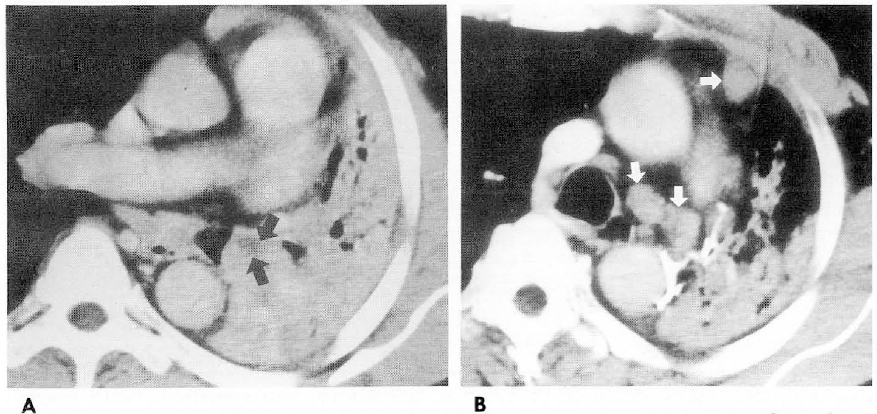
False-positive CT diagnosis for mediastinal nodes in a 47-year- old man with central squamaous cell carcinoma causing atelectasis of the left lung. A. CT scan obtained at the subcar- inal level shows central tumor (black arrow) obstructing the left main bronchus. Peripherally, atelectasis of the left lung is noted. B. CT scan obtained at the level of azygos arch shows enlarged lymph nodes (white arrows) in the ipsil- ateral mediastinum. Pathological diagnosis confirmed no metastasis in these mediastinal nodes.

Table 1.
Factors Influencing the Evaluation of Mediastinal Nodal Metastases with CT


 PDF
PDF ePub
ePub Citation
 Print
Print


 XML Download