Journal List > J Korean Med Sci > v.30(5) > 1022907

Lee, Kim, Jang, Lee, Kim, Bang, and Jun: Increasing Prevalence of Trichophyton rubrum Identified through an Analysis of 115,846 Cases over the Last 37 Years

Abstract

Trichophyton rubrum is the most common dermatophyte in the world with the highest prevalence in Korea. There are few reports about epidemiological and mycological characteristics of T. rubrum based on long-term, large-scale studies. The purpose of this study was to investigate the clinical and epidemiological characteristics of T. rubrum infections in Korea. We retrospectively investigated with patients' records about the epidemiological and mycological status of 115,846 cases with T. rubrum infection that was mycologically diagnosed at Catholic Skin Disease Clinic from 1979 to 2013. Direct microscopy in 15% KOH solution and culture was done in each case. The annual incidence of patients with T. rubrum infection had been increasing during the period; and of 131,122 patients with dermatophytosis, 115,846 patients (88.35%) had T. rubrum infection. Disease was most prevalent among patients in their twenties in the 1970s and 1980s; in their thirties in the 1990s; in their forties in the 2000s; and in their fifties in the 2010s. The sex ratio was 1.5:1. T. rubrum infection was most commonly seen in summer and was found predominantly in patients living in urban areas. Toe webs were most frequently involved, followed by toenails and groin. This epidemiologic findings provide useful information for prevention of T. rubrum infection and future dermatophytosis prospects.

Graphical Abstract

jkms-30-639-ab001

INTRODUCTION

Fungal infections are influenced by interactions between the host, the infectious agents, and the environment (1). During the past 80 yr, there have been dramatic changes in Korean dermatophytosis, including common causative agents of dermatophytosis and clinical characteristics (2,3,4,5,6,7,8). These have resulted from changes in socioeconomic status, lifestyle, and population, as well as the development of new antifungal drugs and diagnostic methods. Therefore, an epidemiologic study will provide useful information for prevention of infection and future dermatophytosis prospects.
Trichophyton rubrum, T. mentagrophytes, Microsporum canis, Epidermophyton floccosum, T. verrucosum, M. gypseum, M. ferrugineum, T. tonsurans, T. schoenleinii, and T. violaceum have been isolated in Korea (9,10,11,12,13,14,15,16,17,18). T. rubrum has been known to be a main causative agent in dermatophytosis in Korea since the 1960s (2). T. tonsurans was newly introduced to Korea in the 1980s (10). Conversely, T. schenleinii has disappeared from Korea (8). Similarly, T. mentagrophytes and M. canis have been decreasing in prevalence since the 2000s (19, 20).
We conducted this study to investigate the clinical and epidemiological characteristics of T. rubrum infection in Korea through the retrospective analysis of medical records of 115,846 patients infected with T. rubrum from 1979 to 2013.

MATERIALS AND METHODS

Patients

Using patient records from the Catholic Skin Disease Clinic from 1979 to 2013, we retrospectively investigated the epidemiological and mycological status of 115,846 cases of mycologically diagnosed T. rubrum infections.

Diagnosis of Trichophyton infection

Using their medical records, 115,846 patients with T. rubrum were retrospectively surveyed to determine annual prevalence of infection and the distribution of age, sex, season, involved sites, and place of residence. Samples for fungal examinations were obtained by scraping the lesions with a scalpel, and microscopic examination in 15% KOH solution was performed. Diagnosis of T. rubrum infection was confirmed with fungal culture using potato dextrose agar corn meal Tween 80 media. The cultures were kept at 24-26℃ and examined after 2 to 4 weeks. In addition, microscopic examination was performed with lactophenol cotton blue stain to identify filamentous fungi with conidia.

Ethics statements

This study was approved by the institutional review board of Kyungpook National University Hospital (IRB No. 2014-06-015). Informed consent was waived by the board.

RESULTS

Prevalence

The annual incidence of T. rubrum infection ranged from 1,436 to 5,565 patients between 1979 and 2013 (Fig. 1). The mean annual incidence of T. rubrum infections was 3,310 patients. The incidence was the lowest in 1986 and the highest in 2002. The prevalence of T. rubrum infection increased from 1979 to 2013. Out of 131,122 patients with dermatophytoses during the study period, 115,846 patients (88.35%) had a T. rubrum infection. The proportion of T. rubrum among dermatophytes was the lowest in 1979 (70.39%) and highest in 2013 (94.28%) (Fig. 2). T. rubrum has been the prevailing cause of dermatophytoses, constituting more than 90% of dermatophytoses since 1991.

Age distribution

There were 27,485 patients (23.73%) in their thirties, the largest overall age group. Of the remaining patients, 25,265 (21.81%) were in their forties; 22,711 patients (19.60%) were in their twenties; and 17,414 patients (15.03%) were in their fifties (Fig. 3). Out of the total 115,846 patients, 92,875 patients (80.17%) were 20-59 yr old. In the 1970s and 1980s, infection was most prevalent in patients in their twenties. In the 1990s, infection was most prevalent in patients in their thirties. In the 2000s, infection was most prevalent in patients in their forties, and in the 2010s, infection was most prevalent in patients in their fifties (Table 1). The age of patients with T. rubrum infection has been steadily increasing (Fig. 4).

Sex distribution

Out of 115,846 patients, 69,976 patients (60.41%) were male and 45,870 patients (39.60%) were female. The male:female ratio was 1.5:1. Females had a higher prevalence of infection than males in the 1970s. Over time, the prevalence in males has been increasing compared to prevalence in females (Fig. 5). In the group less than 70 yr old, more than half of T. rubrum infections occurred in males. However, in the group more than 70 yr old, the prevalence of T. rubrum in females was predominant (Fig. 6).

Seasonal distribution

Out of 115,687 patients, 49,681 patients (42.89%) visited our hospital in summer, more than in any other season (28,119 patients [24.27%] in spring; 20,525 patients [17.72%] in fall; and 17,521 patients [15.12%] in winter).

Topographical distribution

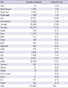
T. rubrum infection developed most commonly in the toe web (36,543 patients, 31.53%), followed by the toenail (25,268 patients, 21.80%), and the groin (20,249 patients, 17.47%) (Table 2). Out of 115,905 patients, 51,466 (44.40%) had tinea pedis; 26,573 patients (22.93%) had onychomycosis; 20,249 patients (17.47%) had tinea cruris; 10,681 patients (9.22%) had tinea corporis; 4,451 patients (3.84%) had tinea manus; 1,779 patients (1.53%) had tinea faciei; and 143 patients (0.1%) had tinea capitis. Tinea pedis was defined as dermatophytosis of the toe web, sole, and dorsal area of the foot. Onychomycosis was defined as dermatophytosis of the toenail and fingernail. Tinea corporis was defined as dermatophytosis of the neck, chest, back, abdomen, axilla, upper and lower extremities, and buttocks. Tinea manus was defined as dermatophytosis of the palm, finger web, and dorsal area of the hand. Additionally, 167 patients had a scrotal infection, 38 patients had a vulvogenital infection, 1 patient had an oral infection, 21 patients had an anal infection, and 151 patients had an ear infection. From the 1970s through the 1990s, tinea pedis was the most common dermatophytosis, followed by tinea cruris, onychomycosis, and tinea corporis. From the 2000s through the 2010s, onychomycosis was the second most common dermatophytosis, followed by tinea cruris and tinea corporis (Table 3). Tinea pedis and onychomycosis increased in prevalence with patient age. The prevalence of patients with tinea capitis was much higher in winter than any other T. rubrum infection subtypes.

Inhabitancy distribution

Out of 115,846 patients, 97,892 (84.50%) lived in an urban area. A 17,954 (15.50%) lived in a rural area.

DISCUSSION

The spectrum of dermatophytes isolated from skin lesions has changed over the last 80 yr (3). T. rubrum has been the most common dermatophyte since the 1960s, accounting for 80%-90% of all isolated species, followed by T. mentagrophytes (22). This study also showed that T. rubrum has been the most common dermatophyte in Korea since the data collection began. The number of patients with T. rubrum infection among dermatophytoses has been continuously increasing since the late 1980s. Out of all patients with dermatophytoses during this study, 88.35% had a T. rubrum infection. The prevalence of T. rubrum among dermatophytes was highest in 2013, accounting for 94.28% of cases. A temporary decrease in the prevalence of T. rubrum infection in the 1980s was caused by a surge of M. canis infection (19).
This evolution of prevailing T. rubrum infection is connected with the parallel increase in the prevalence of tinea pedis and onychomycosis. T. rubrum is one of the anthropophilic dermatophytes that has been isolated from tinea corporis, tinea cruris, tinea pedis, and onychomycosis worldwide (23). In this study, the proportion of tinea pedis (44.40%) and onychomycosis (22.93%) among dermatophytoses caused by T. rubrum was 67.33%. The prevalence of onychomycosis in the United States is also estimated to be at least 12% (24). Sigurgeirsson and Baran reported that T. rubrum was the most common fungal cause of onychomycosis and was cultured on average in 44.9% of cases (25). Tinea pedis is also caused by T. rubrum (1).
A major risk factor for developing a T. rubrum infection in the feet is modern occlusive footwear (26). In addition, incidence of tinea pedis is higher among those using communal baths, showers, or pools. A family history of tinea pedis and onychomycosis and advanced age are also risk factors (27). Because T. rubrum is an anthropophilic dermatophyte, it can be transmitted from infected to healthy persons. It is transmitted via direct contact with infected skin or hair retained in clothing, combs, caps, socks, and towels. The present study revealed that the prevalence of T. rubrum infection has historically been highest among patients in their thirties. However, the age with the highest prevalence has recently advanced to patients in their fifties. Host factors, such as immune responses and peripheral circulation, play a determining role in the acquisition of tinea pedis. Additional factors, such as urbanization and obesity, have also contributed to the increase in T. rubrum infections.
An analysis of the frequency and distribution of tinea pedis in different occupations and leisure-time activities as well as the routes of infection were reported. The spreading of T. rubrum infections in most developed countries represents a considerable economic problem, especially since it was accompanied by a parallel increase in the frequency of onychomycosis.
An epidemiological survey of dermatomycoses and their causative fungi in Japan was conducted (28). Dermatophytosis was the most prevalent cutaneous fungal infection. Among dermatophytoses, tinea pedis was the most frequent, followed by onychomycosis, tinea corporis, tinea cruris, tinea manuum, and tinea capitis. This distribution was similar to that seen in our study. Tinea pedis and onychomycosis incidence increased in the summer season, suggesting that hot and humid weather is one of the main causes of dermatophytoses.
As the causative dermatophyte species, T. rubrum was the most frequently isolated among all dermatophyte infections, as was seen in our study. Specific areas, such as the scrotum and mucosal area, were involved in infection. T. rubrum was isolated from the whole body, including the face and scalp (29, 30). In patients with tinea capitis, zoophilic dermatophytes, such as M. canis and T. verrucosum, were isolated most frequently (31, 32). Recently, Lee et al. (19) also reported that the prevalence of M. canis infection decreased markedly in the 2000s. The prevalence of T. mentagrophytes has decreased in Korea since 2005 (20). Choe et al. (33) reported that the prevalence of T. verrucosum infection decreased much more in the 2000s than in the late 1900s. In addition, the prevalence of T. schoenleinii, T. violaceum, M. gypseum, M. ferrugineum, and E. floccosum is also decreasing, and prevalence of T. tonsurans infection has shown a recent decrease (34). The observed decreases in dermatophytoses infections, except for T. rubrum, may have been caused by public health education, improved hospital accessibility, environmental improvement, and increased lifestyle diversity.
In conclusion, while dermatophytoses pathogen, including T. mentagrophytes and M. canis, are decreasing in prevalence, T. rubrum infections are increasingly prevalent in Korea. We can obtain useful information for T. rubrum infection prevention and future prospects from this epidemiologic study.

Figures and Tables

Fig. 1

Annual number of patients with T. rubrum infection.

jkms-30-639-g001
Fig. 2

Prevalence of T. rubrum infection in all dermatophytoses.

jkms-30-639-g002
Fig. 3

Age distribution of patients with T. rubrum infection.

jkms-30-639-g003
Fig. 4

Age distribution of patients with T. rubrum infection by decade.

jkms-30-639-g004
Fig. 5

Proportion of male to female patients with T. rubrum infection by decade.

jkms-30-639-g005
Fig. 6

Proportion between male and female by age.

jkms-30-639-g006
Table 1

Age distribution (%) of patients with T. rubrum infection by decade

jkms-30-639-i001
Age (yr) 1970s 1980s 1990s 2000s 2010s
< 10 3.02 2.40 1.84 0.94 0.73
10-19 11.59 10.20 6.19 3.67 3.39
20-29 45.44 40.74 19.56 13.08 9.07
30-39 20.82 22.15 31.03 21.93 15.79
40-49 12.61 13.83 19.63 26.84 23.26
50-59 4.22 7.03 12.50 17.37 24.71
60-69 1.63 2.72 6.78 11.28 13.75
70-79 0.48 0.86 2.21 4.27 7.76
80-89 0.12 0.07 0.24 0.57 1.33
≥ 90 0.06 0.00 0.01 0.05 0.19
Total (%) 100.00 100.00 100.00 100.00 100.00
Table 2

Involved site distribution of patients with T. rubrum

jkms-30-639-i002
Site Number of patients Proportion (%)
Palm 25 1.20
Hand dorsum 1,265 1.09
Finger nail 1,305 1.13
Finger web 1,798 1.55
Sole 12,701 10.96
Foot dorsum 2,222 1.92
Toe nail 25,268 21.80
Toe web 36,543 31.53
Scalp 143 0.12
Face 1,779 1.53
Neck 477 0.41
Chest 318 0.27
Back 430 0.37
Abdomen 603 0.52
Axilla 225 0.19
Arm 1,008 0.87
Leg 1,429 1.23
Hip 6,191 5.34
Groin 20,249 17.47
Scrotum 167 0.14
Vagina 25 0.02
Vulvae 13 0.01
Oral mucosa 1 0.00
Anus 21 0.02
Ear 151 0.13
Others 185 0.16
Total 115,905 100
Table 3

Involved site percentage (%) of T. rubrum patients by decade

jkms-30-639-i003
Disease 1970s 1980s 1990s 2000s 2010s
Tinea pedis 43.38 40.48 39.54 46.02 54.90
Onychomycosis 10.50 11.73 18.29 31.02 25.40
Tinea cruris 24.75 27.58 22.20 11.94 9.71
Tinea corporis 9.26 9.11 12.48 7.56 7.14
Tiena manus 8.90 7.64 4.70 2.06 1.74
Tinea faciei 2.20 2.22 2.18 0.99 0.78
Tinea capitis 0.53 0.31 0.11 0.05 0.08
Others 0.47 0.93 0.49 0.37 0.26
Total (%) 100.00 100.00 100.00 100.00 100.00

ACKNOWLEDGEMENT

The authors thank Ji Yeong Kim at Department of Dermatology, Kyungpook National University School of Medicine for data filing.

Notes

This study was supported by the Korean Dermatological Association (2014).

DISCLOSURE The authors have no conflict of interest to declare.

AUTHOR CONTRIBUTION Conception and coordination of the study: Lee WJ. Design of ethical issues: Jang YH, Lee SJ, Kim DW. Acquisition of data: Bang YJ, Jun JB. Data review: Lee WJ, Kim SL, Jun JB. Statistical analysis: Lee WJ, Kim SL. Manuscript preparation: Manuscript approval: all authors.

References

1. Havlickova B, Czaika VA, Friedrich M. Epidemiological trends in skin mycoses worldwide. Mycoses. 2008; 51:2–15.
2. Kim HS. The statistical and mycological survey on superficial dermatomycoses. Korean J Dermatol. 1971; 9:1–4.
3. Rhim KJ, Kim JH, Shin S. A clinical and mycological study of superficial dermatophytoses. Korean J Dermatol. 1978; 16:435–442.
4. Min BK, Chung BS, Choi KC, Kim HK. Clinical and mycological studies on dermatophytosis. Korean J Dermatol. 1984; 22:604–609.
5. Lee HK, Seo SJ, Kim MN, Hong CK, Ro BI. A clinical and mycological study of superficial fungal diseases(vii). Korean J Dermatol. 1993; 31:559–566.
6. Moon HJ, Lee JB, Kim SJ, Lee SC, Won YH. Clinical and mycological studies on dermatomycosis (1991-2000). Korean J Med Mycol. 2002; 7:78–85.
7. Lee DK, Moon KC, Koh JK. Clinical and mycological studies on superficial fungal infection. Korean J Med Mycol. 2006; 11:54–63.
8. Lee YW, Yun SJ, Lee JB, Kim SJ, Lee SC, Won YH. Clinical and mycological studies on dermatomycosis (2001-2010). Korean J Med Mycol. 2013; 18:30–38.
9. Kim BS, Suh SB. Mycological and clinical observation on dermatophytosis. Korean J Dermatol. 1976; 14:325–334.
10. Suh SB, Kim SW, Oh SH, Choi SK, Bang YJ. A case of block dot ringworm caused by Trichophyton tonsurans. Korean J Dermatol. 1998; 36:918–923.
11. Sung SY, Kim HY, Kim HU, Ihm CW. Trichophyton tonsurans infection in Wrestlers and a child. Korean J Dermatol. 1998; 36:732–736.
12. Kim JC, Choi JS, Kim KH, Suh SB. Mycological features of Trichophyton verrucosum isolated in Taegu area. Korean J Dermatol. 1992; 30:761–768.
13. Kim YP, Chun IK, Kim SH. A case of kerion celsi caused by Trichophyton verrucosum and its epidemiologic study. Korean J Dermatol. 1986; 24:687–691.
14. Jun JB, Suh SB. Clinical and mycological studies on microsporum gypseum infection. Korean J Dermatol. 1980; 18:369–381.
15. Lee DS, Cho GY, Kim YH, Houh W. A case of tinea capitis due to microsporum gypseum. Korean J Dermatol. 1984; 22:643–646.
16. Lee H, Lee ES, Kang WH, Lee SN. An unusual clinical manifestation of tinea corporis caused by microsporum ferrugineum. Korean J Dermatol. 1987; 25:383–388.
17. Kim HU, Choi CJ, Yun SK. Three cases of tinea capitis caused by microsporum ferrugineum. Korean J Dermatol. 1993; 31:760–764.
18. Kim YA, Lee KH, Lee JB, Suh SB. A case of fungal granuloma caused by Trichophyton violaceum. Korean J Dermatol. 1989; 27:304–307.
19. Lee WJ, Song CH, Lee SJ, Kim do W, Jun JB, Bang YJ. Decreasing prevalence of microsporum canis infection in Korea: through analysis of 944 cases (1993-2009) and review of our previous data (1975-1992). Mycopathologia. 2012; 173:235–239.
20. Lee WJ, Park KH, Kim MS, Lee SJ, Kim DW, Bang YJ, Jun JB. Decreasing incidence of Trichophyton mentagrophytes in Korea: analysis of 6,250 cases during the last 21-year-period (1992-2012). J Korean Med Sci. 2014; 29:272–276.
21. Seebacher C. The change of dermatophyte spectrum in dermatomycoses. Mycoses. 2003; 46:42–46.
22. Seebacher C, Bouchara JP, Mignon B. Updates on the epidemiology of dermatophyte infections. Mycopathologia. 2008; 166:335–352.
23. Aly R. Ecology and epidemiology of dermatophyte infections. J Am Acad Dermatol. 1994; 31:S21–S25.
24. Scher RK, Rich P, Pariser D, Elewski B. The epidemiology, etiology, and pathophysiology of onychomycosis. Semin Cutan Med Surg. 2013; 32:S2–S4.
25. Sigurgeirsson B, Baran R. The prevalence of onychomycosis in the global population - a literature study. J Eur Acad Dermatol Venereol. 2014; 28:1480–1491.
26. Katoh T. Dermatomycosis and environment. Nihon Ishinkin Gakkai Zasshi. 2006; 47:63–67.
27. Ilkit M, Durdu M. Tinea pedis: the etiology and global epidemiology of a common fungal infection. Crit Rev Microbiol. 2014; doi: 10.3109/1040841X.2013.856853.
28. Sei Y. 2006 Epidemiological survey of dermatomycoses in Japan. Med Mycol J. 2012; 53:185–192.
29. Tchernev G, Cardoso JC, Ali MM, Patterson JW. Primary onychomycosis with granulomatous tinea faciei. Braz J Infect Dis. 2010; 14:546–547.
30. Nenoff P, Mügge C, Herrmann J, Keller U. Tinea faciei incognito due to Trichophyton rubrum as a result of autoinoculation from onychomycosis. Mycoses. 2007; 50:20–25.
31. Mirmirani P, Tucker LY. Epidemiologic trends in pediatric tinea capitis: a population-based study from Kaiser Permanente Northern California. J Am Acad Dermatol. 2013; 69:916–921.
32. Monod M, Fratti M, Mignon B, Baudraz-Rosselet F. Dermatophytes transmitted by pets and cattle. Rev Med Suisse. 2014; 10:749–753.
33. Choe YS, Park BC, Lee WJ, Jun JB, Suh SB, Bang YJ. The clinical observation of Trichophyton verrucosum infections during the last 19 years (1986-2004). Korean J Med Mycol. 2006; 11:45–53.
34. Oh SJ, Lee SY, Lee JS. A clinical and mycological study of dermatophytoses in Chungcheongnam-do Province (2008~ 2012). Korean J Med Mycol. 2013; 18:39–47.
TOOLS
ORCID iDs

Weon Ju Lee
https://orcid.org/http://orcid.org/0000-0001-5708-1305

Sang Lim Kim
https://orcid.org/http://orcid.org/0000-0003-4144-2772

Yong Hyun Jang
https://orcid.org/http://orcid.org/0000-0003-1706-007X

Seok-Jong Lee
https://orcid.org/http://orcid.org/0000-0002-6131-632X

Do Won Kim
https://orcid.org/http://orcid.org/0000-0001-6632-1374

Yong Jun Bang
https://orcid.org/http://orcid.org/0000-0001-7242-9306

Jae Bok Jun
https://orcid.org/http://orcid.org/0000-0003-4834-4526

Similar articles