Journal List > Ann Lab Med > v.39(4) > 1117051

Gasparyan, Ayvazyan, Mukanova, Yessirkepov, and Kitas: The Platelet-to-Lymphocyte Ratio as an Inflammatory Marker in Rheumatic Diseases

Abstract

The platelet-to-lymphocyte ratio (PLR) has emerged as an informative marker revealing shifts in platelet and lymphocyte counts due to acute inflammatory and prothrombotic states. PLR has been extensively examined in neoplastic diseases accompanied by immune suppression and thrombosis, which can be predicted by combined blood cell counts and their ratios. Several large observational studies have demonstrated the value of shifts in PLR in evaluating the severity of systemic inflammation and predicting infections and other comorbidities, in inflammatory rheumatic diseases. The value of PLR as an inflammatory marker increases when its fluctuations are interpreted along with other complementary hematologic indices, particularly the neutrophil-to-lymphocyte ratio (NLR), which provides additional information about the disease activity, presence of neutrophilic inflammation, infectious complications, and severe organ damage in systemic lupus erythematosus. PLR and NLR have high predictive value in rheumatic diseases with predominantly neutrophilic inflammation (e.g., Behçet disease and familial Mediterranean fever). High PLR, along with elevated platelet count, is potentially useful in diagnosing some systemic vasculitides, particularly giant-cell arteritis. A few longitudinal studies on rheumatic diseases have demonstrated a decrease in PLR in response to anti-inflammatory therapies. The main limitations of PLR studies are preanalytical faults, inadequate standardization of laboratory measurements, and inappropriate subject selection. Nonetheless, accumulating evidence suggests that PLR can provide valuable information to clinicians who encounter multisystem manifestations of rheumatic diseases, which are reflected in shifts in platelet, lymphocyte, neutrophil, or monocyte counts. Interpretation of PLR combined with complementary hematologic indices is advisable to more accurately diagnose inflammatory rheumatic diseases and predict related comorbidities.

INTRODUCTION

Blood cell interactions are essential in the pathophysiology of inflammation, immune responses, hemostasis, and oncogenesis. These interactions are multifaceted, and it is often difficult to distinguish primary triggering signals and the specific roles of each cell type in the development and progression of disease states.
Platelets are rich in proinflammatory agents and are capable of releasing highly active microparticles, and they are intimately involved in the development and perpetuation of various inflammatory rheumatic diseases, which often manifest with arthritis and may be complicated by cardiovascular, metabolic, infectious, and lymphoproliferative, among many other, comorbidities [123]. Fluctuations in platelet counts in rheumatic diseases reflect nonspecific inflammatory thrombopoiesis, with abundant release of reactive cells from the bone marrow to the bloodstream, their migration to and excessive consumption at inflammatory sites, and their destruction via binding to anti-platelet antibodies. Platelet counts correlate with platelet volume and reactivity and may indicate autoimmune disease activity, response to anti-inflammatory therapies, and presence of various comorbidities [456].
Observational studies have demonstrated that in rheumatoid arthritis (RA), platelet counts gradually increase with radiological disease progression, whereas leukocyte and erythrocyte count variations are unremarkable [78]. RA patients with increased platelet counts (>400×109/L) appear to show more substantial improvements in disease activity score in 28 joints (DAS28) and acute-phase reactants in response to biological agents, such as tocilizumab, than do RA patients with “normal” platelet counts (<400×109/L) [9].
In systemic lupus erythematosus (SLE), thrombocytopenia is more prevalent (15–27%) than leuco- (14%) or erythrocytopenia (5%) [510]. Platelet counts negatively correlate with lupus activity, whereas thrombocytopenia, which is often mild (70−150×109/L), is associated with arthritis, neurological disturbances, and lupus nephritis. Moderate and severe thrombocytopenia in SLE may manifest with major hemorrhage (25% and 76%, respectively), resulting in death due to blood loss or infection [11]. Although there is no direct evidence of an association of autoimmune markers with hematologic parameters, the anticipated low platelet count in SLE is likely due to disease-specific disturbances, resulting in excessive production of anti-platelet antibodies and intensified platelet destruction in the spleen (hypersplenism) [10].
A high platelet count is of diagnostic value in elderly patients with temporal artery inflammation suggestive of giant-cell arteritis (GCA) [12]. Accordingly, a recent study suggested that a high platelet count predicts diagnosis of GCA even in the absence of characteristic temporal artery involvement [13].
Interestingly, in children with systemic vasculitides, particularly Kawasaki disease (KD), platelet counts may predict disease outcomes at various stages. An increase in platelet count accompanied by an increase in lymphocyte count, a decrease in neutrophil count, and amelioration of immunoglobulin G, M, and A levels suggests a good response to intravenous immunoglobulin and aspirin therapy at the early, in-hospital stages of convalescence in KD [14]. However, persistently high platelet counts may indicate a complicated course and the development of coronary aneurysms in KD [15]. A high platelet count is also associated with renal involvement in Henoch-Schönlein purpura, another common systemic vasculitis in childhood [16].
In-vitro and in-vivo studies have elucidated the origin of activated circulating platelets, which display numerous membrane receptors and release multiple biologically active substances from their granules that can regulate cellular interactions and contribute to immune, inflammatory, and thrombotic diseases [171819]. Circulating platelets can interact with erythrocytes, neutrophils, and lymphocytes in the vessel lumen at sites of vascular damage [2021]. The interaction of platelets with T-lymphocytes, mediated by P-selectin, reduces lymphocyte proliferation, resulting in a decrease in proinflammatory cytokines, such as interferon-alpha (IFN-α), tumor necrosis factor-alpha (TNF-α), and interleukin (IL)-17, and an increase in anti-inflammatory cytokines, such as IL-10 [22]. Drug-induced modulation of such intercellular interactions and subsequent cytokinergic reactions, which depend on platelet and lymphocyte counts, dampens immune inflammation in the synovial space of patients with RA, and can be monitored by employing combined parameters of hemograms, such as the platelet-to-lymphocyte ratio (PLR).
Platelets of healthy subjects modulate the activities of neutrophils and monocytes by responding to stimulation of their platelet-bound Toll-like receptors (TLR) in a dose-dependent manner. Low doses of TLR agonists, such as lipopolysaccharide and fibroblast-stimulating lipopeptide-1, reduce CD66b expression on neutrophils and related granulocyte elastase secretion in platelet-neutrophil coculture [23]. Platelets activated by fibroblast-stimulating lipopeptide-1 in coculture increase IL-6 and IL-10 and reduce TNF-α production.
Activated platelets release microparticles that can interact with neutrophils via the expression of platelet-type lipoxygenase and activation of the eicosanoid pathway [24]. Synovial neutrophils of patients with RA internalize platelet microparticles and thereby intensify synovial inflammation. Currently available anti-cytokinergic drugs, such as anti-TNFα, restrict the ability of platelets to bind to and activate leukocytes in RA [25], which may decrease the risk of thrombotic events [26].
Several in-vitro studies have revealed that platelets regulate chemotactic and cytotoxic activities of neutrophils via CD62P (P-selectin) expression on their surface and subsequent platelet-neutrophil conjugate formation [2728]. Notably, P-selectin expression cascades intercellular interactions in response to anti-citrullinated protein antibodies (ACPA) and may induce inflammatory polyarthritis [29]. Recombinant human vimentin has been shown to bind to P-selectin with subsequent inhibition of neutrophils and disruption of their interactions with endothelial cells and platelets under shear stress [30]. However, preliminary evidence suggests that the P-selectin pathway of platelet-neutrophil interactions does not play a role in SLE and lupus-related organ damage; instead, vascular cell adhesion molecule 1 (VCAM-1) may mediate the adhesion of neutrophils to lymphocytes with subsequent exacerbation of lupus nephritis [31].
Based on the available evidence of the roles of platelet interactions with other blood cells, we attempted to explore the value of PLR as a potential marker of inflammation in rheumatic diseases. Given the importance of neoplastic, prothrombotic, and metabolic states in inflammatory rheumatic diseases, we also analyzed PLR in these potentially comorbid states.

ANALYSIS OF PUBLICATION ACTIVITY

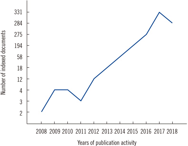
The global interest in the value of PLR in laboratory diagnostics of a wide variety of diseases is reflected in a snapshot Scopus-based bibliographic analysis of relevant publication activity. As of October 30, 2018, 1,186 articles were tagged with the term “platelet lymphocyte ratio” in the title, abstract, or keywords, with the publication date ranging from 1972 to 2018. The analysis revealed some of the closely related keywords, such as “neutrophil lymphocyte ratio” (tagged in 778 articles [66%]), “cancer prognosis” (426, 36%), “overall survival” (403, 34%), “inflammation” (321, 27%), and “cancer staging” (306, 26%). A relatively small number of articles were related to inflammatory rheumatic diseases. Thirteen articles were tagged with the terms “platelet lymphocyte ratio” and “rheumatoid arthritis,” six with “systemic lupus erythematosus,” two with “systemic sclerosis,” two with “spondyloarthritis,” two with “psoriatic arthritis,” 10 with “Behçet disease,” three with “familial Mediterranean fever,” four with “vasculitis,” one with “giant cell arteritis,” and two with “Takayasu arteritis”. The exponential annual increase in the number of articles tagged with “platelet lymphocyte ratio” started in 2008 (two articles) and peaked in 2017 (331 articles) (Fig. 1).
The top five journals covering PLR were Oncotarget (50 articles), Medicine (United States; 35), Angiology (29), Annals of Surgical Oncology (21), and Oncotargets and Therapy (20). Of the indexed rheumatology journals, only Archives of Rheumatology (5), Clinical Rheumatology (4), Egyptian Rheumatologist (2), International Journal of Rheumatic Diseases (2), and Modern Rheumatology (2) published tagged articles. Turkey was the leading country in this field, with 382 articles, closely followed by China (373), the United States (76), Japan (69), and Korea (65). A substantial number of the articles were systematic reviews with meta-analyses (59), predominantly cross-linked to the terms “overall survival” (48), “cancer prognosis” (43), “cancer survival” (25), “cancer staging” (17), and “colorectal cancer” (13).
Ten articles in the field were cited 141–450 times, with overviews of the values of PLR, neutrophil-to-lymphocyte ratio (NLR), and other inflammatory markers for predicting survival in colorectal and other types of cancer topping the list [32]. Finally, 45 articles were tagged with the keyword “platelet lymphocyte ratio” and cited at least 45 times (h-index=45).

PLR IN NEOPLASTIC, PROTHROMBOTIC, AND METABOLIC DISEASES

Over the past decade, PLR has emerged as a universal laboratory marker for predicting various neoplastic, prothrombotic, and metabolic diseases [3334353637]. PLR fluctuations can be interpreted in the context of the underlying multifaceted immune-inflammatory reactions. Shifts in this parameter correlate positively with other markers of systemic inflammation, particularly with NLR. PLR better predicts clinical outcomes in patients with systemic inflammation than either platelet or lymphocyte count. Basically, the magnitude of stress-induced hypercortisolemia with subsequent release of platelets into the bloodstream and transient lymphopenia influence the degree of elevation of PLR across numerous proinflammatory and prothrombotic disease states [38]. Such a nonspecific mechanism of PLR elevation can be counteracted by intensified platelet destruction or consumption at the sites of immune inflammation and thrombosis, necessitating cross-checks of all blood cell counts and other inflammatory and immune markers [39].
In colorectal cancer, PLR often increases along with NLR and platelet volume, and decreases after tumor resection [40]. High PLR is associated with poor cancer-specific survival, as determined by ROC curve analysis [41]. The significance of PLR as an inflammatory marker of cancer has been extensively examined; one exemplary study on patients with colorectal cancer (N=200) suggested both PLR and NLR as predictors of the outcome of surgical intervention and PLR as an independent predictor of overall survival (hazard ratio [HR], 1.971, 95% confidence interval [CI], 1.102–3.335) [42].
Importantly, based on the largest analysis of medical records of patients with colorectal cancer (N=1,868), high PLR and NLR have prognostic value at only advanced stages [43]. Nonetheless, the predictive value of high PLR in terms of overall survival is greater in cancer patients with comorbidities, particularly those with metabolic syndrome, which doubles the risk of cancer-related mortality [44].
In a cohort of 1,646 patients with stable coronary artery disease who underwent coronary angiography, PLR correlated with the Gensini score (r=0.37, P<0.001), which reflects the severity of coronary atherosclerosis, and C-reactive protein (CRP) (r=0.31, P<0.001) [45]. It was also independently associated with severe coronary atherosclerosis in multivariate logistic regression (odds ratio [OR], 1.043, 95% CI, 1.036–1.049) [45]. In another study involving 1,146 patients from cardiology outpatient clinics (539 patients with and 607 without metabolic syndrome), platelet count, CRP, and PLR were all significantly elevated in the metabolic syndrome group [46]. The highest PLR values were noted for patients with several components of metabolic syndrome, and PLR, but not platelet or lymphocyte count, independently predicted the presence of metabolic syndrome in multivariate logistic regression (OR, 1.121, 95% CI, 1.113–1.135) [46].
A recent analysis of eight pooled cohorts with a total of 6,627 patients with acute coronary syndrome demonstrated that high PLR (>150) doubles the risk of in-hospital all-cause and cardiovascular mortality (pooled relative risk, 2.15, 95% CI, 1.73–2.67 and 1.95, 95% CI, 1.30–2.91, respectively) [47].
Some evidence suggests that high PLR is associated with venous thrombosis. In particular, large cohort studies have revealed links between high PLR and cerebral vein thromboses [4849]. The association was strong in cohorts of cancer patients in whom high PLR predicted venous thromboembolism (OR, 2.757, 95% CI, 1.655–3.862) [5051].
Finally, in a cohort of postmenopausal women (N=211), PLR, but not platelet count or NLR, was significantly elevated in women with low bone mineral density (BMD) than in women with normal BMD (P<0.05); PLR was also negatively correlated with femoral BMD (r=−0.21, P<0.002) [52]. A potential mechanism of the association between PLR and bone loss may be related to persistent systemic inflammation with impaired calcium transport and vitamin D metabolism, leading to osteoporosis and bone fractures [52]. Clinical implications of such a mechanism in postmenopausal women with and without chronic inflammatory diseases remain to be elucidated in specifically designed prospective studies.

PLR IN INFLAMMATORY RHEUMATIC DISEASES

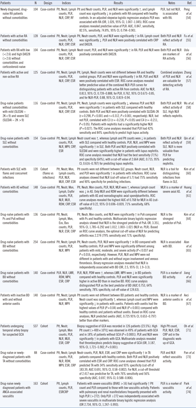
We comprehensively analyzed research reports on PLR across inflammatory rheumatic diseases, employing Scopus database searches and following recommendations on comprehensive and systematic searches of evidence-based data [53]. Large adult-cohort studies on newly-diagnosed and drug-naïve rheumatic patients reported in English-language journals were processed without time limits. The synthesized qualitative data are summarized in Table 1.
Shifts in PLR were examined in four large retrospective cohorts of patients with RA [54555657]. In all four studies, laboratory analyses were carried out in single centers. Patients with cardiovascular, endocrine, hematologic, neoplastic, gastrointestinal, autoimmune comorbidities, as well as those on corticosterioids were reportedly excluded to concentrate on potentially specific associations between PLR and rheumatoid activity. Only one study pointed to an exclusive association of PLR with RA [54], whereas the remaining studies [555657] considered PLR in combination with NLR as potentially valuable in the accurate evaluation of inflammatory activity. Combined analysis of shifts in PLR and NLR suggested an increase in platelet and neutrophil counts and a decrease in lymphocyte count during the active stage of RA. Shifts in separate blood cell counts in RA were moderate and within normal ranges in all these studies. PLR and NLR values in patients with RA differed from those in non-RA controls and correlated positively with laboratory (erythrocyte sedimentation rate [ESR], CRP) and clinical (DAS28) markers of rheumatoid activity. ROC curve analyses suggested that combined PLR-NLR curves more accurately distinguish patients with active RA from healthy controls. The same trend was observed when non-active RA patients were compared with healthy controls [57].
In adult SLE cohorts, blood cell count ratios appeared more informative than blood cell counts per se because of a tendency toward pancytopenia and thrombocytopenia [585960]. In a cohort consisting of 116 patients, SLE disease activity index (SLEDAI) positively and significantly correlated with NLR (r=0.312) and PLR (r=0.298); further, NLR, but not PLR, significantly correlated with C3 (r=−0.218) and C4 complement fractions (r=−0.211) [58]. In the same study, patients with nephritis had higher NLR than those without nephritis (P=0.027). In another study involving 154 patients, both PLR and NLR levels were significantly higher in patients with nephritis than in those without nephritis (P=0.03 and P<0.01, respectively) [59]. However, ROC curve analysis revealed that high NLR was predictive of lupus nephritis (cut-off value of 2.664, 71% sensitivity, 64% specificity, area under the curve [AUC], 0.715, 95% CI, 0.616–0.787) [59]. Finally, in a cohort of 120 lupus patients with and without concurrent infections, NLR, with a cut-off value of 5.7 and an AUC of 0.872, had high sensitivity (75%) and specificity (90%) for predicting infections, whereas individual blood cell counts and PLR were less informative [60]. Overall, high levels of both PLR and NLR were found to reflect lupus activity, whereas only high NLR was predictive of lupus nephritis and concurrent infections [585960].
A recent study extensively examined blood cell counts and ratios in 148 patients with ankylosing spondylitis (AS) [61]. Although various cell ratios significantly differed between patients and healthy controls and positively correlated with inflammatory markers (P<0.05), only the monocyte-to-lymphocyte ratio (MLR) differed significantly (P<0.05) between patients with and without radiographic sacroiliitis, a feature of severe disease. In addition, ROC curve analyses indicated that MLR yielded a higher AUC (0.768) than PLR and NLR. Thus, MLR, but not PLR or NLR, was suggested as a marker of severe axial spondyloarthritis. Indirect evidence from two other cohort studies in patients with AS with sensorineural hearing loss and cardiovascular diseases corroborated that neither PLR nor NLR reflects inflammatory activity and severity in this disease [6263].
An analysis of hemograms of patients with psoriasis (N=111) and psoriatic arthritis (N=25) also demonstrated increases in neutrophil, monocyte, and platelet counts in psoriatic disease, and NLR, not PLR, was the strongest predictor of the presence of spondyloarthritis (OR, 3.351, 95% CI, 1.785–6.292) [64].
A series of specifically designed adult-cohort studies have examined the values of blood cell ratios in systemic vasculitides, focusing on their associations with clinical manifestations. Three studies have examined shifts in Behçet disease (BD) [656667], which is an autoinflammatory disease with neutrophilic inflammation, predominantly venous thromboses, and other manifestations resembling those of spondyloarthritides and systemic vasculitides [6869]. PLR was not associated with joint, eye, central nervous system, large vessel, or gastrointestinal involvement in BD [65]. Neither PLR nor NLR was associated with venous thromboses. However, based on ROC curve analyses, NLR was a more informative predictor of anterior uveitis than PLR (AUC, 0.725, 95% CI 0.653–0.797, P<0.001, and AUC, 0.6, 95% CI, 0.523–0.676, P=0.012, respectively) [67]. These results are partly in line with the results of another study that high-lighted the value of NLR as a marker of BD-related inflammatory activity, but not thrombosis [70].
Interestingly, a high platelet count and PLR may aid in confirming diagnosis in patients with large-vessel vasculitides, particularly in those with GCA. Analysis of a large number of hemograms and temporal artery biopsies of 537 patients with suspected GCA between 1992 and 2015 revealed that positive biopsies were twice more likely in patients with thrombocytosis, and that a high platelet count and PLR were more valuable for predicting positive results on a temporal artery biopsy than were NLR and other inflammatory markers [71]. Reportedly, 49% of patients with GCA, but only 18% of patients without GCA, had an elevated platelet count (OR, 4.36, 95% CI, 2.61–7.30) [71]. Likewise, an elevated PLR was found in 44% of patients with GCA but only in 19.4% of those without GCA (OR 3.34, 95% CI, 2.00–5.58) [71]. These results are in line with a population-based study that pointed out thrombocytosis as an informative clue in the differential diagnosis of GCA [72].
In patients with Takayasu arteritis (TA), both PLR and NLR are predictive of vasculitis, albeit with low sensitivity and specificity [73]. One study that examined solely PLR in anti-neutrophil cytoplasmic antibody (ANCA)-associated vasculitis revealed that pulmonary and renal manifestations were associated with high PLR values (above the cut-off of 272) and that values above the cut-off independently predicted severe vasculitis at diagnosis (OR 2.7) [74].
Additional information on diagnostic and predictive values of blood cell ratios can be derived from reports from pediatric vasculitis cohorts. In patients with KD, small- and medium-size arteritis with platelet count fluctuations is a hallmark of disease activity and coronary artery dilation [7576]. The combination of PLR and NLR was predictive of a severe course, intravenous immunoglobulin resistance, and coronary artery aneurysm development in KD [77787980]. A retrospective analysis of hemograms of 217 patients with KD from the period 2004–2014 pointed out an association of both low (≤300×109/L) and high (≥550×109/L) platelet counts with a severe course, resulting in treatment resistance and coronary artery aneurysms, respectively [80].
Finally, two studies analyzed PLR, NLR, and other inflammatory markers in cohorts of pediatric patients with familial Mediterranean fever (FMF) [8182], an autoinflammatory disease with characteristic periodic attacks of polyserositis due to neutrophilic inflammation and diverse rheumatic manifestations and vascular comorbidities [83]. Despite some discrepancies, both studies examined hemograms of children with FMF regularly treated with colchicine (0.5–2 mg/daily) and did not recommend PLR as a predictor of inflammation at attacks and during attack-free periods [8182]. NLR performed better as an inflammatory marker of attacks in these studies.

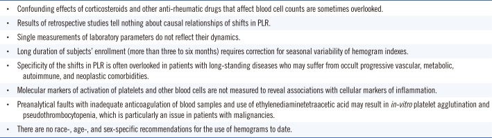
LIMITATIONS OF STUDIES ON PLR

The reliability of the specificity of shifts in blood cell ratios, and particularly, PLR in laboratory studies is highly dependent on several confounding factors (Box 1). These factors are sometimes overlooked and not reported. The most important one is perhaps the enrollment of patients with high-grade inflammatory diseases with history of drug therapies affecting the maturation of blood cells in the bone marrow and their release into the bloodstream. In this regard, information on the use of corticosteroids and manifestations of hypercortisolemia, associated with decreased lymphocyte and increased neutrophil counts, should be derived from the patients' medical records to correct for these acute effects [84]. There can be also diurnal fluctuations in blood cell counts. Patients with immune-mediated inflammatory diseases chronically treated with oral methylprednisolone experience transient acute lymphopenia within eight hours of corticosteroid administration, followed by morning lymphocytosis [85].
Lymphocyte and platelet counts can vary in response to short- and long-term therapies with methotrexate, a frontline drug for the treatment of RA, other rheumatic diseases, and malignancies. A single high dose of methotrexate (50 mg/m2 intramuscular injection) rarely causes thrombocytopenia [86]. However, chronic exposure to regular therapeutic doses of the drug results in fluctuations in lymphocyte counts that may trigger lymphoproliferative diseases in some cases [87]. Recovery of the absolute lymphocyte count in methotrexate-induced immunodeficiency with lymphoproliferative diseases takes from two to 76 weeks after cessation of the therapy [8889].
The cross-sectional, single-center design of and single measurements of laboratory parameters in the absolute majority of the studies on PLR limit their clinical significance. One can ask in what way and to what extent the observed associations are helpful in routine practice. Are there validated cut-offs one could rely on to suspect a severe course or a complication of an inflammatory disease? Cut-offs derived from a single study are not generalizable.
Ideally, PLR values should be reported along with values for NLR and other inflammatory markers, which may facilitate drawing the whole picture of inflammatory diseases and their infectious, thrombotic, or neoplastic complications. Associations between PLR and the course of an inflammatory disease can be more accurately explored in prospective studies with repeated measurements of laboratory parameters and information on morbidity and mortality in follow-up reports. There currently are only two small trial reports on PLR in inflammatory rheumatic diseases treated with anti-inflammatory drugs. A recent study on 38 patients with RA reported a significant decrease in PLR along with a drop in DAS28 and markers of systemic inflammation in response to six months of therapy with rituximab [90]. Likewise, 12-month-apart repeated measurements of NLR, PLR, and CRP in patients with psoriatic arthritis (N=50) revealed decreases in all these inflammatory markers in the course of therapies with infliximab, adalimumab, and ustekinumab [91]. However, repeated measurements of platelet and other blood counts in a period longer than three to six months require controlling for seasonal variations and other confounding factors, such as progressive cardiovascular, metabolic, autoimmune, hematologic, and neoplastic comorbidities. Both of the above studies [9091] had limitations related to the lack of controls and corrections for confounders.
Although a single-center design limits the generalizability of results, multicenter studies may have their own drawbacks, including variable standardization of blood processing techniques among laboratories [92].
To some extent, the issue of nonspecific shifts in hemograms relates to the so-called pseudothrombocytopenia and is primarily due to the anticoagulation agent EDTA and in-vitro-mediated platelet agglutination [93]. Although the incidence of pseudothrombocytopenia is generally low (0.07–2%), and sometimes relates to technical faults, its likelihood increases in patients with cancer [94].
Finally, numerous observational studies have analyzed ROC curves and reported predictive cut-off thresholds of PLR and other blood cell ratios. The use of any of such cut-off values is limited, given the linear origin of inflammatory markers and the necessity to correct for race-, age-, and sex-specific confounders [959697].

CONCLUDING REMARKS

Accumulating evidence points to potentially great diagnostic and prognostic values of complementary components of complete blood count, and particularly, blood cell count ratios. Numerous observational studies have suggested that PLR is an inflammatory marker of immune-mediated, metabolic, prothrombotic, and neoplastic diseases. Bibliographic searches suggested that the absolute majority of articles in this field are related to the value of PLR in cancer. The value of PLR as a predictor of severe course and development of concurrent infections and aneurismal coronary artery disease has been also highlighted in large cohort studies on various inflammatory rheumatic diseases [6077787980], although the number of related articles is relatively small. To date, the major rheumatology journals have rarely covered PLR and other hematologic indices, which are readily available clinical data.
Although available studies on PLR are heterogenous in terms of background disease, race, gender, patient age and enrollment, and analytical methods, we can draw some general conclusions. The majority of these studies, analyzed in this article, focus on the association of PLR with other laboratory markers of inflammation. The main finding is that combined evaluation of shifts in PLR and NLR is advisable for predicting the severity of inflammation, infectious complications, and other comorbidities. The recording of other blood cell ratios is also desirable as immune inflammation is multifaceted and may lead to predominant involvement of certain blood cells (e.g., monocytes in spondyloarthritis, and neutrophils in BD and FMF). Indirect evidence from these studies suggests that monitoring of PLR and other blood cell ratios may aid in the diagnosis of comorbidities of rheumatic diseases developing in the course of long-term anti-inflammatory and immunosuppressive therapies. In particular, hemograms may point to overt lymphoproliferative and other neoplastic comorbidities. However, the value of PLR in the early detection of these comorbidities remains uncertain [43], and properly designed, large longitudinal studies with serial laboratory measurements are warranted. Such longitudinal studies may also shed light on the potential clinical significance of reported cut-off thresholds of PLR, NLR, and other complementary markers.
Additional information can be obtained from future analyses of PLR in combination with the closely related mean platelet volume (MPV) and red cell distribution width (RDW). These parameters can be easily calculated from hemograms based on related indices. Some of the observational studies analyzed in this article have indirectly reflected on shifts in MPV and RDW in inflammation and thrombosis, without drawing conclusions on their predictive values [596165666773]. The main issue is that MPV and platelet distribution width (PDW) are more variable than platelet and other blood cell counts and related ratios [9899], limiting their attractiveness for prospective studies. Nonetheless, the interpretation of combined hematologic indices may uncover patients at increased risk of vascular comorbidities and thrombotic events in rheumatic diseases, particularly in light of the closely related pathobiological shifts in platelets and erythrocytes in high-grade inflammatory diseases [20]. A few preliminary studies have already examined the abovementioned parameters in combination. In a recent study, an increased MPV-to-lymphocyte ratio reliably distinguished patients with diabetic nephropathy from control subjects [100]. An observational study of pregnant subjects with and without preeclampsia indicated that elevation of PLR, NLR, MPV, and RDW predicted arterial hypertension and high-risk pregnancy [101]. Finally, a pilot study in RA patients (N=57), which was not included in our analysis, demonstrated elevated NLR, MPV, and PDW, but not PLR, in patients with active rheumatoid disease; MPV and PDW negatively correlated with ESR [102]. These results are partly in line with previous studies on RA and SLE that reported an association between high disease activity and low MPV [103104]. However, a major limitation of the above pilot study was that the patients were on corticosteroids, methotrexate, and other anti-inflammatory drugs [102], making it difficult to draw a conclusion.
In summary, PLR is an emerging inflammatory marker that, in combination with NLR and other hematologic indices, can help in the diagnosis and assessment of the activity and severity of several rheumatic diseases, in early detection of various comorbidities at a subclinical stage, and in monitoring the response to anti-inflammatory therapies. More research is warranted to elucidate the role of serial measurements of PLR in the diagnosis and monitoring of proinflammatory and prothrombotic disease states. The design of future studies should take into account confounding effects of numerous clinical, drug-related, and preanalytical factors that affect the variability of PLR and other indices. Prospective enrollment of subjects in these studies and standardized measurements of all laboratory parameters at a single time point are advisable. Race, age, and sex of the subjects, among many other confounders, should be considered when interpreting results.

Notes

Authors' Disclosures of Potential Conflicts of Interest: The authors have no conflicts of interest to declare.

References

1. Gasparyan AY, Stavropoulos-Kalinoglou A, Mikhailidis DP, Douglas KM, Kitas GD. Platelet function in rheumatoid arthritis: arthritic and cardiovascular implications. Rheumatol Int. 2011; 31:153–164. PMID: 20390282.
2. Olumuyiwa-Akeredolu OO, Pretorius E. Rheumatoid arthritis: notable biomarkers linking to chronic systemic conditions and cancer. Curr Pharm Des. 2016; 22:918–924. PMID: 26648464.
3. Scherlinger M, Guillotin V, Truchetet ME, Contin-Bordes C, Sisirak V, Duffau P, et al. Systemic lupus erythematosus and systemic sclerosis: all roads lead to platelets. Autoimmun Rev. 2018; 17:625–635. PMID: 29635077.
4. Gasparyan AY, Sandoo A, Stavropoulos-Kalinoglou A, Kitas GD. Mean platelet volume in patients with rheumatoid arthritis: the effect of anti-TNF-α therapy. Rheumatol Int. 2010; 30:1125–1129. PMID: 20066426.
5. Abdel Galil SM, Edrees AM, Ajeeb AK, Aldoobi GS, El-Boshy M, Hussain W. Prognostic significance of platelet count in SLE patients. Platelets. 2017; 28:203–207. PMID: 27590999.
6. Lood C, Tydén H, Gullstrand B, Nielsen CT, Heegaard NHH, Linge P, et al. Decreased platelet size is associated with platelet activation and anti-phospholipid syndrome in systemic lupus erythematosus. Rheumatology (Oxford). 2017; 56:408–416. PMID: 28031442.
7. Milovanovic M, Nilsson E, Järemo P. Relationships between platelets and inflammatory markers in rheumatoid arthritis. Clin Chim Acta. 2004; 343:237–240. PMID: 15115702.
8. Zha Q, He Y, Lu Y, Lu A. Relationship between platelet counts and cartilage erosion in 436 cases of rheumatoid arthritis. Clin Chim Acta. 2006; 371:194–195. PMID: 16626675.
9. Matsuno H. Remarkable efficacy of tocilizumab for treating rheumatoid arthritis in patients with high platelet counts. Mod Rheumatol. 2015; 25:38–42. PMID: 25529071.
10. Üsküdar Cansu D, Üsküdar Teke H, Musmul A, Korkmaz C. Is thrombocytosis always an indicator of autosplenectomy in patients with systemic lupus erythematosus. Rheumatol Int. 2018; 38:239–247. PMID: 29119304.
11. Li J, Pan Z, Liu H, Ding F, Shu Q, Li X. Retrospective analysis of the risk of hemorrhage associated with moderate and severe thrombocytopenia of 173 patients with systemic lupus erythematosus. Medicine (Baltimore). 2018; 97:e11356. PMID: 29979417.
12. El-Dairi MA, Chang L, Proia AD, Cummings TJ, Stinnett SS, Bhatti MT. Diagnostic algorithm for patients with suspected giant cell arteritis. J Neuroophthalmol. 2015; 35:246–253. PMID: 25802967.
13. Bornstein G, Barshack I, Koren-Morag N, Ben-Zvi I, Furie N, Grossman C. Negative temporal artery biopsy: predictive factors for giant cell arteritis diagnosis and alternate diagnoses of patients without arteritis. Clin Rheumatol. 2018; 37:2819–2824. PMID: 29549493.
14. Han JW, Oh JH, Rhim JW, Lee KY. Correlation between elevated platelet count and immunoglobulin levels in the early convalescent stage of Kawasaki disease. Medicine (Baltimore). 2017; 96:e7583. PMID: 28723797.
15. Maric LS, Knezovic I, Papic N, Mise B, Roglic S, Markovinovic L, et al. Risk factors for coronary artery abnormalities in children with Kawasaki disease: a 10-year experience. Rheumatol Int. 2015; 35:1053–1058. PMID: 25429794.
16. Elmas AT. Platelet counts in children with Henoch-Schonlein purpura—relationship to renal involvement. J Clin Lab Anal. 2016; 30:71–74. PMID: 25385472.
17. Cognasse F, Hamzeh-Cognasse H, Lafarge S, Chavarin P, Cogné M, Richard Y, et al. Human platelets can activate peripheral blood B cells and increase production of immunoglobulins. Exp Hematol. 2007; 35:1376–1387. PMID: 17656005.
18. Koupenova M, Clancy L, Corkrey HA, Freedman JE. Circulating platelets as mediators of immunity, inflammation, and thrombosis. Circ Res. 2018; 122:337–351. PMID: 29348254.
19. Yeung J, Li W, Holinstat M. Platelet signaling and disease: targeted therapy for thrombosis and other related diseases. Pharmacol Rev. 2018; 70:526–548. PMID: 29925522.
20. Olumuyiwa-Akeredolu OO, Pretorius E. Platelet and red blood cell interactions and their role in rheumatoid arthritis. Rheumatol Int. 2015; 35:1955–1964. PMID: 26059943.
21. Łukasik ZM, Makowski M, Makowska JS. From blood coagulation to innate and adaptive immunity: the role of platelets in the physiology and pathology of autoimmune disorders. Rheumatol Int. 2018; 38:959–974. PMID: 29492586.
22. Zamora C, Cantó E, Nieto JC, Bardina J, Diaz-Torné C, Moya P, et al. Binding of platelets to lymphocytes: a potential anti-inflammatory therapy in rheumatoid arthritis. J Immunol. 2017; 198:3099–3108. PMID: 28250158.
23. Hally KE, La Flamme AC, Harding SA, Larsen PD. Platelets regulate leucocyte responses to Toll-like receptor stimulation. Clin Transl Immunology. 2018; 7:e1036. PMID: 30065836.
24. Duchez AC, Boudreau LH, Naika GS, Bollinger J, Belleannée C, Cloutier N, et al. Platelet microparticles are internalized in neutrophils via the concerted activity of 12-lipoxygenase and secreted phospholipase A2-IIA. Proc Natl Acad Sci U S A. 2015; 112:E3564–E3573. PMID: 26106157.
25. Bunescu A, Seideman P, Lenkei R, Levin K, Egberg N. Enhanced Fcgamma receptor I. alphaMbeta2 integrin receptor expression by monocytes and neutrophils in rheumatoid arthritis: interaction with platelets. J Rheumatol. 2004; 31:2347–2355. PMID: 15570633.
26. Manfredi AA, Baldini M, Camera M, Baldissera E, Brambilla M, Peretti G, et al. Anti-TNFα agents curb platelet activation in patients with rheumatoid arthritis. Ann Rheum Dis. 2016; 75:1511–1520. PMID: 26819099.
27. Kornerup KN, Salmon GP, Pitchford SC, Liu WL, Page CP. Circulating platelet-neutrophil complexes are important for subsequent neutrophil activation and migration. J Appl Physiol (1985). 2010; 109:758–767. PMID: 20558756.
28. Maugeri N, Rovere-Querini P, Evangelista V, Godino C, Demetrio M, Baldini M, et al. An intense and short-lasting burst of neutrophil activation differentiates early acute myocardial infarction from systemic inflammatory syndromes. PLoS One. 2012; 7:e39484. PMID: 22761804.
29. Habets KL, Trouw LA, Levarht EW, Korporaal SJ, Habets PA, de Groot P, et al. Anti-citrullinated protein antibodies contribute to platelet activation in rheumatoid arthritis. Arthritis Res Ther. 2015; 17:209. PMID: 26268317.
30. Lam FW, Da Q, Guillory B, Cruz MA. Recombinant human vimentin binds to p-selectin and blocks neutrophil capture and rolling on platelets and endothelium. J Immunol. 2018; 200:1718–1726. PMID: 29335256.
31. Cheng Q, Hoi A, Hickey MJ, Morand EF. Lymphocytes from systemic lupus erythematosus patients display increased spreading on VCAM-1, an effect associated with active renal involvement. Lupus. 2012; 21:632–641. PMID: 22345121.
32. Roxburgh CS, McMillan DC. Role of systemic inflammatory response in predicting survival in patients with primary operable cancer. Future Oncol. 2010; 6:149–163. PMID: 20021215.
33. Gary T, Pichler M, Belaj K, Hafner F, Gerger A, Froehlich H, et al. Platelet-to-lymphocyte ratio: a novel marker for critical limb ischemia in peripheral arterial occlusive disease patients. PLoS One. 2013; 8:e67888. PMID: 23874462.
34. Templeton AJ, Ace O, McNamara MG, Al-Mubarak M, Vera-Badillo FE, Hermanns T, et al. Prognostic role of platelet: lymphocyte ratio in solid tumors: a systematic review and meta-analysis. Cancer Epidemiol Biomarkers Prev. 2014; 23:1204–1212. PMID: 24793958.
35. Tan D, Fu Y, Su Q, Wang H. Prognostic role of platelet: lymphocyte ratio in colorectal cancer: a systematic review and meta-analysis. Medicine (Baltimore). 2016; 95:e3837. PMID: 27310960.
36. Li W, Liu Q, Tang Y. Platelet to lymphocyte ratio in the prediction of adverse outcomes after acute coronary syndrome: a meta-analysis. Sci Rep. 2017; 7:40426. PMID: 28071752.
37. Wang Q, Ma J, Jiang Z, Ming L. Prognostic value of neutrophil-to-lymphocyte ratio and platelet-to-lymphocyte ratio in acute pulmonary embolism: a systematic review and meta-analysis. Int Angiol. 2018; 37:4–11. PMID: 28541022.
38. Mischler K, Fischer JE, Zgraggen L, Kudielka BM, Preckel D, von Känel R. The effect of repeated acute mental stress on habituation and recovery responses in hemoconcentration and blood cells in healthy men. Life Sci. 2005; 77:1166–1179. PMID: 15978266.
39. Aktar F, Tekin R. Mean platelet volume, neutrophil to lymphocyte ratio and platelet: lymphocyte ratio in determining the diagnosis or outcome in children with snakebite. Arch Argent Pediatr. 2017; 115:576–580. PMID: 29087117.
40. Kilincalp S, Çoban Ş, Akinci H, Hamamcı M, Karaahmet F, Coşkun Y, et al. Neutrophil/lymphocyte ratio, platelet/lymphocyte ratio, and mean platelet volume as potential biomarkers for early detection and monitoring of colorectal adenocarcinoma. Eur J Cancer Prev. 2015; 24:328–333. PMID: 25304028.
41. Ozawa T, Ishihara S, Nishikawa T, Tanaka T, Tanaka J, Kiyomatsu T, et al. The preoperative platelet to lymphocyte ratio is a prognostic marker in patients with stage II colorectal cancer. Int J Colorectal Dis. 2015; 30:1165–1171. PMID: 26049902.
42. Kwon HC, Kim SH, Oh SY, Lee S, Lee JH, Choi HJ, et al. Clinical significance of preoperative neutrophil-lymphocyte versus platelet-lymphocyte ratio in patients with operable colorectal cancer. Biomarkers. 2012; 17:216–222. PMID: 22424597.
43. Kim JH, Lee JY, Kim HK, Lee JW, Jung SG, Jung K, et al. Prognostic significance of the neutrophil-to-lymphocyte ratio and platelet-to-lymphocyte ratio in patients with stage III and IV colorectal cancer. World J Gastroenterol. 2017; 23:505–515. PMID: 28210087.
44. You J, Zhang H, Shen Y, Chen C, Liu W, Zheng M, et al. Impact of platelet to lymphocyte ratio and metabolic syndrome on the prognosis of colorectal cancer patients. Onco Targets Ther. 2017; 10:2199–2208. PMID: 28458563.
45. Akboga MK, Canpolat U, Yayla C, Ozcan F, Ozeke O, Topaloglu S, et al. Association of platelet to lymphocyte ratio with inflammation and severity of coronary atherosclerosis in patients with stable coronary artery disease. Angiology. 2016; 67:89–95. PMID: 25922197.
46. Akboga MK, Canpolat U, Yuksel M, Yayla C, Yilmaz S, Turak O, et al. Platelet to lymphocyte ratio as a novel indicator of inflammation is correlated with the severity of metabolic syndrome: a single center large-scale study. Platelets. 2016; 27:178–183. PMID: 26196312.
47. Li H, Zhou Y, Ma Y, Han S, Zhou L. The prognostic value of the platelet: lymphocyte ratio in acute coronary syndrome: a systematic review and meta-analysis. Kardiol Pol. 2017; 75:666–673. PMID: 28394006.
48. Akboga YE, Bektas H, Anlar O. Usefulness of platelet to lymphocyte and neutrophil to lymphocyte ratios in predicting the presence of cerebral venous sinus thrombosis and in-hospital major adverse cerebral events. J Neurol Sci. 2017; 380:226–229. PMID: 28870575.
49. Artoni A, Abbattista M, Bucciarelli P, Gianniello F, Scalambrino E, Pappalardo E, et al. Platelet to lymphocyte ratio and neutrophil to lymphocyte ratio as risk factors for venous thrombosis. Clin Appl Thromb Hemost. 2018; 24:808–814. PMID: 29088921.
50. Yang W, Liu Y. Platelet-lymphocyte ratio is a predictor of venous thromboembolism in cancer patients. Thromb Res. 2015; 136:212–215. PMID: 25533129.
51. Tham T, Rahman L, Persaud C, Olson C, Costantino P. Venous thromboembolism risk in head and neck cancer: significance of the preoperative platelet-to-lymphocyte ratio. Otolaryngol Head Neck Surg. 2018; 159:85–91. PMID: 29406795.
52. Koseoglu SB. Bone loss and platelet-to-lymphocyte ratio. Biomark Med. 2017; 11:5–10. PMID: 27917655.
53. Gasparyan AY, Ayvazyan L, Blackmore H, Kitas GD. Writing a narrative biomedical review: considerations for authors, peer reviewers, and editors. Rheumatol Int. 2011; 31:1409–1417. PMID: 21800117.
54. Peng YF, Cao L, Zeng YH, Zhang ZX, Chen D, Zhang Q, et al. Platelet to lymphocyte ratio and neutrophil to lymphocyte ratio in patients with rheumatoid arthritis. Open Med (Wars). 2015; 10:249–253. PMID: 28352702.
55. Fu H, Qin B, Hu Z, Ma N, Yang M, Wei T, et al. Neutrophil- and platelet-to-lymphocyte ratios are correlated with disease activity in rheumatoid arthritis. Clin Lab. 2015; 61:269–273. PMID: 25974992.
56. Uslu AU, Küçük A, Şahin A, Ugan Y, Yılmaz R, Güngör T, et al. Two new inflammatory markers associated with Disease Activity Score-28 in patients with rheumatoid arthritis: neutrophil-lymphocyte ratio and platelet-lymphocyte ratio. Int J Rheum Dis. 2015; 18:731–735. PMID: 25900081.
57. Zhang Y, Yin Y, Kuai S, Shan Z, Pei H, Wang J. Combination of neutrophil to lymphocyte ratio and platelet to lymphocyte ratio as diagnostic biomarker for rheumatoid arthritis. Int J Clin Exp Med. 2016; 9:22076–22081.
58. Wu Y, Chen Y, Yang X, Chen L, Yang Y. Neutrophil-to-lymphocyte ratio (NLR) and platelet-to-lymphocyte ratio (PLR) were associated with disease activity in patients with systemic lupus erythematosus. Int Immunopharmacol. 2016; 36:94–99. PMID: 27111516.
59. Qin B, Ma N, Tang Q, Wei T, Yang M, Fu H, et al. Neutrophil to lymphocyte ratio (NLR) and platelet: lymphocyte ratio (PLR) were useful markers in assessment of inflammatory response and disease activity in SLE patients. Mod Rheumatol. 2016; 26:372–376. PMID: 26403379.
60. Kim HA, Jung JY, Suh CH. Usefulness of neutrophil-to-lymphocyte ratio as a biomarker for diagnosing infections in patients with systemic lupus erythematosus. Clin Rheumatol. 2017; 36:2479–2485. PMID: 28840341.
61. Huang Y, Deng W, Zheng S, Feng F, Huang Z, Huang Q, et al. Relationship between monocytes to lymphocytes ratio and axial spondyloarthritis. Int Immunopharmacol. 2018; 57:43–46. PMID: 29471252.
62. Bozan N, Alpaycı M, Aslan M, Cankaya H, Kıroglu AF, Turan M, et al. Mean platelet volume, red cell distribution width, platelet-to-lymphocyte and neutrophil-to-lymphocyte ratios in patients with ankylosing spondylitis and their relationships with high-frequency hearing thresholds. Eur Arch Otorhinolaryngol. 2016; 273:3663–3672. PMID: 27034281.
63. Seng JJB, Kwan YH, Low LL, Thumboo J, Fong WSW. Role of neutrophil to lymphocyte ratio (NLR), platelet to lymphocyte ratio (PLR) and mean platelet volume (MPV) in assessing disease control in Asian patients with axial spondyloarthritis. Biomarkers. 2018; 23:335–338. PMID: 29307233.
64. Kim DS, Shin D, Lee MS, Kim HJ, Kim DY, Kim SM, et al. Assessments of neutrophil to lymphocyte ratio and platelet to lymphocyte ratio in Korean patients with psoriasis vulgaris and psoriatic arthritis. J Dermatol. 2016; 43:305–310. PMID: 26381893.
65. Alan S, Tuna S, Türkoğlu EB. The relation of neutrophil-to-lymphocyte ratio, platelet-to-lymphocyte ratio, and mean platelet volume with the presence and severity of Behçet's syndrome. Kaohsiung J Med Sci. 2015; 31:626–631. PMID: 26709224.
66. Jiang Y, Zang M, Li S. Serum PLR and LMR in Behçet's disease: can they show the disease activity? Medicine (Baltimore). 2017; 96:e6981. PMID: 28538403.
67. Avci A, Avci D, Erden F, Ragip E, Cetinkaya A, Ozyurt K, et al. Can we use the neutrophil: lymphocyte ratio, platelet: lymphocyte ratio, and mean platelet volume values for the diagnosis of anterior uveitis in patients with Behcet's disease? Ther Clin Risk Manag. 2017; 13:881–886. PMID: 28769565.
68. La Regina M, Gasparyan AY, Orlandini F, Prisco D. Behçet's disease as a model of venous thrombosis. Open Cardiovasc Med J. 2010; 4:71–77. PMID: 20360979.
69. Cocco G, Gasparyan AY. Behçet's disease: an insight from a cardiologist's point of view. Open Cardiovasc Med J. 2010; 4:63–70. PMID: 20360978.
70. Balkarli A, Kucuk A, Babur H, Erbasan F. Neutrophil/lymphocyte ratio and mean platelet volume in Behçet's disease. Eur Rev Med Pharmacol Sci. 2016; 20:3045–3050. PMID: 27460734.
71. Oh LJ, Wong E, Andrici J, McCluskey P, Smith JEH, Gill AJ. Full blood count as an ancillary test to support the diagnosis of giant cell arteritis. Intern Med J. 2018; 48:408–413. PMID: 29236347.
72. Walvick MD, Walvick MP. Giant cell arteritis: laboratory predictors of a positive temporal artery biopsy. Ophthalmology. 2011; 118:1201–1204. PMID: 21232803.
73. Pan L, Du J, Li T, Liao H. Platelet-to-lymphocyte ratio and neutrophil-to-lymphocyte ratio associated with disease activity in patients with Takayasu's arteritis: a case-control study. BMJ Open. 2017; 7:e014451.
74. Park HJ, Jung SM, Song JJ, Park YB, Lee SW. Platelet to lymphocyte ratio is associated with the current activity of ANCA-associated vasculitis at diagnosis: a retrospective monocentric study. Rheumatol Int. 2018; 38:1865–1871. PMID: 30088046.
75. Shin J, Lee H, Eun L. Verification of current risk scores for Kawasaki disease in Korean children. J Korean Med Sci. 2017; 32:1991–1996. PMID: 29115081.
76. Ha KS, Jang GY, Lee J, Lee KC, Son CS. Laboratory markers in incomplete Kawasaki disease according to coronary artery outcome. Korean Circ J. 2018; 48:287–295. PMID: 29625511.
77. Kawamura Y, Takeshita S, Kanai T, Yoshida Y, Nonoyama S. The combined usefulness of the neutrophil-to-lymphocyte and platelet-to-lymphocyte ratios in predicting intravenous immunoglobulin resistance with Kawasaki disease. J Pediatr. 2016; 178:281.e1–284.e1. PMID: 27526622.
78. Takeshita S, Kanai T, Kawamura Y, Yoshida Y, Nonoyama S. A comparison of the predictive validity of the combination of the neutrophil-to-lymphocyte ratio and platelet-to-lymphocyte ratio and other risk scoring systems for intravenous immunoglobulin (ivig)-resistance in Kawasaki disease. PLoS One. 2017; 12:e0176957. PMID: 28542183.
79. Yuan YD, Sun J, Li PF, Wei CL, Yu YH. Values of neutrophil-to-lymphocyte ratio and platelet-to-lymphocyte ratio in predicting sensitivity to intravenous immunoglobulin in Kawasaki disease. Zhongguo Dang Dai Er Ke Za Zhi. 2017; 19:410–413. PMID: 28407827.
80. Chantasiriwan N, Silvilairat S, Makonkawkeyoon K, Pongprot Y, Sittiwangkul R. Predictors of intravenous immunoglobulin resistance and coronary artery aneurysm in patients with Kawasaki disease. Paediatr Int Child Health. 2018; 38:209–212. PMID: 29768976.
81. Özer S, Yılmaz R, Sönmezgöz E, Karaaslan E, Taşkın S, Bütün İ, et al. Simple markers for subclinical inflammation in patients with familial Mediterranean fever. Med Sci Monit. 2015; 21:298–303. PMID: 25615955.
82. Kelesoglu FM, Aygun E, Okumus NK, Ersoy A, Karapınar E, Saglam N, et al. Evaluation of subclinical inflammation in familial Mediterranean fever patients: relations with mutation types and attack status: a retrospective study. Clin Rheumatol. 2016; 35:2757–2763. PMID: 27106545.
83. Ozçakar ZB, Yalçınkaya F. Vascular comorbidities in familial Mediterranean fever. Rheumatol Int. 2011; 31:1275–1281. PMID: 21437693.
84. Tatsi C, Boden R, Sinaii N, Keil M, Lyssikatos C, Belyavskaya E, et al. Decreased lymphocytes and increased risk for infection are common in endogenous pediatric Cushing syndrome. Pediatr Res. 2018; 83:431–437. PMID: 29211058.
85. Bromberg L, Roufosse F, Pradier O, Delporte C, Van Antwerpen P, De Maertelaer V, et al. Methylprednisolone-induced lymphocytosis in patients with immune-mediated inflammatory disorders. Am J Med. 2016; 129:746–752. PMID: 26968468.
86. Song T, Kim MK, Kim ML, Jung YW, Yun BS, Seong SJ. Single-dose versus two-dose administration of methotrexate for the treatment of ectopic pregnancy: a randomized controlled trial. Hum Reprod. 2016; 31:332–338. PMID: 26701971.
87. Harigai M. Lymphoproliferative disorders in patients with rheumatoid arthritis in the era of widespread use of methotrexate: a review of the literature and current perspective. Mod Rheumatol. 2018; 28:1–8. PMID: 28758827.
88. Inui Y, Matsuoka H, Yakushijin K, Okamura A, Shimada T, Yano S, et al. Methotrexate-associated lymphoproliferative disorders: management by watchful waiting and observation of early lymphocyte recovery after methotrexate withdrawal. Leuk Lymphoma. 2015; 56:3045–3051. PMID: 25721751.
89. Takanashi S, Aisa Y, Ito C, Arakaki H, Osada Y, Amano Y, et al. Clinical characteristics of methotrexate-associated lymphoproliferative disorders: relationship between absolute lymphocyte count recovery and spontaneous regression. Rheumatol Int. 2017; 37:1629–1633. PMID: 28676912.
90. Sargin G, Senturk T, Yavasoglu I, Kose R. Relationship between neutrophil-lymphocyte, platelet : lymphocyte ratio and disease activity in rheumatoid arthritis treated with rituximab. Int J Rheum Dis. 2018; 21:2122–2127. PMID: 30338636.
91. Asahina A, Kubo N, Umezawa Y, Honda H, Yanaba K, Nakagawa H. Neutrophil-lymphocyte ratio, platelet: lymphocyte ratio and mean platelet volume in Japanese patients with psoriasis and psoriatic arthritis: response to therapy with biologics. J Dermatol. 2017; 44:1112–1121. PMID: 28493493.
92. Gasparyan AY, Ayvazyan L, Mikhailidis DP, Kitas GD. Mean platelet volume: a link between thrombosis and inflammation? Curr Pharm Des. 2011; 17:47–58. PMID: 21247392.
93. Uslu AU, Inal S, Balta S. Assessment of platelet-lymphocyte ratio based on EDTA-dependent pseudothrombocytopenia. Angiology. 2016; 67:96–97. PMID: 26124494.
94. Ohashi-Fukuda N, Inokuchi R, Sato H, Nakamura K, Iwagami M, Wada T, et al. Poorer prognosis with ethylenediaminetetraacetic acid-dependent pseudothrombocytopenia: a single-center case-control study. Medicine (Baltimore). 2015; 94:e674. PMID: 25881844.
95. Kweon OJ, Lee MK, Kim HJ, Chung JW, Choi SH, Kim HR. Neutropenia and neutrophil : lymphocyte ratio in a healthy Korean population: race and sex should be considered. Int J Lab Hematol. 2016; 38:308–318. PMID: 27018397.
96. Lee JS, Kim NY, Na SH, Youn YH, Shin CS. Reference values of neutrophil-lymphocyte ratio, lymphocyte-monocyte ratio, platelet-lymphocyte ratio, and mean platelet volume in healthy adults in South Korea. Medicine (Baltimore). 2018; 97:e11138. PMID: 29952958.
97. Nah EH, Kim S, Cho S, Cho HI. Complete blood count reference intervals and patterns of changes across pediatric, adult, and geriatric ages in Korea. Ann Lab Med. 2018; 38:503–511. PMID: 30027692.
98. Crawford VL, McNerlan SE, Stout RW. Seasonal changes in platelets, fibrinogen and factor VII in elderly people. Age Ageing. 2003; 32:661–665. PMID: 14600009.
99. Noris P, Melazzini F, Balduini CL. New roles for mean platelet volume measurement in the clinical practice? Platelets. 2016; 27:607–612. PMID: 27686008.
100. Kocak MZ, Aktas G, Erkus E, Duman TT, Atak BM, Savli H. Mean platelet volume to lymphocyte ratio as a novel marker for diabetic nephropathy. J Coll Physicians Surg Pak. 2018; 28:844–847. PMID: 30369376.
101. Gogoi P, Sinha P, Gupta B, Firmal P, Rajaram S. Neutrophil-to-lymphocyte ratio and platelet indices in pre-eclampsia. Int J Gynaecol Obstet. 2019; 144:16–20. PMID: 30362112.
102. Sağ S, Sağ MS, Tekeoğlu I, Kamanlı A, Nas K, Acar BA. Relationship of hematologic markers with IL-17 and IL-1 beta in patients with rheumatoid arthritis. J Back Musculoskelet Rehabil. 2018; 31:703–707. PMID: 29578474.
103. Tekeoğlu İ, Gürol G, Harman H, Karakeçe E, Çiftçi İH. Overlooked hematological markers of disease activity in rheumatoid arthritis. Int J Rheum Dis. 2016; 19:1078–1082. PMID: 26620362.
104. Xie S, Chen X. Red blood cell distribution width-to-platelet ratio as a disease activity-associated factor in systemic lupus erythematosus. Medicine (Baltimore). 2018; 97:e12342. PMID: 30278511.
Fig. 1

Number of Scopus-indexed articles tagged with the term “platelet lymphocyte ratio” in 2008–2018 (as of October 30, 2018).

alm-39-345-g001
Table 1

Studies on PLR and other laboratory indices in adults with inflammatory rheumatic diseases

alm-39-345-i001
Patients N Design Indices Results Notes Ref.
Newly diagnosed, drug-naïve RA patients without comorbidities 104 Case-control Plt, Neutr, Lymph counts, PLR, NLR, CRP, RF Plt and Neutr counts, PLR, and NLR were significantly ↑, and Lymph count was significantly ↓ in patients with RA compared with healthy controls. In an adjusted stepwise logistic regression analysis PLR was associated with RA (OR, 1.026, 95% CI, 1.007–1.045). ROC curve analyses revealed predictive value of PLR: AUC, 0.847, sensitivity, 82.5%, sensitivity, 74.8%, 95% CI, 0.794–0.901. PLR, but not NLR, is associated with RA. Peng et al. [54]
Patients with active RA without comorbidities 128 Case-control Plt, Neutr, Lymph counts, PLR, NLR, CRP, ESR Plt, Neutr counts, PLR, and NLR were significantly ↑ in RA. Lymph count was significantly ↓. PLR and NLR were positively correlated with CRP and DAS28. Both PLR and NLR reflect activity of RA. Fu et al. [55]
Patients with RA with low (<2.6) and high DAS28 (>2.6) without comorbidities and corticosteroid-naive 104 Case-control Plt, Neutr, Lymph counts, PLR, NLR, CRP, ESR Neutr count, PLR, and NLR were significantly ↑ in RA. PLR and NLR were positively correlated with DAS28. Both PLR and NLR are markers of RA activity. Uslu et al. [56]
Patients with active and non-active RA 125 Case-control Plt, Neutr, Lymph counts, PLR, NLR, CRP, ESR Plt, Lymph, Neutr counts were not different between RA and healthy control groups. PLR and NLR were significantly ↑ in RA. PLR and NLR were positively correlated with ESR. ROC curve analyses revealed better predictive value of the combined NLR-PLR curves for distinguishing patients with active RA from controls: AUC for NLR, 0.813, 95% CI, 0.739–0.888, for PLR, 0.762, 95% CI 0.673–0.85, and for NLR-PLR, 0.88, 95% CI, 0.819–0.94. Combined analyses of PLR and NLR are valuable for detecting activity of RA. Zhang et al. [57]
Drug-naïve patients without comorbidities with mild (SLEDAI − 2K<9) and severe SLE (SLEDAI − 2K >9) 116 Case-control Plt, Neutr, Lymph counts, PLR, NLR, CRP, ESR Plt, Neutr, Lymph counts were significantly ↓, whereas PLR and NLR were significantly ↑ in patients with SLE compared with healthy controls. Both PLR and NLR were positively correlated with SLEDAI-2K (r=0.298, P<0.001 and r=0.312, P<0.001, respectively). NLR, but not PLR, correlated with C3 (r=-0.218, P<0.05) and C4 (r=-0.211, P<0.05). Patients with nephritis had a significantly higher NLR (P=0.027). The ROC curve analyses revealed that PLR had 42% sensitivity and 84% specificity to predict high lupus activity. Both PLR and NLR reflect activity of SLE. High NLR reflects nephritis. Wu et al. [58]
Drug-naive patients with SLE without comorbidities 154 Case-control Plt, Neutr, Lymph counts, PLR, NLR, MPV, CRP, ESR Neutr count was ↑, whereas Lymph and Plt counts were ↓ in patients with SLE compared with healthy controls. PLR, NLR, and MPV were significantly ↑ in SLE. Both PLR and NLR positively correlated with SLEDAI and were significantly higher in patients with lupus nephritis. ROC curve analysis revealed that NLR had the best sensitivity (71%) and specificity (64%), with a cut-off value of 2.664 (AUC, 0.715, 95% CI, 0.616–0.787) for predicting lupus nephritis. Both PLR and NLR reflect activity of SLE. NLR is more valuable than PLR for predicting nephritis. Qin et al. [59]
Patients with SLE with flares and concurrent infections 120 Cohort (flares vs infections) Plt, Neutr, Mon, Lymph counts, PLR, NLR, MLR, CRP, ESR Plt counts were not different between SLE groups. Neutr count, NLR, and PLR were significantly ↑ in patients with infections. ROC curve analyses showed that NLR (cut-off value of 5.7) had 75% sensitivity and 90% specificity for predicting infections. NLR is a tool for distinguishing infections from flares in SLE. Kim et al. [60]
Patients with AS without comorbidities 148 Case-control Plt, Neutr, Mon, Lymph, Erythr counts, PLR, NLR, MLR, RDW, CRP, ESR Plt, Neutr, Mon counts, PLR, NLR, MLR were ↑, whereas Lymph count was ↓ in AS. Only MLR and RDW were significantly different between patients with AS and nonradiographic axial spondyloarthritis. ROC curve analyses revealed the highest AUC of 0.768 for MLR in AS (cut-off value of 0.22, 95% CI 0.696–0.839, 71% sensitivity, 68% specificity). MLR is a marker of severe axial AS. Huang et al. [61]
Drug-naïve patients with Ps and PsA without comorbidities 136 Case-control Plt, Neutr, Lymph, Mon, Eos, Bas counts, PLR, NLR, ESR, CRP Plt, Neutr, Mon counts, and NLR were significantly ↑ in PsA compared with Ps and healthy controls. Multivariate binary logistic regression analysis showed that NLR is the strongest predictor of PsA: OR, 3.351, 95% CI, 1.785–6.292 and 1.012, 1.003–1.021 (NLR vs. PLR). Based on ROC curve analysis, the optimal cut-off value of NLR for predicting arthritis was 2.274 (79% sensitivity and 71% specificity). NLR is the strongest predictor of PsA. Kim et al. [64]
Drug-naïve patients with BD without comorbidities 254 Case-control Plt, Neutr, Lymph counts, PLR, NLR, MPV Plt, Neutr counts, PLR, NLR were significantly ↑ in BD compared with healthy controls. PLR and MPV were significantly different among patients with mild, moderate, and severe activity (P<0.037 and P<0.016, respectively). However, PLR, NLR and MPV were not different in patients with and without organ involvement and venous thrombosis. Binary regression analyses revealed that NLR is independently associated with BD (OR, 2.5, 95% CI, 1.9–3.3). NLR is associated with BD. Alan et al. [65]
Drug-naive patients with BD without comorbidities 140 Case-control PLR, NLR, LMR, RDW, MPV, ESR, CRP PLR, NLR, PDW were ↑, whereas LMR, MPV were ↓ in BD patients compared with healthy controls. PLR and NLR were significantly higher in active BD than in inactive BD. ROC curve analysis distinguished PLR as the best predictor of BD (AUC 0.753, 64% sensitivity, 78% specificity, cut-off value of 124.63). PLR is a marker of BD activity. Jiang et al. [66]
Patients with inactive BD with and without anterior uveitis 140 Case-control Plt, Neutr, Lymph counts, PLR, NLR, MPV Plt counts were not different between BD patients and healthy controls. Neutr count was significantly ↑, whereas Lymph count and MPV were significantly ↓ in patients with uveitis. Patients with uveitis had the highest values of PLR (P=0.04) and NLR (P<0.001) compared with healthy controls and patients without uveitis. Based on ROC curve analyses, NLR predicted uveitis (AUC, 0.725 95% CI, 0.653–0.797) better than other markers . NLR is a marker of anterior uveitis in BD. Avci et al. [67]
Patients undergoing temporal artery biopsy for suspected GCA 537 Cohort Plt, Neutr, Lymph, Mon counts, PLR, NLR, MLR, ESR, CRP Biopsy suggestive of GCA was recorded in 126 patients (23.5%). High Plt count (>400×109/L) was observed in 49% of patients with GCA and in 18% of patients without GCA (P<0.0001). PLR and NLR were significantly ↑ in patients with GCA. Multivariate analysis revealed that thrombocytosis predicts biopsy suggestive of GCA (OR, 3.187, 95% CI, 1.721–5.902, P<0.001). High Plt count, PLR, NLR, ESR, CRP aid in the diagnostic work-up of GCA. Oh et al. [71]
Drug-naïve or newly- diagnosed patients with TA without comorbidities 88 Case-control Plt, Neutr, Lymph counts, PLR, NLR, RDW, MPV, ESR, CRP Neutr counts, PLR, NLR, ESR, and CRP were significantly ↑ in TA patients compared with healthy controls. Both PLR and NLR positively correlated with ESR and CRP. ROC curve analyses showed that PLR predicted TA with 34% sensitivity and 93% specificity (cut-off of 183.39, AUC 0.691, 95% CI, 0.58–0.802). For NLR, a cut-off threshold of 2.417 was predictive for TA, with 76% sensitivity and 56% specificity (AUC, 0.697, 95% CI, 0.588–0.806). PLR and NLR reflect vasculitis activity. Pan et al. [73]
Drug-naive newly diagnosed patients with ANCA-associated vasculitis 163 Cohort Plt, Lymph counts, PLR, ESRCRP Patients with severe vasculitis (BVAS >16) had significantly ↑ Plt count and PLR compared to those with low vasculitis activity. Patients with pulmonary and renal manifestations frequently presented with high PLR (>272). Only PLR ≥272 was independently associated with severe vasculitis in multivariate binary logistic regression analysis (OR 2.734, 95% CI, 1.247–5.993). PLR is a marker of vasculitis activity. Park et al. [74]

Abbreviations: PLR, platelet-to-lymphocyte ratio; RA, rheumatoid arthritis; Plt; platelet; Neutr, neutrophil; Lymph, lymphocyte; NLR, neutrophil-to-lymphocyte ratio; ↑, increased; ↓, decreased; CRP, C-reactive protein; RF, rheumatoid factor; OR, odds ratio; AUC, area under the curve; CI, confidence interval; ESR, erythrocyte sedimentation rate; DAS28, disease activity score in 28 joints; SLE, systemic lupus erythematosus; SLEDAI-2K, SLE disease activity index 2000; Mon, monocyte; MLR, monocyte-to-lymphocyte ratio; AS, ankylosing spondylitis; Erythr, erythrocyte; RDW, red cell distribution width; Ps, psoriasis; PsA, psoriatic arthritis; Eos, eosinophil; Bas, basophil; BD, Behçet disease; MPV, mean platelet volume; LMR, lymphocyte-to-monocyte ratio; PDW, platelet distribution width; GCA, giant-cell arteritis; TA, Takayasu arteritis; ANCA, anti-neutrophil cytoplasmic antibodies; BVAS, Birmingham vasculitis activity score.

Box 1

Main limitations of studies on the platelet-to-lymphocyte ratio

alm-39-345-i002
• Confounding effects of corticosteroids and other anti-rheumatic drugs that affect blood cell counts are sometimes overlooked.
• Results of retrospective studies tell nothing about causal relationships of shifts in PLR.
• Single measurements of laboratory parameters do not reflect their dynamics.
• Long duration of subjects' enrollment (more than three to six months) requires correction for seasonal variability of hemogram indexes.
• Specificity of the shifts in PLR is often overlooked in patients with long-standing diseases who may suffer from occult progressive vascular, metabolic, autoimmune, and neoplastic comorbidities.
• Molecular markers of activation of platelets and other blood cells are not measured to reveal associations with cellular markers of inflammation.
• Preanalytical faults with inadequate anticoagulation of blood samples and use of ethylenediaminetetraacetic acid may result in in-vitro platelet agglutination and pseudothrombocytopenia, which is particularly an issue in patients with malignancies.
• There are no race-, age-, and sex-specific recommendations for the use of hemograms to date.
TOOLS
Similar articles