

 PDF
PDF ePub
ePub Citation
 Print
Print


Abstract
Purpose
A case of a transient visual field defect and a change in spectral-domain optical coherence tomography (SD-OCT) after an overdose of sildenafil citrate is described.
Case summary
A 67-year-old male with no previous medical history presented with a bluish tinge and visual field defect in both eyes. He had consumed eight tablets of sildenafil citrate (800 mg) 3 days before the visit. His best-corrected visual acuity was 14/20 in the right eye and 20/20 in the left eye. No specific finding was noted on slit-lamp examination. Fundus examination and fundus photography revealed focal foveal hypopigmentation in both eyes. He underwent SD-OCT imaging with the Cirrus HD-OCT (Carl Zeiss Meditec, Oberkochen, Germany), and thickening of the ellipsoid zone and choroid was revealed by SD-OCT scans. He was advised not to take any more sildenafil citrate and was followed for 1 week after the first visit. Central scotomas of both eyes were revealed by a visual field test, and thickening of the ellipsoid zone and choroid remained. His eyes were re-evaluated 1 and 3 months after the first visit, and although the symptoms nearly disappeared, abnormalities in the visual field test and on SD-OCT remained, albeit with some degree of improvement. He revisited us 4 months after the first visit, at which time the visual field test and SD-OCT scans showed results within normal ranges.
REFERENCES
1). Laties AM. Vision disorders and phosphodiesterase type 5 inhibitors: a review of the evidence to date. Drug Saf. 2009; 32:1–18.
2). Marmor M. Sildenafil (Viagra) and ophthalmology. Arch Ophthalmol. 1999; 117:518–9.

3). Donahue SP, Taylor RJ. Pupil-sparing third nerve palsy associated with sildenafil citrate (Viagra). Am J Ophthalmol. 1998; 126:476–7.
4). Fraunfelder FW, Pomeranz HD, Egan RA. Nonarteritic anterior ischemic optic neuropathy and sildenafil. Arch Ophthalmol. 2006; 124:733–4.
5). Tripathi A, O'Donnell NP. Branch retinal artery occlusion; another complication of sildenafil. Br J Ophthalmol. 2000; 84:934–5.
6). Jang YS, Ahn GS, Kim SD. Retinal hemorrhage associated with Viagra (sildenafil citrate). J Korean Ophthalmol Soc. 2002; 43:1340–4.
7). Jung YH, Lee NY, Yim HB. A case of inferior rectus muscle enlargement after taking sildenafil citrate. J Korean Ophthalmol Soc. 2013; 54:382–6.
8). Lee WJ, Seong M. Bilateral simultaneous acute angle closure glaucoma following sexual intercourse aided by sildenafil citrate. J Korean Ophthalmol Soc. 2011; 52:1123–7.
9). Kim P, Kim SY. A case of transient color anomaly and persistent visual field defect after sildenafil citrate overdose. J Korean Ophthalmol Soc. 2015; 56:1473–8.
10). Coscas F, Coscas G, Zucchiatti I, et al. Optical coherence tomography in tadalafil-associated retinal toxicity. Eur J Ophthalmol. 2012; 22:853–6.
11). Allibhai ZA, Gale JS, Sheidow TS. Central serous chorioretinopathy in a patient taking sildenafil citrate. Ophthalmic Surg Lasers Imaging. 2004; 35:165–7.
12). Murata M, Ideta H, Kawasaki T, Noda Y. A case of central serous chorioretinopathy after sildenafil(Viagra). Kyushu Ganka Gakkai. 2000; 42:727–30.
13). Quiram P, Dumars S, Parwar B, Sarraf D. Viagra-associated serous macular detachment. Graefes Arch Clin Exp Ophthalmol. 2005; 243:339–44.
14). Kim DY, Silverman RH, Chan RV, et al. Measurement of choroidal perfusion and thickness following systemic sildenafilI(Viagra (R)). Acta Ophthalmol. 2013; 91:183–8.
15). Bonini Filho M, Witkin A. Outer retinal layers as predictors of vision loss. Review of Ophthalmology. 2015; 4:78–83.
16). Laties A, Ellis P, Koppiker N, et al. Visual function testing in patients and healthy volunteers receiving Viagra. Ophthalmic Res. 1998; 30(Suppl 1):177.
17). Laties AM, Ellis P, Mollon JD. The effects of sildenafil citrate(Viagra (R)) on color discrimination in volunteers and patients with erectile dysfunction. Invest Ophthalmol Vis Sci. 1999; 40:S693.
18). Schachat AP, Wilkinson CP, Hinton DR, et al. Ryan's Retina. 6th. Philadelphia: Elsevier Health Sciences;2017. p. 41.
19). Curcio C, Allen KA, Sloan KR, et al. Distribution and morphology of human cone photoreceptors stained with anti-blue opsin. J Comp Neurol. 1991; 312:610–24.
Figure 1.
Wide fundus photography at the first visit. Hypopigmentation was noted on his right (A) and left (B) eyes.
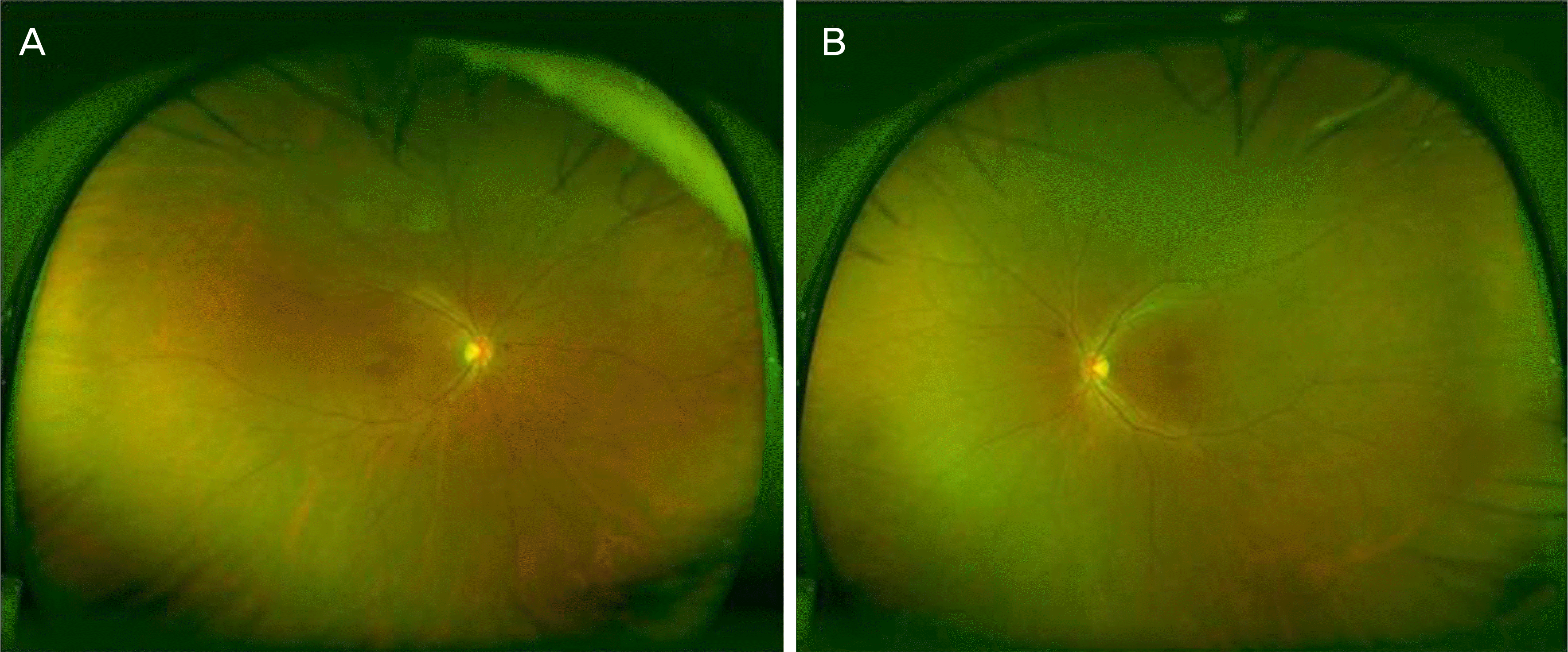
Figure 2.
Optical coherence tomography scans at the first visit (A, B), enhanced depth imaging spectral domain-optical coherence tomography scans 1 week after the first visit (C, D), 1 month after the first visit (E, F), and 4 months after first visit (G, H). Red line in each figure represents choroidal thickness of the B-scan (A-H). Thickening with increased signal and some irregularity of ellipsoid zone (EZ) and thickening of choroid (320 μm in the right eye and 301 μm in the left eye) was noted at the first visit (A, B). Improved but remaining thickened EZ of both eyes and worsened thickening of choroid in the right eye (384 μm in the right eye and 319 μm in the left eye) were noted at 1 week after the first visit (C, D). Improved but remaining thickened EZ and improved thickening of choroid in both eyes (298 μm in the right eye and 307 μm in the left eye) were noted 1 month after the first visit (E, F). Almost complete resolution of thickening of EZ and choroid (262 μm in the right eye and 235 μm in the left eye) were noted at scans on the day of 4 month after the first visit (G, H).

Figure 3.
Visual field test at 1 week after the first visit (A, B), 1 month [after the first visit (C, D), 2 months after the first visit (E, F) and 4 months after the first visit (G, H). Central scotomas on both eyes was noted on the day of 1 week after the first visit (A, B). After 4 months, central scotoma was nearly invisible and the test result was almost within normal range (G, H). POS = positive; NEG = negative; ASB = apostilb; SITA = Swedish Interactive Threshold Algorithm; RX = prescription; DS = diopter sphere; DC = dipoter cylinder; GHT = glaucoma hemifield test; MD = mean deviation; PSD = pattern standard deviation.

 XML Download