

 PDF
PDF ePub
ePub Citation
 Print
Print


Abstract
Purpose
We aimed to analyze the clinical features of herpetic endotheliitis and to investigate the risk factors for recurrence of herpetic endotheliitis and corneal decompensation.
Methods
The medical records of 58 patients (58 eyes) who were diagnosed with herpetic endotheliitis were retrospectively reviewed. Patients with a follow-up period of less than 6 months and a previous history of ocular inflammation were excluded from this study. We recorded visual acuity, intraocular pressure, central corneal thickness, and endothelial cell density on both initial and final visit, and recorded clinical findings of the anterior and posterior segments of the eye only on initial visit. Factors affecting the recurrence of herpetic endotheliitis and corneal decompensation were also analyzed.
Results
Among the 58 patients, 45 patients had disciform type endotheliitis, 11 patients had diffuse type, and 2 patients had linear type. There were no significant differences between final clinical manifestations according to subtype. 14 patients exhibited recurrence of herpetic endotheliitis. High intraocular pressure and high-grade anterior chamber cells were associated with the recurrence of herpetic endotheliitis. On multivariate analysis, the only risk factor for the recurrence of herpetic endotheliitis was high intraocular pressure. We found that 8 patients exhibited corneal decompensation, and high intraocular pressure, high anterior chamber cell grade, and a history of cataract surgery were associated with corneal decompensation. On multivariate analysis, the risk factors for corneal decompensation were high anterior chamber cell grade and cataract surgery history.
Conclusions
For herpetic endotheliitis, the subtype did not affect the final records of clinical manifestation, and the only risk factor for the recurrence of herpetic endotheliitis was high intraocular pressure. Additionally, the risk factors of corneal decompensation were found to be high-grade anterior chamber cells and a history of cataract surgery. Initial examinations of clinical manifestation are important for the successful treatment of herpetic endotheliitis.
Figures and Tables
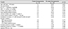
Table 1
Baseline characteristics of herpetic endotheliitis patients according to the type
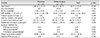
Values are presented as mean ± standard deviation or n.
BCVA = best corrected visual acuity; logMAR = logarithm of the minimum angle of resolution; IOP = intraocular pressure; HTN = hypertension; DM = diabetes mellitus; CVA = cerebrovascular accident; PCR = polymerase chain reaction.
*Student-t test; †Fisher's exact test.
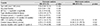
Table 2
Clinical outcomes of herpetic endotheliitis patients according to the type at the final visit
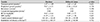
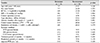
Table 4
Factors affecting recurrence in herpetic endotheliitis by univariate and multivariate analysis
References
1. Liesegang TJ. Herpes simplex virus epidemiology and ocular importance. Cornea. 2001; 20:1–13.
2. Williams LE, Nesburn AB, Kaufman HE. Experimental induction of disciform keratitis. Arch Ophthalmol. 165; 73:112–114.
3. Power WJ, Hillery MP, Benedict-Smith A, Collum LM. Acyclovir ointment plus topical betamethasone or placebo in first episode disciform keratitis. Br J ophthalmol. 1992; 76:711–713.
4. Mannis MJ, Holland EJ. Cornea. 4th ed. Vol. 1. Amsterdam: Elsevier;2017. p. 909–941.
5. Kaye S, Choudhary A. Herpes simplex keratitis. Prog Retin Eye Res. 2006; 25:355–380.
6. Hwang JS, Wee WR, Lee JH, Kim MK. Clinical analysis of herpetic keratitis in Korea. J Korean Ophthalmol Soc. 2007; 48:1212–1219.
7. The Herpetic Eye Disease Study Group. A controlled trial of oral acyclovir for iridocyclitis caused by herpes simplex virus. Arch Ophthalmol. 1996; 114:1065–1072.
8. The Herpetic Eye Disease Study Group. Acyclovir for the prevention of recurrent herpes simplex virus eye disease. N Engl J Med. 1998; 339:300–306.
9. I GU, Lee JH. Corticosteroid therapy in herpes simplex virus keratitis. J Korean Ophthalmol Soc. 1986; 27:493–496.
10. Jabs DA, Nussenblatt RB, Rosenbaum JT. Standardization of Uveitis Nomenclature (SUN) Working Group. Stanbdardization of uveitis nomenclature for reporting clinical data Results of the First International Workshop. Am J Ophthalmol. 2005; 140:509–516.
11. White ML, Chodosh J. Herpes simplex virus keratitis: a treatment guideline. Ocular Microbiology and Immunology Group and American Academy of Ophthalmology;2014. 32–69. Accessed January 20, 2017. http://www.hbky.com/uploadfile/2016/0124/20160124817259.pdf.
12. Ohashi T, Yamamoto S, Nishida K, et al. Demonstaration of herpes simplex virus DNA in idiopathic corneal endotheliopathy. Am J Ophthalmol. 1991; 112:419–423.
13. Carrillo-Arroyo I, Gutiérrez-Díaz E, Mencía-Gutiérrez E, et al. Herpetic endotheliitis and trabeculitis with delayed corneal involvement. Arch Soc Esp Oftalmol. 2012; 87:47–49.
14. Amano S, Oshika T, Kaji Y, et al. Herpes simeplex virus in the trabeculum of an eye with corneal endotheliitis. Am J ophthalmol. 1999; 127:721–722.
15. Reijo A, Antti V, Jukka M. Endothelial cell loss in herpes zoster keratouveitis. Br J Ophthalmol. 1983; 67:751–754.
16. Linebarger EJ, Hardten DR, Shah GK, Lindstrom RL. Phacoemulsification and modern cataract surgery. Surv Ophthalmol. 1999; 44:123–147.
17. Hwang HB, Lyu B, Yim HB, Lee NY. Endothelial cell loss after phacoemulsification according to different anterior chamber depths. J Ophthalmol. 2015; 2015:210716.
18. Díaz-Valle D, Benítez Del, Toledano N, et al. Endothelial morphological and functional evaluation after cataract surgery. Eur J Ophthalmol. 1996; 6:242–245.
19. Choi JH, Oh HJ, Yoon KC. Long-term results after cataract surgery in patients with low corneal endothelial cell density. J Korean Ophthalmol Soc. 2013; 54:602–609.
 XML Download