

 PDF
PDF ePub
ePub Citation
 Print
Print


Abstract
Purpose
The purpose of this study is to investigate the effect of unilateral medial rectus muscle resection (UMR) for the treatment of recurrent intermittent exotropia after bilateral lateral rectus muscle recession (BLR).
Methods
Medical records of 121 subjects who underwent UMR for the treatment of recurrent intermittent exotropia after BLR with more than six months of follow-up were reviewed. Patients were classified into two groups, the 4-mm group who underwent 4-mm UMR and the 5-mm group who underwent 5-mm UMR. Successful postoperative motor alignment was defined as within 10 prism diopters (PD) of exotropia and four PD of esotropia.
Results
The mean time interval between the primary surgery and recurrence was 13.55 ± 20.78 months (1-120 months). Average follow-up period after secondary surgery was 27.42 ± 15.98 months (6-48 months). Cumulative success rate at six months after UMR was 87.1% in the 4-mm group and 88.2% in the 5-mm group, respectively, and that at 24 months was 72.7% in the 4-mm group and 50.0% in the 5-mm group (p = 0.132). The average effect of resection was 4.87 ± 0.91 PD/mm in the 4-mm group and 4.73 ± 0.84 PD/mm in the 5-mm group (p = 0.374).
Conclusions
Because of recurrent intermittent exotropia, less time is required for surgery in UMR after BLR, and patients and parents are more likely to accept a secondary surgery because of single muscle surgery. Therefore, UMR may be an effective surgical method for patients with 16-30 PD of recurrent intermittent exotropia.
References
1. Zibrandtsen P, Rindziunski E, Gregersen E. Ten years follow-up of surgery for intermittent exotropia. Acta Ophthalmol (Copenh). 1986; 64:374–8.

2. Scott WE, Keech R, Mash AJ. The postoperative results and stability of exodeviations. Arch Ophthalmol. 1981; 99:1814–8.
3. Kim SJ, Choi DG. The clinical analysis after reoperation for recurrent intermittent exotropia. J Korean Ophthalmol Soc. 2007; 48:321–7.
4. Yang HK, Hwang JM. Bilateral vs unilateral medial rectus resection for recurrent exotropia after bilateral lateral rectus recession. Am J Ophthalmol. 2009; 148:459–65.
5. Kim SC, Kim MM. The efficacy of unilateral rectus resection in the reoperation of strabismus. J Korean Ophthalmol Soc. 2003; 44:904–10.
6. Yazdian Z, Ghiassi G. Re-recession of the lateral rectus muscles in patients with recurrent exotropia. J AAPOS. 2006; 10:164–7.
7. Suh YW, Seo IH, Cho YA, Kim SH. Analysis of the effects of medial rectus muscle resection for recurrent exotropia. Korean J Ophthalmol. 2011; 25:341–3.
8. Olitsky SE, Kelly C, Lee H, Nelson LB. Unilateral rectus resection in the treatment of undercorrected or recurrent strabismus. J Pediatr Ophthalmol Strabismus. 2001; 38:349–53.
9. Mims JL 3rd. Outcome of 5 mm resection of one medial rectus extraocular muscle for recurrent exotropia. Binocul Vis Strabismus Q. 2003; 18:143–50.
10. Chae SH, Chun BY, Kwon JY. The effect of unilateral medial rectus muscle resection in patients with recurrent exotropia. Korean J Ophthalmol. 2008; 22:174–7.
Figure 1.
Kaplan–Meier survival curve showing the cumulative probability of surgical success following a unilateral lateral rectus muscle resection procedure (p = 0.132, log rank test). UMR = unilateral medial rectus muscle resection.
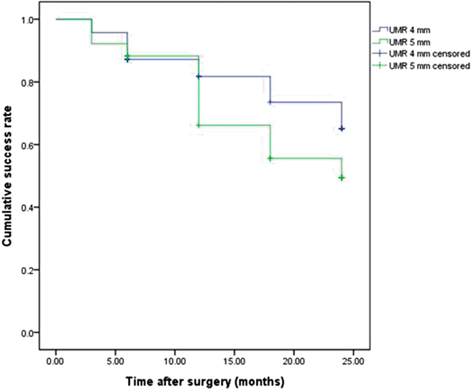
Table 1.
Characteristics of patients
| Factors | Values (n = 121) |
|---|---|
| Sex (M:F) | 60:61 |
| Age at onset (years) | 2.13 ± 1.50 |
| Age at diagnosis (years) | 3.66 ± 2.11 |
| Age at primary surgery (years) | 5.49 ± 2.00 |
| Amount of primary surgery with BLR | |
| 5 mm | 35 (28.9%) |
| 6 mm | 86 (71.1%) |
| Time to recurrence∗ after first surgery (months) | 13.55 ± 20.78 |
| Amount of secondary surgery with UMR | |
| 4 mm | 70 (57.9%) |
| 5 mm | 51 (42.1%) |
| Average postoperative follow-up period after secondary surgery (months) | 27.42 ± 15.98 |
Table 2.
Characteristics of patients in UMR 4-mm group and UMR 5-mm group
| Factors | UMR 4 mm group | UMR 5 mm group | p-value |
|---|---|---|---|
| No | 70 (57.9%) | 51 (42.1%) | |
| Sex (M:F) | 34:36 | 26:25 | 0.794† |
| Age at onset (years) | 2.27 ± 1.37 | 1.94 ± 1.66 | 0.245§ |
| Age at diagnosis (years) | 3.84 ± 2.01 | 3.41 ± 2.23 | 0.269§ |
| Family history of strabismus | 5 (7.1) | 7 (13.7) | 0.232† |
| Spherical equivalent refractive error (D) | |||
| Right eye | 0.27 ± 0.92 | 0.35 ± 0.92 | 0.619§ |
| Left eye | 0.33 ± 0.85 | 0.37 ± 0.90 | 0.772§ |
| Anisometropia (>1.5 D) | 1 (1.4) | 1 (2.0) | 1.000Π |
| Previous treatment of amblyopia | 9 (12.9) | 5 (9.8) | 0.604Π |
| Primary surgery | |||
| Age at primary surgery (years) | 5.64 ± 2.02 | 5.29 ± 1.98 | 0.347§ |
| Preoperative deviation (PD) | 23.21 ± 2.84 | 24.82 ± 2.70 | 0.002§ |
| Recession amount (5 mm:6 mm) | 26:44 | 9:42 | 0.020† |
| Overcorrection∗ at postoperative 1 week | 3 (4.3) | 6 (11.8) | 0.156Π |
| Time to recurrence† (months) | 16.93 ± 23.92 | 8.92 ± 14.46 | 0.024§ |
| Period between primary and secondary surgery (months) | 60.02 ± 34.25 | 50.94 ± 35.58 | 0.159§ |
| Secondary surgery | |||
| Age at secondary surgery (years) | 6.35 ± 2.91 | 5.66 ± 2.73 | 0.190§ |
| Preoperative deviation (PD) | 19.74 ± 1.43 | 25.10 ± 1.62 | <0.001§ |
| Overcorrection† at postoperative 1 week | 5 (7.1) | 2 (3.9) | 0.698Π |
| The average postoperative follow-up (months) | 27.68 ± 16.06 | 27.05 ± 16.02 | 0.832§ |
| Time to recurrence‡ after secondary surgery (months) | 18.12 ± 13.45 | 17.56 ± 12.61 | 0.981§ |
Table 3.
Deviation at preoperative day in UMR 4-mm group and UMR 5-mm group
| PD |
No. of patients |
|
|---|---|---|
| UMR 4-mm group | UMR 5-mm group | |
| <20 | 15 | 0 |
| 20 | 53 | 2 |
| 25 | 2 | 46 |
| >30 | 0 | 3 |
Table 4.
Comparisons of cumulative probability of surgical success rate in UMR 4-mm group and UMR 5- mm group
Table 5.
Surgical effects of medial rectus resection according to the amount of resection
| Amount of resection (mm) | No. of patients | Mean corrected deviation (PD) | Mean effects per millimeter of MR resection (PD/mm)∗ |
|---|---|---|---|
| 4 | 70 | 19.51 ± 3.65 | 4.87 ± 0.91 |
| 5 | 51 | 23.66 ± 4.20 | 4.73 ± 0.84 |
 XML Download