

 PDF
PDF ePub
ePub Citation
 Print
Print


Abstract
Purpose
To compare SLO (scanning laser ophthalmoscope) retromode images with conventional color fundus photography for the detection of drusen.
Methods
We obtained color fundus photography and SLO retromode images of the ten fellow eyes of ten patients with unilateral exudative age-related macular degeneration (AMD) and twenty eyes of 20 patients who had only drusen without exudative AMD. The numbers of druse in each image were compared within the same retinal boundary.
Results
In the fellow eyes of unilateral exudative AMD, an average number of 63.1 ± 81.9 drusen in color fundus photography and an average number of 141.3 ± 124.1 drusen in SLO retromode images were detected (p = 0.005). In the eyes with only drusen, an average number of 57.0 ± 43.9 drusen in color fundus photography and an average number of 112.2 ± 82.0 drusen in SLO retromode images were detected (p = 0.000). In the presence of media opacity like cataract, drusen were better detected in SLO retromode images than they were in color fundus photography.
Conclusions
About twice as many drusen were detected in SLO retromode images than in color fundus photography. Drusen were also better detected in SLO retromode images in cases of media opacity. SLO retromode images might provide more sensitive images for the detection of drusen than does color fundus photography.
References
1. Klein R, Klein BE, Linton KL. Prevalence of age-related maculopathy: the Beaver Dam Eye Study. Ophthalmology. 1992; 99:933–43.
2. Pauleikhoff D, Barondes MJ, Minassian D, et al. Drusen as risk factors in age-related macular disease. Am J Ophthalmol. 1990; 109:38–43.

3. Gass JD. Drusen and disciform macular detachment and degeneration. Arch Ophthalmol. 1973; 90:206–17.
4. Vinding T. Occurrence of drusen, pigmentary changes and exudative changes in the macula with reference to age-related macular degeneration An epidemiological study of 1000 aged individuals. Acta Ophthalmol. 1990; 68:410–4.
5. Oshima Y, Ishibashi T, Murata T, et al. Prevalence of age-related maculopathy in a representative Japanese population: the Hisayama Study. Br J Ophthalmol. 2001; 85:1153–7.
6. Tanaka Y, Shimada N, Ohno-Matsui K, et al. Retromode retinal imaging of macular retinoschisis in highly myopic eyes. Am J Ophthalmol. 2010; 149:635–40.
7. Yamamoto M, Mizukami S, Tsujikawa A, et al. Visualization of cystoid macular oedema using a scanning laser ophthalmoscope in the retromode. Clin Exp Ophthalmol. 2010; 38:27–36.
8. Ishiko S, Akiba J, Horikawa Y, Yoshida A. Detection of drusen in the fellow eye of Japanese patients with age-related macular degeneration using scanning laser ophthalmoscopy. Ophthalmology. 2002; 109:2165–9.
9. Friedman DS, Katz J, Bressler NM, et al. Racial differences in the prevalence of age-related macular degeneration. The Baltimore Eye Survey. Ophthalmology. 1999; 106:1049–55.
10. Schachat AP, Hyman L, Leske MC, et al. Features of age-related macular degeneration in a black population. The Barbados Eye Study Group. Arch Ophthalmol. 1995; 113:728–35.
11. Klein R, Clegg L, Cooper LS, et al. Prevalence of age-related maculopathy in the Atherosclerosis Risk in Communities Study. Arch Ophthalmol. 1999; 117:1203–10.
Figure 1.
A schematic mechanism of SLO (scanning laser ophthalmoscopeopacity) retromode. The retromode use a modified aperture technique consisted with ring aperture and laterally deviated aperture. With the ring aperture, the direct reflected light from retina is blocked (central stop). With the laterally deviated aperture, the one side of scattered light from deep retina or choroid is passed through the aperture but the other side of scattered light is not. So, a light blocking material like drusen can be visualized.
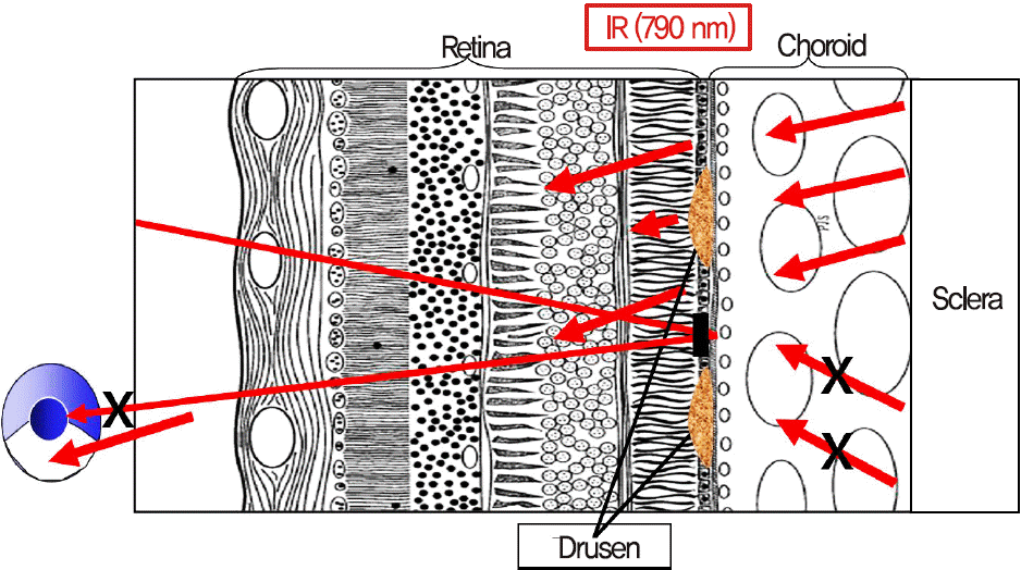
Figure 2.
An example in the fellow eyes of unilateral exudative age-related macular degeneration patients. Soft drusen and hard drusen mixed. (A) Fundus photography. (B) Scanning laser ophthalmoscopeopacity (SLO) retromode. (C) Optical coherence tomography (OCT). The drusen in SLO retromode image are seen pseu-do-3-dimensional and clearer than in fundus photography. The OCT finding is RPE elevation which is compatible for the drusen.
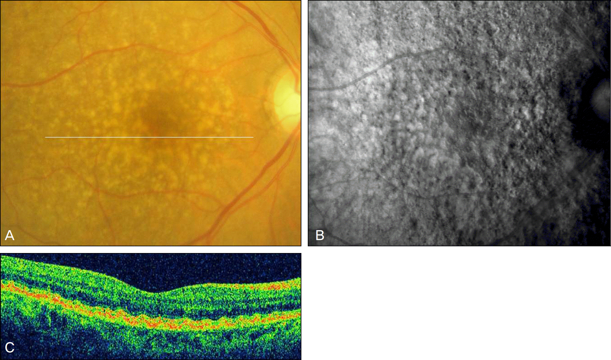
Figure 3.
An example in the eyes with only drusen without exudative age-related macular degeneration. Hard drusen. (A) Fundus photography. (B) Scanning laser ophthalmoscope (SLO) retromode.(C) Optical coherence tomography (OCT). The drusen indicated by OCT scan (arrow) are not seen in fundus photography but seen well in SLO retromode image.
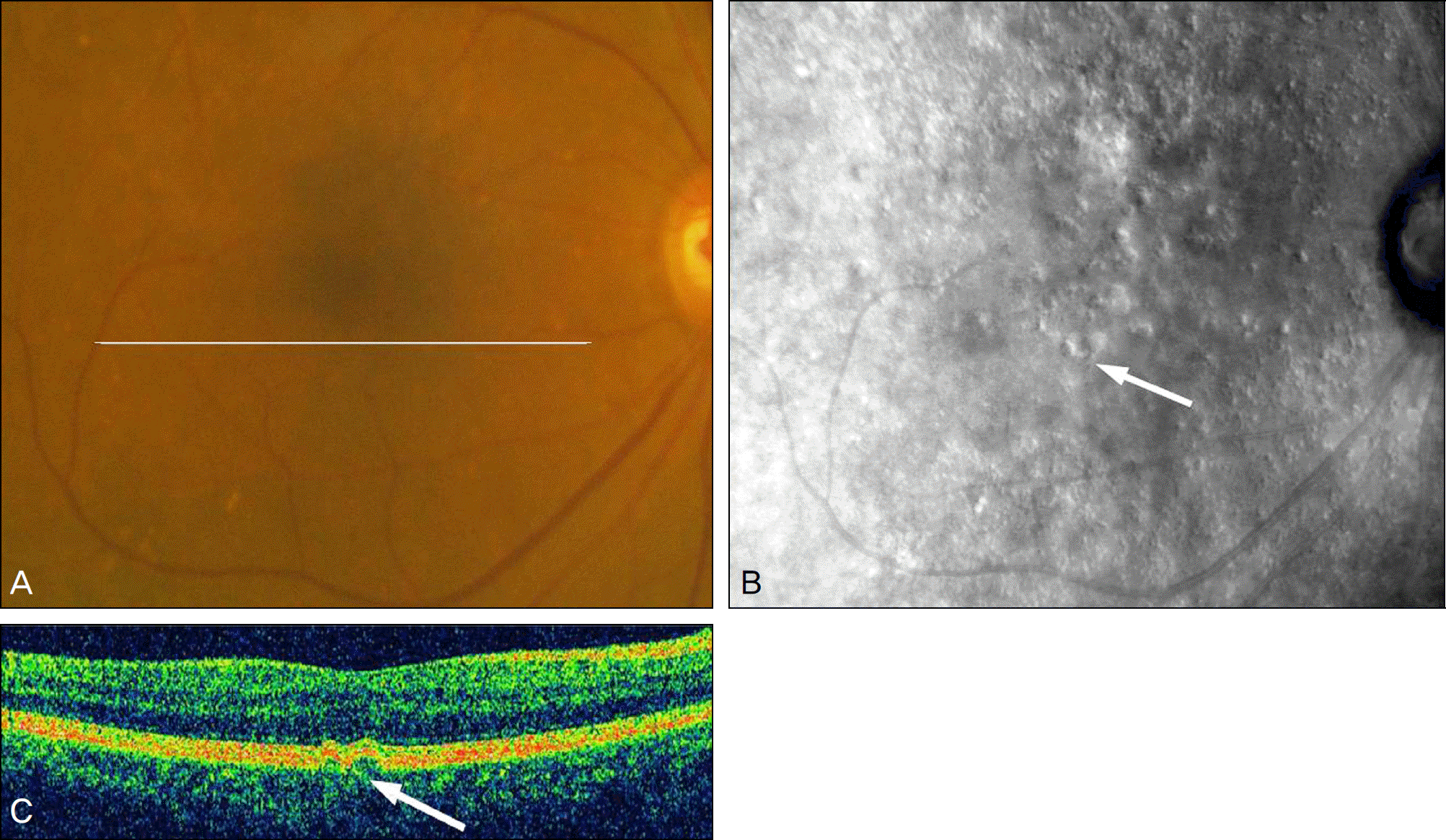
Figure 4.
An example in the eyes with only drusen without exudative age-related macular degeneration. Hard drusen. (A) Fundus photography. (B) Scanning laser ophthalmoscope (SLO) retromode.(C) Optical coherence tomography (OCT). The outline of hard drusen is seen clearer than the outline of soft drusen.
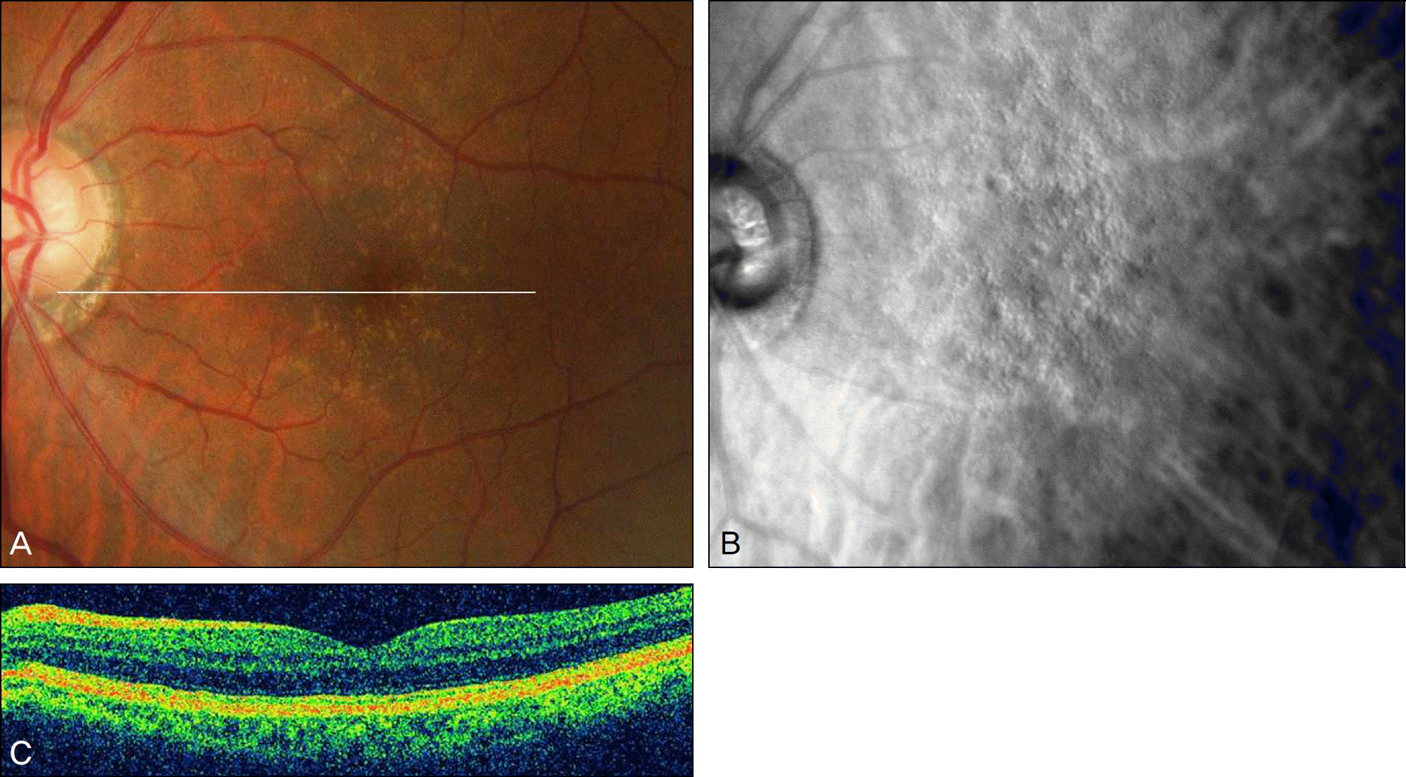
Figure 5.
Drusen with the media opacity. (A) Fundus photography. (B) Scanning laser ophthalmoscope (SLO) retromode. In the media opacity like cataract, drusen are detected better in SLO retromode image than in color fundus photography.
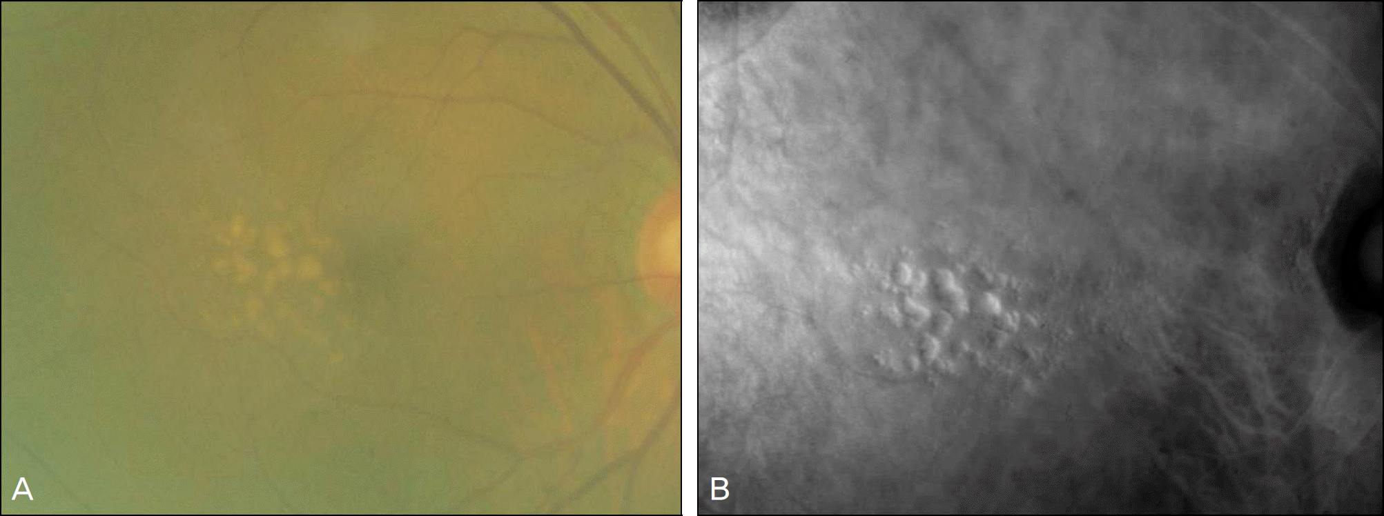
Table 1.
The number of drusen detected in each patient with unilateral exudative age-related macular degeneration
| Patient | 1 | 2 | 3 | 4 | 5 | 6 | 7 | 8 | 9 | 10 |
|---|---|---|---|---|---|---|---|---|---|---|
| Fundus photography | 22 | 11 | 42 | 13 | 99 | 7 | 1 | 21 | 13 | 232 |
| SLO* retromode | 271 | 56 | 46 | 97 | 226 | 37 | 3 | 67 | 97 | 245 |
Table 2.
The number of drusen detected in each patient with only drusen without exudative age-related macular degeneration
| Patient | 1 | 2 | 3 | 4 | 5 | 6 | 7 | 8 | 9 | 10 | 11 | 12 | 13 | 14 | 15 | 16 | 17 | 18 | 19 | 20 |
|---|---|---|---|---|---|---|---|---|---|---|---|---|---|---|---|---|---|---|---|---|
| Fundus photography | 10 | 45 | 32 | 91 | 7 | 30 | 121 | 38 | 35 | 94 | 105 | 31 | 24 | 162 | 102 | 5 | 24 | 64 | 92 | 28 |
| SLO* retromode | 52 | 78 | 56 | 185 | 31 | 78 | 275 | 52 | 102 | 98 | 221 | 60 | 51 | 249 | 251 | 14 | 61 | 97 | 178 | 55 |
 XML Download