Abstract
Purpose
To compare the intraoperative and short term postoperative outcomes of cataract surgery performed with custom pulse mode of OZil® and hyperpulse mode of conventional phacoemulsification
Methods
The patients underwent cataract surgery using the OZil® custom pulse mode phacoemulsification (Group 1, n=30) or the conventional phaco Hyperpulse mode one (Group 2, n=36) of Infiniti Vision System. Mean ultrasound time, CDE (Cumulated dissipated energy) and total BSS volume were measured during operation. Best corrected visual acuity, central corneal thickness and endothelial cell counts were checked on 1 day, 1 momth, 2 months after surgery.
Results
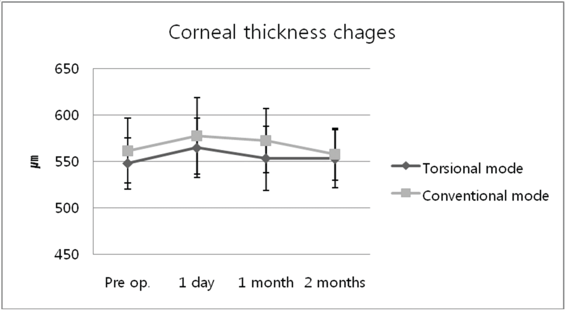
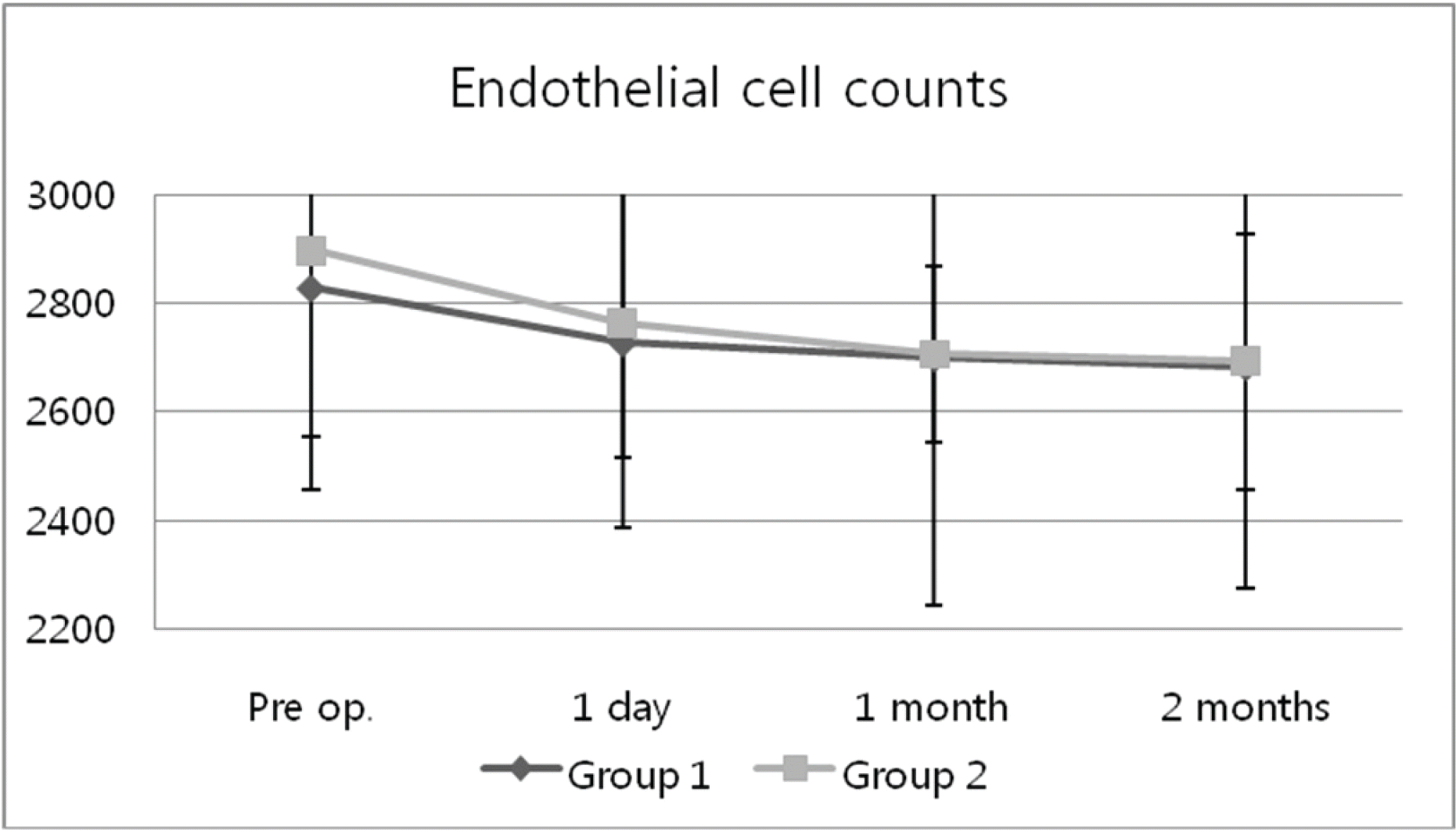
CDE was decreased significantly in group 1 (p<0.05). But, there were no significant differences in two groups of the mean ultrasound time and the BSS volume (p>0.05). At 2 months, the endothelial thickness was 552.94±27.95 μ m in group 1, and 568.00±31.22 μ m in group 2 and the endothelial cell counts was 2683.0±235.6 cell/mm2 and 2694.2±407.9 cell/mm2, respectatively. There were no significant differences in two groups.
References
1. Vargas LG, Holzer MP, Solomon KD, et al. Endothelial cell integrity after phacoemulsification with 2 different handpieces. J Cataract Refract Surg. 2004; 30:478–82.

2. O'Brien PD, Fitzpatrick P, Kilmartin DJ, Beatty S. Risk factors for endothelial cell loss after phacoemulsification surgery by a junior resident. J Cataract Refract Surg. 2004; 30:839–43.
3. Rho CR, Kim SY, Joo CK. Clinical Result of Cataract Operation using Custom Control Software. J Korean Ophthalmol Soc. 2006; 47:735–39.
4. Liu Y, Zeng M, Liu X, et al. Torsional mode versus conventional ultrasound mode phacoemulsification: randomized comparative clinical study. J Cataract Refract Surg. 2007; 33:287–92.
5. Chylack LT Jr, Leske MC, McCarthy D. Lens Opacities Classification System II (LOCS II). Arch Ophthalmol. 1989; 107:991–7.

6. Vasavada V, Vasavada V, Raj SM, Vasavada AR. Intraoperative performance and postoperative outcomes of microcoaxial phacoemulsification: Observational study. J Cataract Refract Surg. 2007; 33:1019–24.
7. Kelman CD. Phaco-emulsification and aspiration. A new technique of cataract removal. A preliminary report. Am J Ophthalmol. 1967; 64:23–35.
8. Polack FM, Sugar A. The phacoemulsification procedure Ⅲ corneal complications. Invest Ophthalmol Vis Sci. 1977; 16:39–46.
9. Gwin RM, Warren JK, Samuelson DA, Gum GG. Effects of phacoemulsification and extracapsular lens removal on corneal thickness and endothelialcell density in the dog. Invest Ophthalmol Vis Sci. 1983; 24:227–36.
10. Strobel J, Jacobi KW. Phacoemulsification and planed ECCE: Intraoperative differences in intraocular heating. Eur J Implant Refract Surg. 1991; 3:135–8.
11. Gil SY, Kang SB, Lee SH, Chung SK. The Effect of Phaco-emulsification with Oscillation Device on the Cornea and Lens Opcatiy. J Korean Ophthalmol Soc. 2007; 47:1948–53.
12. Kim HJ, Kim JH, Lee DH. Coaxial phacoemulsification, Corneal endothelial cell damage, Microincision cataract surgery. J Korean Ophthalmol Soc. 2007; 48:19–26.
13. Hoffman RS, Fine IH, Packer M, Brown LK. Comparison of sonic and ultrasonic phacoemulsification using the Staar Sonic Wave system. J Cataract Refract Surg. 2002; 28:1581–4.

14. Mackool RJ, Brint SF. AquaLase: a new technology for cataract extraction. Curr Opin Ophthalmol. 2004; 15:40–3.

15. Vasavada AR, Raj SM, Lee YC. NeoSoniX ultrasound versus ultrasound alone for phacoemulsification; randomized clinical trial. J Cataract Refract Surg. 2004; 30:2332–5.
16. Seibel RS. Phacodynamics: Mastering the tools and techniques of phacoemulsification sergery. 4th ed.Los Angeles: SLACK;2005. p. 122–3.
17. Braga-Mele R. Thermal effect of microburst and hyperpulse settings during sleeveless bimanual phacoemulsification with advanced power modulations. J Cataract Refract Surg. 2006; 32:639–42.

18. Alio J, Rodriguez-Prats JL, Galal A, Ramzy M. Outcomes of microincision cataract versus coaxial phacoemulsification. Ophthalmology. 2005; 112:1997–2003.
Figure 1.
Corneal thickness in 2 groups. there is no statistically significant difference between the two groups (p>0.05). Group 1=OZil® custom pulse mode phacoemulsification; Group 2=Hyperpulse mode phaco-emulsification.

Figure 2.
Postoperative Changes in endothelial cell count (cell/mm2). Group 1=OZil® custom pulse mode phacoemulsification; Group 2=Hyperpulse mode phaco-emulsification.

Figure 3.
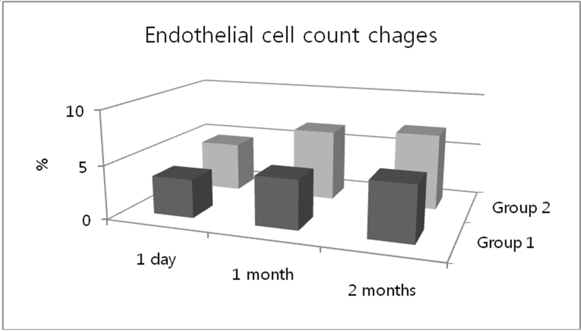
Endothelial cell count change in two groups. The mean endothelial cell counts decreased 5.14% in torsional phacoemulsification and 7.07% in conventional phacoemulsification at 60 days, respectively. Group 1=OZil® custom pulse mode phacoemulsification; Group 2=Hyperpulse mode phacoemulsification.

Table 1.
Surgical parameters used in the study with Infiniti phacoemulsification platform
| Group 1* | Group 2† | |
|---|---|---|
| Incision size (mm) | 3.0 mm | 3.0 mm |
| Capsulorrhexis diameter (mm) | 5.5 mm | 5.5 mm |
| Phacoemulsification tip | 30° | 30° |
| Prechopping | No | No |
| Settings | ||
| Amplitude/Power (Linear control) | 45% (phaco) | 45% |
| /100% (torsional oscillation) | ||
| Aspiration flow | 45 cm3/min | 45 cm3/min |
| Vacuum | 450 mmHg | 450 mmHg |
| Anterior chamber pressure | 105 cm3-H2 O | 105 cm3-H2 O |
| Mode | Custom pulse | Hyperpulse |
| phaco: 100 ms on, 10 ms off | (30 pps, 40% time on) | |
| ultrasonic oscillations: 100 ms on, 10 ms off | ||
| I/A of cortical remnants | 450 mmHg | 450 mmHg |
| I/A of viscoelastic | 150 mmHg | 150 mmHg |
Table 2.
Patient characteristics (N=66)
| Group 1* | Group 2† | p-values‡ | |
|---|---|---|---|
| Number (eye) | 30 | 36 | |
| Gender (M/F) | 8/13 | 10/17 | |
| Age | 67.56±6.87 | 62.67±8.56 | 0.12 |
| Mean Nucleus density | 2.53±0.57 | 2.49±0.62 | 0.45 |
Table 3.
Comparison of mean surgical parameters between OZil® mode and hyperpulse mode
| Group 1† | Group 2‡ | p value§ | |
|---|---|---|---|
| Mean phaco time (sec) | 16.04±7.79 | 16.22±16.76 | 0.216 |
| CDE* (sec) | 3.63±2.32 | 4.92±2.48 | 0.005 |
| Total BSS used (cc) | 54.26±8.12 | 56.2±4.68 | 0.571 |
Table 4.
Postoperative changes in corneal thickness (µm)
| Group 1* | Group 2† | p value‡ | |
|---|---|---|---|
| Pre-op | 548.10±35.04 | 561.87±27.61 | 0.526 |
| POD op. # 1 day | 564.84±41.37 | 577.6±32.05 | 0.793 |
| POD op. # 1 month | 553.52±34.84 | 572.46±34.60 | 0.193 |
| POD op. # 2 months | 552.94±27.95 | 568.00±31.22 | 0.904 |


 PDF
PDF ePub
ePub Citation
 Print
Print


 XML Download