Abstract
Purpose
To investigate the effect of preoperative part-time occlusion therapy on long-term surgical success in early-onset exotropia.
Methods
The medical records of patients who underwent surgery for exotropia with onset before the first year of age and who were followed for ≥3 years were reviewed. Patients were divided into two groups according to the degree of compliance with part-time occlusion therapy: the good compliance group (>50% adherence rate) and the poor compliance group (≤50% adherence rate). Surgical success was defined as orthophoria to exodeviation less than 10 prism diopters both at distance and near. The level of postoperative stereopsis was compared between the two study groups among total enrolled patients and among those with constant exotropia.
Results
Of the 51 patients, 26 were assigned to the good compliance group and the remaining 25 patients to the poor compliance group. The surgical success rate was significantly higher in the good compliance group than in the poor compliance group (80.8% vs. 52.0%, p = 0.040). Among 24 constant exotropia patients (12 patients for each group), the success rate was insignificantly higher in the good compliance group than in the poor compliance group (75.0% vs. 58.3%, p = 0.448). The good compliance group had a better level of stereopsis than the poor compliance group (p = 0.045 for all 44 patients, p = 0.020 for 19 patients with constant exotropia).
Exotropia in healthy children younger than 1 year of age is rare, and several studies have been conducted on this disease entity [1234567]. Occlusion therapy alone is not sufficiently effective in reducing exodeviation, and surgery is the most promising treatment for early-onset exotropia as well as intermittent exotropia [8].
Although non-surgical treatments, including occlusion therapy, have limitations as a single treatment for intermittent exotropia [9], surgery in combination with preoperative occlusion therapy has been found to more effective in reducing exodeviation and has shown a superior success rate compared to that of surgery alone [10].
Occlusion therapy is frequently recommended for patients with exotropia to prevent amblyopia or/and suppression [11]. However, no studies have investigated whether preoperative occlusion therapy is effective in reducing recurrence after surgery for early-onset exotropia. Therefore, we investigated the effect of preoperative part-time occlusion therapy on long-term outcome after surgery for early-onset exotropia.
The study was approved by the ethics committee of Gachon University Gil Hospital, in Korea. We conducted a retrospective review of the medical records of 51 consecutive patients who had undergone surgery for early-onset exotropia in our clinic (Department of Pediatric Ophthalmology, Gachon University Gil Hospital) between March 1998 and December 2010, and who were followed postoperatively for at least 3 years. In this study, early-onset exotropia was defined as either constant or intermittent exotropia with onset prior to 1 year of age. Patients with exotropia who had history of prematurity (defined as a gestational age <37 weeks), trauma or neurological problems (such as developmental delay, seizures, or cerebral palsy), ocular diseases that could affect vision or ocular alignment, history of previous ocular muscle surgery, and follow-up duration <3 years after primary exotropia surgery were excluded. All patients were referred to a pediatrician to exclude neurological disorders [1].
Patients data of gender, age at onset, age at diagnosis, preoperative and postoperative angles of deviation, intermittent versus constant phase before surgery, age at surgery for exotropia, follow-up period after surgery, preoperative refractive status, associated inferior oblique muscle weakening procedure performed simultaneously with surgery for exotropia, amblyopia, postoperative stereopsis at the final visit, and compliance with part-time occlusion therapy were recorded.
All patients underwent a full initial ophthalmologic and orthoptic examination before surgery and were strongly advised to undergo daily part-time occlusion therapy using adhesive patches during a preoperative follow-up period of at least 6 months. Occlusion therapy was prescribed as follows: monocular patching in the dominant eye or alternate patching in the absence of a dominant eye. Eye dominance was determined using results from the cover–uncover test on each eye and the hole-in-the card test performed repeatedly. For the hole-in-the-card test, patients were instructed to view a distant object through the hole with both eyes open. Then, the examiner determined which eye the patient was using to see through the hole. Occlusion time was decided in consideration with the children's ages (1.5 to 2 hours for children younger than 3 years to minimize the risk of occlusion amblyopia and 3 hours for children older than 3 years). The occlusion therapy was advised to be performed at home under the surveillance of parents. Using self-report calendar logs and clinical interviews, compliance with occlusion therapy was calculated as the ratio of the number of hours when occlusion was actually performed (that were estimated by the parents) against the number of hours for which it had been prescribed. Compliance was graded as the following: “1” (for no compliance), “2” (for 1%–25%), “3” (for 26%–50%), “4” (for 51%–75%), or “5” (for 76%–100%). Patients with early-onset exotropia were followed preoperatively every 3 months, and the compliance rate was calculated at every follow-up session and averaged at the end of follow-up.
Age at onset was defined as the age at which a family member first observed ocular misalignment. These estimates were confirmed using previous patient photographs or video records. The angle of deviation was measured using an alternate prism cover test at distance (6 m) and at near (1/3 m) with an accommodative target with spectacles. The modified Krimsky test was applied in a few patients in whom the alternate prism cover test had not been possible. Constant exotropia was defined as a clinic control score ≥5, in accordance with the revised Newcastle control score [12]. A score ≥5 was determined when exotropia manifested spontaneously both in near and distant viewing conditions or when exotropia occurred spontaneously in one condition and remained manifest after dissociation/prolonged fixation in the other condition. The preoperative refractive status was defined as the mean spherical equivalent of both eyes measured by cycloplegic refraction with topical administration of 1% tropicamide (Mydriacyl; Alcon, Puurs, Belgium) and 1% cyclopentolate hydrochloride (Cyclogyl; Alcon Laboratories, Fort Worth, TX, USA). Amblyopia was determined using results from recognition acuity tests or regarded as being present when a preverbal child consistently appeared uncooperative in fixation behavior while covering of one eye. Near stereopsis was measured using the Titmus stereoacuity test (Stereo Optical, Chicago, IL, USA) and was graded into four categories: stereopsis of 100 arcsec or better, 140 to 400 arcsec, 800 to 3,000 arcsec, and nil stereopsis.
The surgical procedure, which was performed by the senior surgeon (HJP), was primarily bilateral lateral rectus muscle recession, and surgical dosages were applied using a standard table [13]. When there was associated inferior oblique muscle overaction (with/without V pattern) or dissociated vertical deviation, an inferior oblique muscle weakening procedure was performed simultaneously. Among the enrolled patients in this study, there were no cases of surgery on the vertical rectus or superior oblique muscles.
Surgical outcome was regarded as being successful when ocular alignment was measured as orthophoria to exodeviation less than 10 prism diopters (PD) both at distance and near. Reoperation was recommended when a cosmetically objectionable deviation (usually exotropia exceeding 20 PD or esotropia exceeding 15 PD) was observed. When consecutive esotropia was observed, patients were followed up for at least 6 months before reoperation was recommended.
In order to investigate the effect of preoperative occlusion therapy on the long-term surgical outcome of early-onset exotropia after surgery, we compared the long-term surgical success/recurrence/consecutive esodeviation and the level of postoperative stereopsis between patients with good compliance with part-time occlusion therapy (>50% adherence rate, grades 4 and 5) and those with poor compliance (≤50% adherence rate, grades 1, 2, and 3). Additionally, in order to investigate the effect of preoperative occlusion therapy on patients with constant exotropia, we compared these factors between patients with good compliance and patients with poor compliance in the constant exotropia patients.
We conducted the Fisher's exact test, Mann-Whitney U-test, and Kaplan-Meier survival analysis using SPSS ver. 17.0 (SPSS Inc., Chicago, IL, USA). The p-values less than 0.05 were considered statistically significant.
In all 51 early-onset exotropia patients who underwent surgery during the study period, the mean duration of preoperative occlusion therapy was 10.2 ± 5.4 months (range, 6 to 28 months). The mean follow-up duration after surgery for exotropia was 78.0 ± 28.1 months (range, 36 to 135 months). Overall, the final success rate of surgery for early-onset exotropia was 66.7%. Five patients (9.8%) showed persisting consecutive esotropia and eventually underwent surgical correction for these consecutive esotropia at a mean age of 18.8 months (range, 8 to 40 months) after the primary surgery for exotropia. Meanwhile, 12 patients (23.5%) showed recurrence of exotropia greater than 10 PD, and four patients (7.8%) underwent surgical correction for recurrent exotropia during the follow-up period after the initial surgery.
With the exception of three patients who experienced both 1.5 to 2 hours of patching and 3 hours of patching due to continued occlusion treatment around 36 months of age, 38 patients were younger than 3 years and were prescribed 1.5 to 2 hours of patching (compliance rate, 2.7 ± 1.6), and 10 patients were older than 3 years and prescribed 3 hours of pathing (compliance rate, 3.1 ± 1.5). The compliance rates were not different according to patching hours (p = 0.626). Fig. 1 depicts the compliance rates of the 51 enrolled patients. Twenty-six patients fulfilled part-time occlusion therapy with >50% adherence rate (grades 4 and 5) and were assigned to the good compliance group. The remaining 25 patients who underwent part-time occlusion therapy with a ≤50% adherence rate (grades 1, 2, and 3) were assigned to the poor compliance group. The demographic and clinical data of these 51 patients are presented in Table 1. There were no significant differences between the two study groups except for the improvement-to-dete-rioration ratio of the angle of deviation during the preoperative follow-up period. Improvement in the angle of deviation of more than 10 PD was shown in 10 patients. Among these patients, nine completed the occlusion therapy with a >50% adherence rate. Meanwhile, no patients in the good compliance group showed deterioration.
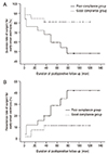
The success rate of surgery was significantly higher in the good compliance group (21 of 26 patients, 80.8%) than in the poor compliance group (13 of 25 patients, 52.0%) (p = 0.040). There were also significant differences between the two study groups in the undercorrection/recurrence rate (p = 0.049). The recurrence rate was higher in the poor compliance group (nine of 25 patients, 36.0%) than in the good compliance group (three of 26 patients, 11.5%). The Kaplan-Meier estimates also showed significant differences in the success rate (p = 0.049) (Fig. 2A) and the competing risk analysis for undercorrection/recurrence (p = 0.046) (Fig. 2B). The overcorrection/consecutive exodeviation rates were similar between the good compliance group (two of 26 patients, 7.7%) and the poor compliance group (three of 25 patients, 12.0%) (p = 0.614).
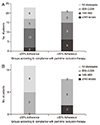
Postoperative stereopsis could be measured in 44 of the 51 patients in our cohort. The good compliance group had a better level of stereopsis than the poor compliance group (p = 0.045) (Fig. 3A).
When comparing the success rate according to compliance with part-time occlusion therapy among the 24 patients who had surgery for constant exotropia, the success rate was greater in the good compliance subgroup (nine of 12 patients, 75.0%) than in the poor compliance subgroup (seven of 12 patients, 58.3%). However, this result was not statistically significant (p = 0.448). Meanwhile, the level of stereopsis was significantly better in the good compliance subgroup than in the poor compliance subgroup in the 19 patients who had early-onset constant exotropia and who underwent stereoacuity testing (p = 0.020) (Fig. 3B).
Several studies have investigated the factors influencing surgical outcomes of exotropia in patients younger than 1 year of age. However, the factors that predict the long-term surgical success of early-onset exotropia have not been well established [34]. Although smaller preoperative deviation was found to be associated with more favorable out-comes at 6 weeks after surgery, smaller preoperative deviation did not guarantee the long-term outcome at 1 year following operation, according to results from a study by Yam et al. [3] and Park and Kim [4] also reported that no single preoperative factor affected the surgical outcome of early-onset exotropia at 1 year after surgery. Among patients who showed exotropia within the first year of life, age of onset did not influence the surgical outcome (p = 0.488). With 6 months of age at onset as a reference, the number of patients who showed exodeviation before 6 months of age was also not different between groups (nine patients in the poor compliance group and six patients in the good compliance group, p = 0.368).
In this study, preoperative part-time occlusion therapy was suggested to be a possible factor improving long-term surgical outcome. During the preoperative follow-up period, patients with early-onset exotropia were strongly advised to undergo part-time occlusion therapy and asked to record their compliance with part-time occlusion therapy. Then, we identified higher long-term success and lower recurrence rate in the good compliance group than in the poor compliance group. In addition, with respect to sensory outcomes, fulfillment of occlusion therapy was associated with better postoperative levels of stereopsis.
The beneficial effect of occlusion therapy on intermittent exotropia has been reported frequently [1415161718]. These studies have reported that occlusion therapy decreased the angle of exodeviation in intermittent exotropia or even that occlusion therapy postponed surgical intervention by converting exotropia to orthophoria or exophoria. A recent randomized controlled trial also showed less deterioration of intermittent exotropia in patients who underwent part-time occlusion compared to those who did not (6.1% vs. 0.6%, p = 0.004) [11]. Occlusion therapy limits binocular stimulation, avoiding and correcting abnormal retinal correspondence and suppression [19]. Occlusion therapy has both sensory and motor effects on intermittent exotropia by reducing scotoma size and improving fusional vergence amplitude [20]. In addition to these positive effects on intermittent exotropia, part-time occlusion therapy in children with intermittent exotropia helped to solve the concentration problem psychosocially [21]. The results of our study support the previous hypothesis emphasizing the usefulness of occlusion therapy even in early-onset exotropia [101114151617181920].
Among patients with constant exotropia, we failed to find a statistically significant difference in surgical success in the motor aspect (p = 0.448). Meanwhile, there was a statistically significant difference in the level of stereopsis among patients with constant exotropia (p = 0.020). The level of stereopsis was better in the good compliance subgroup than in the poor compliance subgroup (Fig. 3B). The lack of significant difference in the motor aspect between subgroups may be due to small sample size within each subgroup and may indicate the need for larger sample size to compare the surgical success among early-onset constant exotropia patients.
There are several limitations in this study. The design of this study was retrospective in nature. In addition, onset of early-onset exotropia was based on patient history, and this could be affected by recall bias. However, we confirmed these estimates using a number of old photographs or video records provided by patients. As another limitation related with retrospective design, this investigation was an observational study rather than a case-control study. We advised parents to ensure that their children underwent part-time occlusion therapy stringently. Then, we checked the level of compliance with occlusion therapy at every follow-up session and averaged these to obtain more reliable data related to compliance. Third, we assessed and ruled out any children who were suspected to have developmental difficulties that could influence poor compliance and surgical outcomes, with consultation from pediatricians. Although we excluded these patients, a few patients with subtle developmental difficulties may have been included in the enrolled patients, and this might influence the compliance rate with preoperative occlusion therapy and the surgical outcomes of exotropia. One of the biggest limitations was the small number of subjects. However, the small number of subjects in this study is an inherent limitation since exotropia in healthy children younger than 1 year of age is rare [22]. This study included a relatively larger number of subjects compared to previous studies. We suggest that a large, case-control study is needed to confirm the usefulness of preoperative occlusion therapy in early-onset exotropia surgery.
Although the effect of preoperative occlusion therapy on the success rate of constant exotropia was insignificant, preoperative occlusion therapy was useful for improving surgical outcomes among patients who underwent surgery for exotropia. In addition, preoperative occlusion therapy also improved postoperative stereopsis in all patients who underwent surgery for early-onset exotropia and even in those who underwent surgery for constant exotropia. Therefore, preoperative part-time occlusion therapy should be proposed and explained to the family during the preoperative follow-up period to improve surgical outcomes.
Figures and Tables
Fig. 1
The distribution of compliance rates with part-time occlusion therapy in 51 patients who underwent surgery for early-onset exotropia.

Fig. 2
The Kaplan-Meier estimates of surgical outcome in the two study groups according to the degree of compliance with part-time occlusion therapy: (A) success rate (p = 0.049), (B) recurrence rate among a total of 51 enrolled patients (p = 0.046).

Fig. 3
The level of stereopsis in the two study groups according to the degree of compliance with part-time occlusion therapy: (A) among a total of 44 enrolled patients (p = 0.045), (B) among 19 patients with early-onset constant exotropia (p = 0.020).

References
1. Hunter DG, Kelly JB, Buffenn AN, Ellis FJ. Long-term outcome of uncomplicated infantile exotropia. J AAPOS. 2001; 5:352–356.
2. Biglan AW, Davis JS, Cheng KP, Pettapiece MC. Infantile exotropia. J Pediatr Ophthalmol Strabismus. 1996; 33:79–84.
3. Yam JC, Chong GS, Wu PK, et al. Prognostic factors predicting the surgical outcome of bilateral lateral rectus recession surgery for patients with infantile exotropia. Jpn J Ophthalmol. 2013; 57:481–485.
4. Park JH, Kim SH. Clinical features and the risk factors of infantile exotropia recurrence. Am J Ophthalmol. 2010; 150:464–467.e2.
5. Yoo EJ, Kim SH. Optimal surgical timing in infantile exotropia. Can J Ophthalmol. 2014; 49:358–362.
6. Suh SY, Kim MJ, Choi J, Kim SJ. Outcomes of surgery in children with early-onset exotropia. Eye (Lond). 2013; 27:836–840.
7. Paik HJ, Yim HB. Clinical effect of early surgery in infantile exotropia. Korean J Ophthalmol. 2002; 16:97–102.
8. Kraft SP. Special forms of comitant exotropia. In : Hoyt CS, Taylor D, editors. Pediatric ophthalmology and strabismus. 4th ed. New York: Elsevier Saunders;2013. p. 792–800.
9. Buck D, Powell CJ, Rahi J, et al. The improving outcomes in intermittent exotropia study: outcomes at 2 years after diagnosis in an observational cohort. BMC Ophthalmol. 2012; 12:1.
10. Figueira EC, Hing S. Intermittent exotropia: comparison of treatments. Clin Exp Ophthalmol. 2006; 34:245–251.
11. Pediatric Eye Disease Investigator Group. Cotter SA, Mohney BG, et al. A randomized trial comparing part-time patching with observation for children 3 to 10 years of age with intermittent exotropia. Ophthalmology. 2014; 121:2299–2310.
12. Buck D, Clarke MP, Haggerty H, et al. Grading the severity of intermittent distance exotropia: the revised Newcastle Control Score. Br J Ophthalmol. 2008; 92:577.
13. Raab EL, Parks MM. Recession of the lateral recti: early and late postoperative alignments. Arch Ophthalmol. 1969; 82:203–208.
14. Spoor DK, Hiles DA. Occlusion therapy for exodeviations occurring in infants and young children. Ophthalmology. 1979; 86:2152–2157.
15. Iacobucci IL, Archer SM, Giles CL. Children with exotropia responsive to spectacle correction of hyperopia. Am J Ophthalmol. 1993; 116:79–83.
16. Berg PH, Lozano MJ, Isenberg SJ. Long term results of part-time occlusion for intermittent exotropia. Am Orthopt J. 1998; 48:85–89.
17. Suh YW, Kim SH, Lee JY, Cho YA. Conversion of intermittent exotropia types subsequent to part-time occlusion therapy and its sustainability. Graefes Arch Clin Exp Ophthalmol. 2006; 244:705–708.
18. Freeman RS, Isenberg SJ. The use of part-time occlusion for early onset unilateral exotropia. J Pediatr Ophthalmol Strabismus. 1989; 26:94–96.
19. Chutter CP. Occlusion treatment of intermittent divergent strabismus. Am Orthopt J. 1977; 27:80–84.
20. Flynn JT, McKenney S, Rosenhouse M. A method of feating intermittent divergence strabismus (author's transl). Klin Monbl Augenheilkd. 1975; 167:185–190.
21. Kim US, Park S, Yoo HJ, Hwang JM. Psychosocial distress of part-time occlusion in children with intermittent exotropia. Graefes Arch Clin Exp Ophthalmol. 2013; 251:315–319.
22. Biedner B, Marcus M, David R, Yassur Y. Congenital constant exotropia: surgical results in six patients. Binocul Vis Eye Muscle Surg Q. 1993; 8:137–140.


 PDF
PDF ePub
ePub Citation
 Print
Print


 XML Download