

 PDF
PDF ePub
ePub Citation
 Print
Print


Abstract
Purpose
To seek for mechanisms to prevent fixed dilated pupil including Urrets-Zavalia syndrome after intraocular surgery by analyzing and classifying the causes of such cases.
Methods
Medical records and anterior segment photographic images of patients with fixed dilated pupil who underwent pene-trating keratoplasty, lamellar keratoplasty, or cataract surgery were analyzed in a retrospective manner from April, 1984 to February, 2014.
Results
Among 15 cases of postoperative fixed dilated pupil, 8 eyes of keratoconus eyes had received penetrating keratoplasty done and 7 eyes with ocular disorders other than keratoconus underwent intraocular surgeries. In cases 1 and case 2, which received penetrating keratoplasty for keratoconus, dilated pupil with regular pupil border, iris atrophy, and secondary glaucoma oc-curred; these cases were classified as group 1 and diagnosed as Urrets-Zavalia syndrome. Cases from 3 to 8 which also received penetrating keratoplasties due to keratoconus, irregularly dilated pupil, severe iris atrophy, posterior synechiae after moderate to severe inflammation in the anterior chamber, and fibrotic membrane on the anterior capsule occurred; these cases were classified as group 2. Finally, cases 9 to 15, which had mild inflammation, no fibrotic membrane, and regularly fixed dilated pupil after receiving other intraocular surgeries were classified as group 3.
Conclusions
Differences exist between definite Urrets-Zavalia syndrome and postoperative fixed dilated pupil with regards to regularity of pupillary margin, degree of iris atrophy, posterior synechiae, fibrotic membrane, and posterior subcapsular opacity. Therefore, a new classification of fixed dilated pupil after intraocular surgery which addresses these characteristics is required and various trials to prevent the adverse postoperative complications of fixed dilated pupil should be performed. Preventive measures may include careful control of intraocular pressure, restricting atropine use, completely removing of viscoelastics, and minimal air or gas injection.
References
1. Urrets-Zavaliaa A. Fixed, dilated pupil, iris atrophy and secondary glaucoma: a distinct clinical entity following penetrating kerato-plasty in keratoconus. Am J Ophthalmol. 1963; 56:257–65.
2. Srinivasan M, Patnaik L. Fixed dilated pupil (Urrets-Zavalia syndrome) in corneal dystrophies. Cornea. 2004; 23:81–3.

3. Jastaneiah S, Al-Towerki AE, Al-Assiri A. Fixed dilated pupil after penetrating keratoplasty for macular corneal dystrophy and keratoconus. Am J Ophthalmol. 2005; 140:484–9.
4. Spadea L, Viola M, Viola G. Regression of urrets-zavalia syndrome after deep lamellar keratoplasty for keratoconus: a case study. Open Ophthalmol J. 2008; 2:130–1.
5. Minasian M, Ayliffe W. Fixed dilated pupil following deep lamellar keratoplasty (Urrets-Zavalia syndrome). Br J Ophthalmol. 2002; 86:115–6.
6. Maurino V, Allan BD, Stevens JD, Tuft SJ. Fixed dilated pupil (Urrets-Zavalia syndrome) after air/gas injection after deep lamellar keratoplasty for keratoconus. Am J Ophthalmol. 2002; 133:266–8.
7. Niknam S, Rajabi MT. Fixed dilated pupil (urrets-zavalia syndrome) after deep anterior lamellar keratoplasty. Cornea. 2009; 28:1187–90.
8. Fournié P, Ponchel C, Malecaze F, Arné JL. Fixed dilated pupil (urrets-zavalia syndrome) and anterior subcapsular cataract formation after descemet stripping endothelial keratoplasty. Cornea. 2009; 28:1184–6.
9. Yuzbasioglu E, Helvacioglu F, Sencan S. Fixed, dilated pupil after phakic intraocular lens implantation. J Cataract Refract Surg. 2006; 32:174–6.
10. Tan AK, Humphry RC. The fixed dilated pupil after cataract sur-gery--is it related to intraocular use of hypromellose? Br J Ophthalmol. 1993; 77:639–41.
11. Jain R, Assi A, Murdoch IE. Urrets-Zavalia syndrome following trabeculectomy. Br J Ophthalmol. 2000; 84:338–9.
12. Tuft SJ, Buckley RJ. Iris ischaemia following penetrating kerato-plasty for keratoconus (Urrets-Zavalia syndrome). Cornea. 1995; 14:618–22.
13. Davies PD, Ruben M. The paretic pupil: its incidence and aetiology after keratoplasty for keratoconus. Br J Ophthalmol. 1975; 59:223–8.
14. Sharif KW, Casey TA. Penetrating keratoplasty for keratoconus: complications and long-term success. Br J Ophthalmol. 1991; 75:142–6.
15. Tourtas T, Laaser K, Bachmann BO. . Descemet membrane endothelial keratoplasty versus descemet stripping automated endothelial keratoplasty. Am J Ophthalmol. 2012; 153:1082–90.e2.
16. Bourcier T, Laplace O, Touzeau O. . [Urrets-Zavalia syndrome]. J Fr Ophtalmol. 2001; 24:303–8.
17. Kim JH. Intraocular inflammation of denatured viscoelastic substance in cases of cataract extraction and lens implantation. J Cataract Refract Surg. 1987; 13:537–42.
18. Nuyts RM, Breebaart AC, Pels E, Edelhauser HF. Acute corneal decompensation following routine cataract surgery. Invest Ophthalmol Vis Sci. 1989; 30:338.
19. Kim MR, Chung ES. Clinical results of deep anterior lamellar keratoplasty. J Korean Ophthalmol Soc. 2005; 46:1464–70.
20. Bowden B. Keratoconus, keratoplasty and iris atrophy. Trans Ophthalmol Soc Aust. 1966; 25:20–2.
21. Ha CI, Park JI, Choi SK. . Three cases of Urrets-Zavalia syndrome following deep lamellar keratoplasty (DLKP). J Korean Ophthalmol Soc. 2008; 49:1857–61.
Figure 1.
Case 1. Eleven years postoperatively, regularly dilated pupil is sustained with posterior synechiae at 7 o’clock in the left eye.
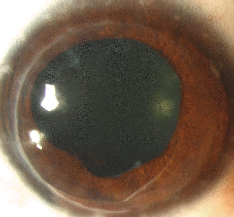
Figure 2.
Case 5. Twelve years postoperatively, the fixed dilated pupil, iris atrophy, and diffuse posterior synechiae is sustained in the left eye.
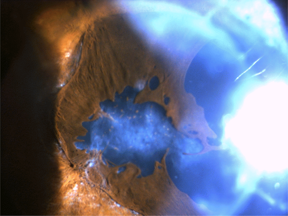
Figure 3.
Case 4. Twelve years postoperatively, fixed dilated pupil, severe iris atrophy, and diffuse posterior synechiae are seen in the left eye.
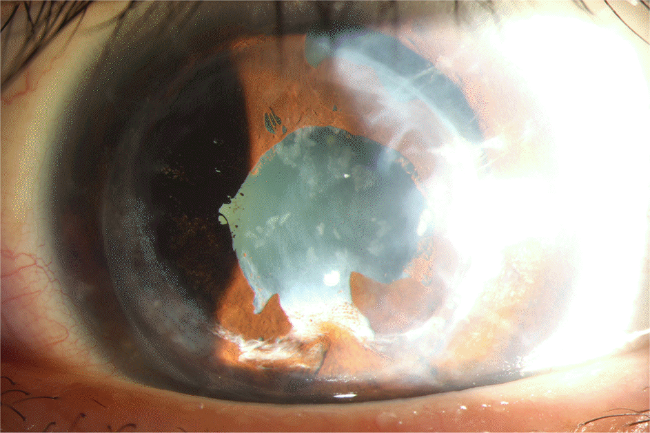
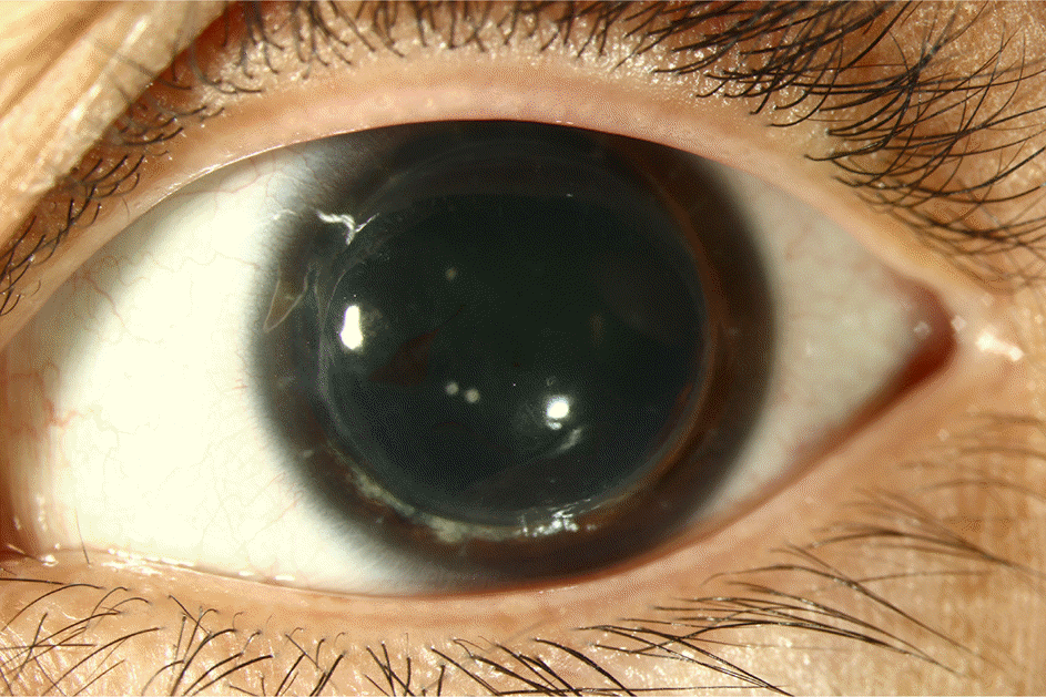
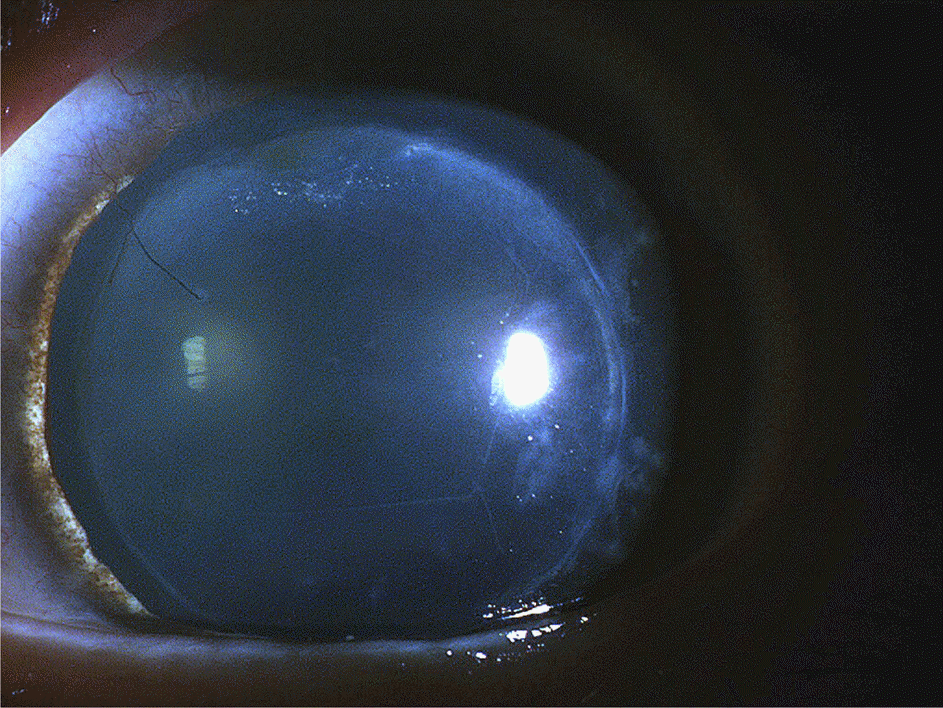
Table 1.
Demographics of each patient with fixed dilated pupil after intraocular surgeries
Dx = diagnosis; Preop = preoperative; BCVA = best corrected visual acuity; IOP = intraocular pressure; ICE syndrome = iridocorneal endothelial syndrome; PRK = photorefractive keratectomy; PKP = penetrating keratoplasty; KP = keratoplasty; Phaco + PCL = phacoemulsification and posterior chamber lens insertion; ECCE + PCL = extracapsular cataract extraction and posterior chamber lens insertion; FC = finger count.
Table 2.
Characteristics of each patient with fixed dilated pupil after intraocular surgeries
| Case | POD#1∗ IOP (mm Hg) | FDP† (days) | Anterior chamber cells | Aqueous flare | Fibrotic memb. | Pupillary margin | Iris atrophy | Posterior synechiae |
|---|---|---|---|---|---|---|---|---|
| 1 | 60 | 1 | + | - | - | Regular | Mild | Localized |
| 2 | 43 | 1 | + | - | - | Regular | Mild | Absent |
| 3 | 34 | 2 | +++ | +++ | + | Irregular | Moderate | Multiple |
| 4 | 29 | 3 | ++++ | +++ | + | Irregular | Severe | Multiple |
| 5 | 38 | 2 | ++++ | ++++ | + | Irregular | Severe | Multiple |
| 6 | 22 | 2 | +++ | ++ | + | Irregular | Moderate | Localized |
| 7 | 42 | 1 | ++++ | ++++ | + | Irregular | Severe | Multiple |
| 8 | 46 | 1 | +++ | +++ | + | Irregular | Moderate | Multiple |
| 9 | 34 | 1 | + | - | - | Regular | Absent | Absent |
| 10 | 35 | 1 | ++ | + | - | Regular | Mild | Absent |
| 11 | 37 | 1 | ++ | + | - | Regular | Mild | Localized |
| 12 | 37 | 1 | + | - | - | Regular | Absent | Absent |
| 13 | 59 | 2 | + | - | - | Regular | Absent | Absent |
| 14 | 33 | 1 | ++ | - | - | Regular | Mild | Localized |
| 15 | 26 | 1 | + | + | - | Regular | Mild | Absent |
Table 3.
Classification of fixed dilated pupil after intraocular surgeries
| Group 1 | Group 2 | Group 3 | |
|---|---|---|---|
| (case 1 and 2) | (case 3-8) | (case 9-15) | |
| Viscoelastic material | Used | Used | Used |
| FDP∗ | 1.00 day | 1.83 days | 1.14 days |
| Pupillary margin | Regular | Irregular | Regular |
| Iris atrophy | Absent or mild | Moderate or severe | Absent or mild |
| Posterior synechiae | Absent or localized | Multiple or localized | Absent or localized |
| Anterior chamber cells | Mild | Severe | Mild |
| Aqueous flare | Nil | Marked or intense | Nil or just detectable |
| Fibrotic membrane | - | + | - |
| Features | Mild A/C cells | Severe A/C cells | Mild A/C cells |
| Fibrotic membrane (-) | Fibrotic membrane (+) | Fibrotic membrane (-) | |
| Regular pupil margin | Irregular pupil margin | Regular pupil margin | |
| Mild iris atrophy | Severe iris atrophy | Mild iris atrophy | |
| D/Dx. point | Atropine use | Postop. severe A/C inflammation | Intra A/C air or gas injection |
| PAS |
 XML Download