Abstract
Purpose
To report a case of corneal ulcer caused by Paecilomyces lilacinus after penetrating keratoplasty.
Case summary
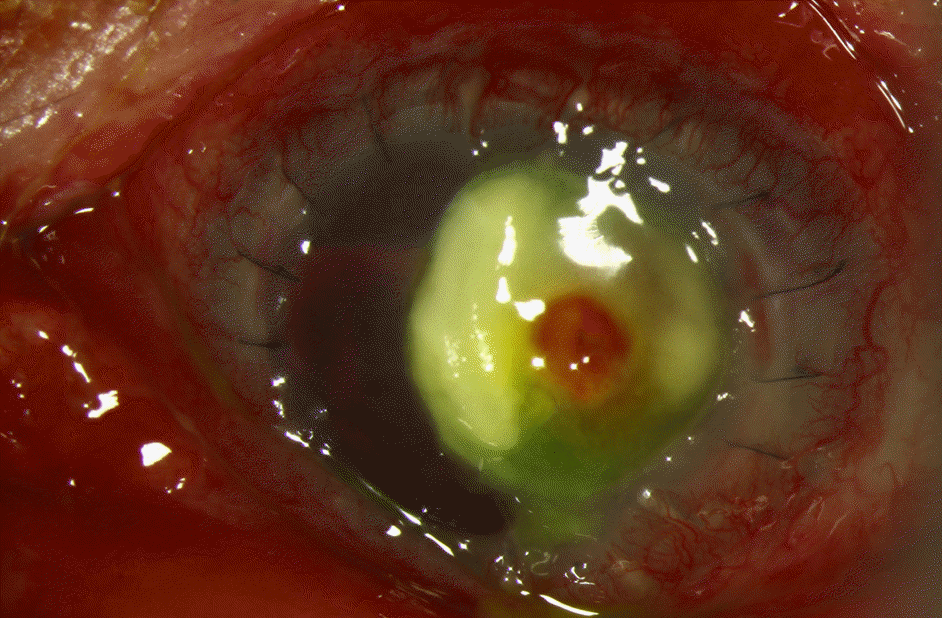
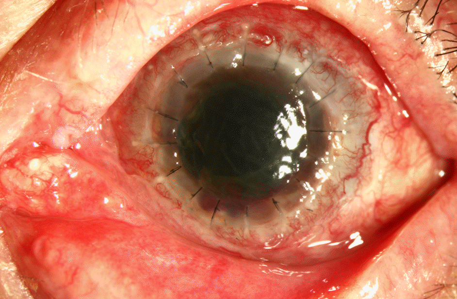
A 67-year-old male with a history of penetrating keratoplasty in the left eye 7 years prior and re-penetrating keratoplasty in the left eye due to graft failure in June 2013, visited our clinic for ocular pain and conjunctival injection in the left eye 3 days in duration. Corneal scrapings were performed for Gram and fungal stains and cultures. The patient was admitted to the hospital for hourly topical fortified ceftazidime and amphotericin B. Despite intensive topical therapy, no improvement was observed. Three days later, fungal culture confirmed Paecilomyces lilacinus and topical voriconazole was prepared from the intravenous formulation and was administered topically and intravenously. Despite medical therapy with voriconazole, perforation occurred requiring a tectonic keratoplasty.
Go to : 
References
1. Lee DC, Lee JW, Chang SD. A case of fusarium keratitis treated with moxifloxacin 0.5% ophthalmic solution. J Korean Ophthalmol Soc. 2012; 53:338–41.

2. Kozarsky AM, Stulting RD, Waring GO 3rd, et al. Penetrating keratoplasty for exogenous Paecilomyces keratitis followed by postoperative endophthalmitis. Am J Ophthalmol. 1984; 98:552–7.

3. Hariprasad SM, Mieler WF, Lin TK, et al. Voriconazole in the treatment of fungal eye infections: a review of current literature. Br J Ophthalmol. 2008; 92:871–8.

5. Whitcher JP, Srinivasan M, Upadhyay MP. Corneal blindness: a global perspective. Bull World Health Organ. 2001; 79:214–21.
6. Oh DH, Kim JC, Chun YS. A case of fusarium deep keratitis following scleral graft. J Korean Ophthalmol Soc. 2010; 51:606–10.

7. O'Day DM, Head WS, Robinson RD, Clanton JA. Corneal penetration of topical amphotericin B and natamycin. Curr Eye Res. 1986; 5:877–82.
8. Hariprasad SM, Mieler WF, Lin TK, et al. Voriconazole in the treatment of fungal eye infections: a review of current literature. Br J Ophthalmol. 2008; 92:871–8.

9. Aydin S, Ertugrul B, Gultekin B, et al. Treatment of two postoperative endophthalmitis cases due to Aspergillus flavus and Scopulariopsis spp. with local and systemic antifungal therapy. BMC Infect Dis. 2007; 7:87.

10. Sabo JA, Abdel-Rahman SM. Voriconazole: a new triazole antifungal. Ann Pharmacother. 2000; 34:1032–43.

11. Jurkunas UV, Langston DP, Colby K. Use of voriconazole in the treatment of fungal keratitis. Int Ophthalmol Clin. 2007; 47:47–59.

12. Johnson LB, Kauffman CA. Voriconazole: a new triazole anti-fungal agent. Clin Infect Dis. 2003; 36:630–7.

13. Deng SX, Kamal KM, Hollander DA. The use of voriconazole in the management of post-penetrating keratoplasty Paecilomyces keratitis. J Ocul Pharmacol Ther. 2009; 25:175–7.

14. Pastor FJ, Guarro J. Clinical manifestations, treatment and outcome of Paecilomyces lilacinus infections. Clin Microbiol Infect. 2006; 12:948–60.

15. Byun DS, Yang HN, Cho HG, Cha YJ. A case of corneal ulcer caused by Paecilomyces in diabetic patient wearing soft contact lens. J Korean Ophthalmol Soc. 1987; 28:667–71.
16. Bunya VY, Hammersmith KM, Rapuano CJ, et al. Topical and oral voriconazole in the treatment of fungal keratitis. Am J Ophthalmol. 2007; 143:151–3.

Go to : 
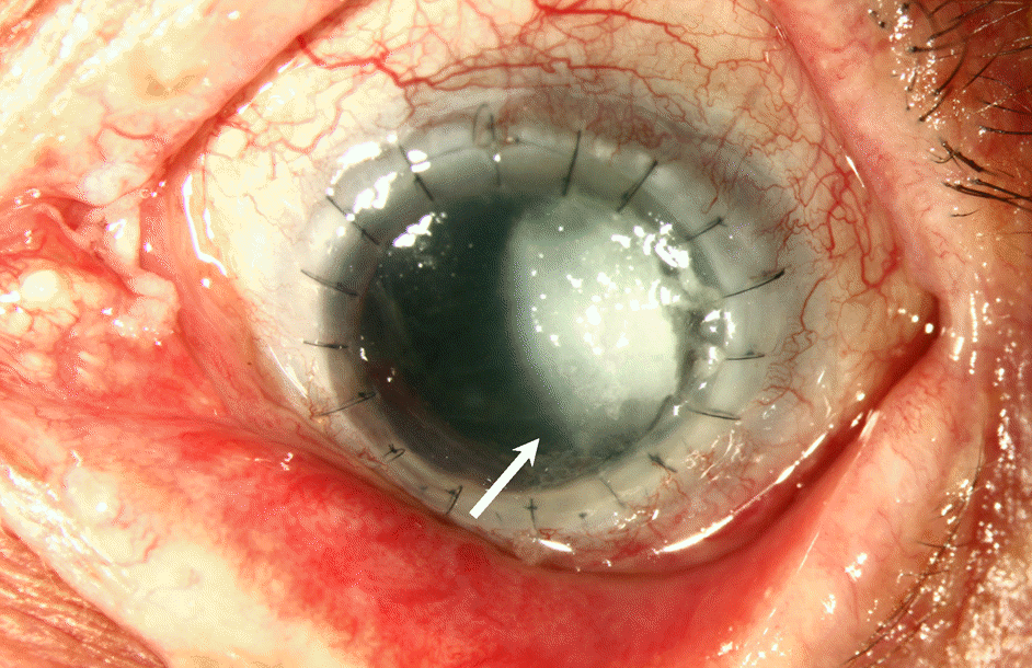
 | Figure 1.Pre-therapeutic photograph. 3.8 × 6 mm sized corneal ulcer with feathery margin (arrow) and deep stromal infiltration were observed on initial presentation. |


 PDF
PDF ePub
ePub Citation
 Print
Print


 XML Download