

 PDF
PDF ePub
ePub Citation
 Print
Print


Abstract
Case summary
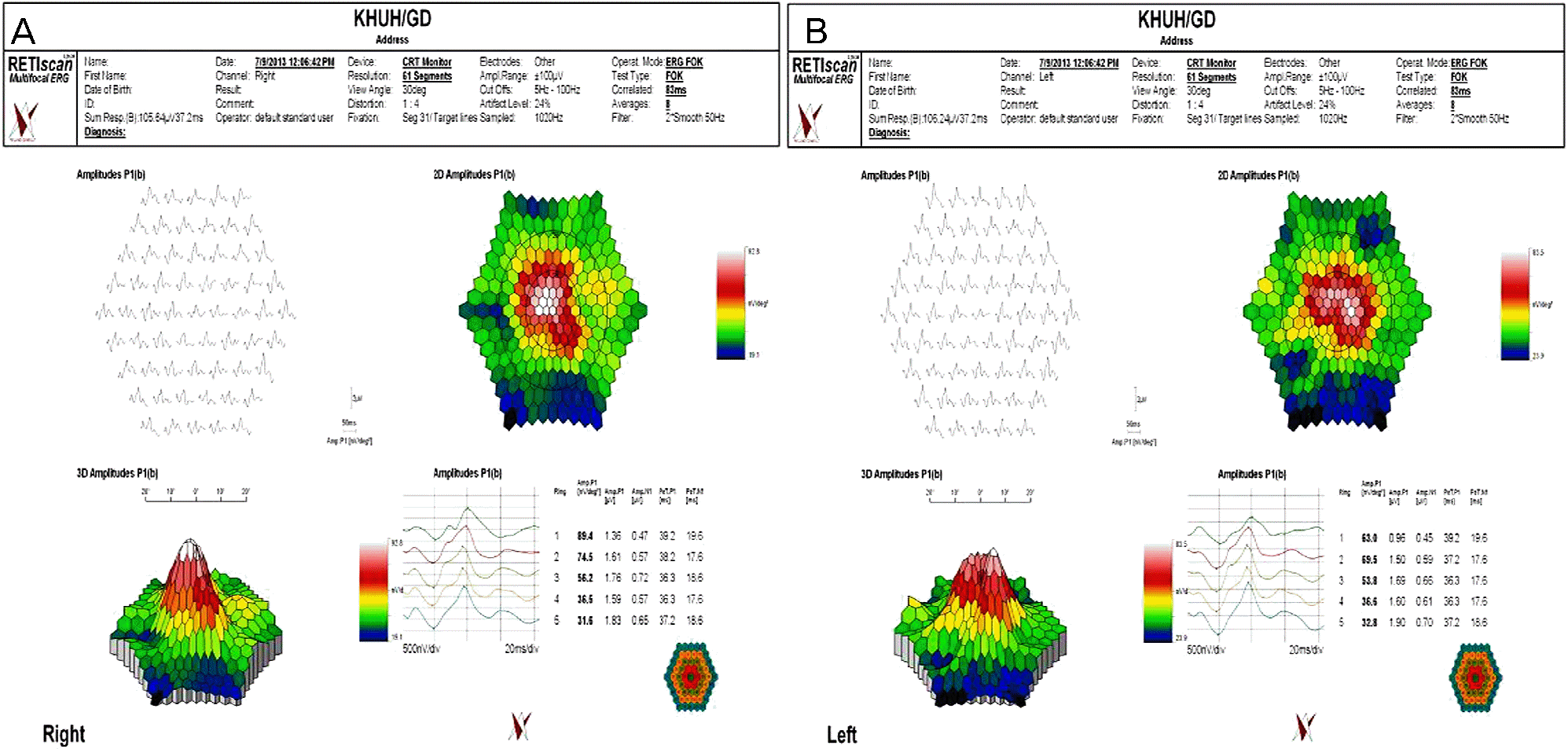
A 31-year-old female was referred to our clinic with blurred vision. The patient had visited Bali, Indonesia approximately 2 weeks prior. Dengue fever was diagnosed at the Division of Infectious Disease because the patient’s serum dengue virus antibodies test was positive for dengue viral IgM antibodies. The patient’s best corrected visual acuity was 0.4 in the right eye and 0.6 in the left eye. Slit lamp examination showed inflammatory cells in the vitreous but not in the anterior segment of both eyes. Fundus examination showed thickening of the retina in both eyes as well as a splinter retinal hemorrhage in left eye. Fluorescein angiography revealed hyperfluorescence of the venule in the perifoveal vascular network of the left macula, and indocyanine green angiography showed early diffuse hyperfluorescence in both eyes. Standard automated perimetry showed an overall reduction of the visual field and an increase in scotoma in both eyes. At 1 week after the initial visit, the macular edema had not improved and the patient’s vision had deteriorated, especially in left eye. To improve the macular edema and ocular inflammation, a subtenon triamcinolone acetonide injection in the left eye was administered. At 5 weeks after treatment, corrected visual acuity improved to 1.0 in both eyes. Ocular findings, such as macular edema and intraretinal hemorrhage were resolved. The patient did not complain of any remaining discomfort. However, standard automated perimetry revealed that a pericentral scotoma was still present in left eye.
References
1. Simmons CP, Farrar JJ, Nguyen vV, Dengue Wills B.N Engl J Med. 2012; 366:1423–32.
2. Martins Sde T, Silveira GF, Alves LR. . Dendritic cell apoptosis and the pathogenesis of dengue. Viruses. 2012; 4:2736–53.

3. Wilder-Smith A.Dengue infections in travellers. Paediatr Int Child Health. 2012; 32(Suppl 1):28–32.
4. Chung MH.Dengue fever. Korean J Med. 2009; 77:165–70.
5. Wichmann O, Hongsiriwon S, Bowonwatanuwong C. . Risk factors and clinical features associated with severe dengue in-fection in adults and children during the 2001 epidemic in Chonburi, Thailand. Trop Med Int Health. 2004; 9:1022–9.
6. Su DH, Bacsal K, Chee SP. et al. Dengue Maculopathy Study Group. Prevalence of dengue maculopathy in patients hospitalized for dengue fever. Ophthalmology. 2007; 114:1743–7.
7. Chan DP, Teoh SC, Tan CS. . Ophthalmic complications of dengue. Emerg Infect Dis. 2006; 12:285–9.
8. Juanarita J, Azmi MN, Azhany Y, Liza-Sharmini AT. Dengue re-lated maculopathy and foveolitis. Asian Pac J Trop Biomed. 2012; 2:755–6.
9. Kim MJ, Oh DH, Oh IM. . A case of dengue fever complicated by retinitis. Infect Chemother. 2012; 44:307–9.
10. Nam KW, Kim JE, Doo EY. . A case of dengue fever with maculopathy. Infect Chemother. 2012; 44:504–7.
11. Gupta A, Srinivasan R, Setia S. . Uveitis following dengue fever. Eye (Lond). 2009; 23:873–6.
12. Sanjay S, Wagle AM, Au Eong KG. Optic neuropathy associated with dengue fever. Eye (Lond). 2008; 22:722–4.
13. Teoh SC, Chee CK, Laude A. et al. Eye Institute Dengue-related Ophthalmic Complications Workgroup. Optical coherence tomog-raphy patterns as predictors of visual outcome in dengue-related maculopathy. Retina. 2010; 30:390–8.
14. Loh BK, Bacsal K, Chee SP. . Foveolitis associated with den-gue Fever: a case series. Ophthalmologica. 2008; 222:317–20.
15. Bacsal KE, Chee SP, Cheng CL, Flores JV.Dengue-associated maculopathy. Arch Ophthalmol. 2007; 125:501–10.
16. Chia A, Luu CD, Mathur R. . Electrophysiological findings in patients with dengue-related maculopathy. Arch Ophthalmol. 2006; 124:1421–6.
17. Lim WK, Mathur R, Koh A. . Ocular manifestations of dengue fever. Ophthalmology. 2004; 111:2057–64.
18. Chee E, Sims JL, Jap A. . Comparison of prevalence of dengue maculopathy during two epidemics with differing predominant serotypes. Am J Ophthalmol. 2009; 148:910–3.
Figure 1.
(A, B) Fundus photography showed intraretinal white lesions along the venules and arterioles of the perifoveolar vascular network and thickening of the retina at the papillomacular bundle in both eyes and a splinter retinal hemorrhage in the left eye at patient’s first visit. (C, D) Six weeks after the first visit, white lesions, thickening of retina and a splinter hemorrhage and macular edema subsided.
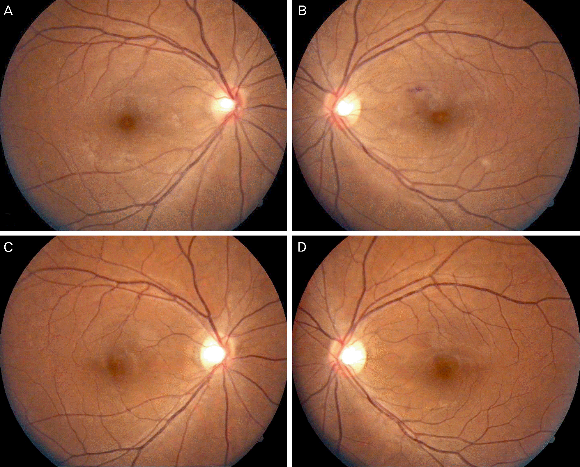
Figure 2.
(A, B) Fluorescein angiography (left column) revealed late hyperfluorescence of the arterioles of the macula in left eye. Indocyanine green angiography (right column) showed early diffuse hyperfluorescence in both eyes at patient’s first visit. The hypofluorescent spots (white arrows) which were corresponding to yellow retinal lesion on fundus photograph were noted. (C, D) Six weeks after the first visit, hyperfluorescence were resolved.
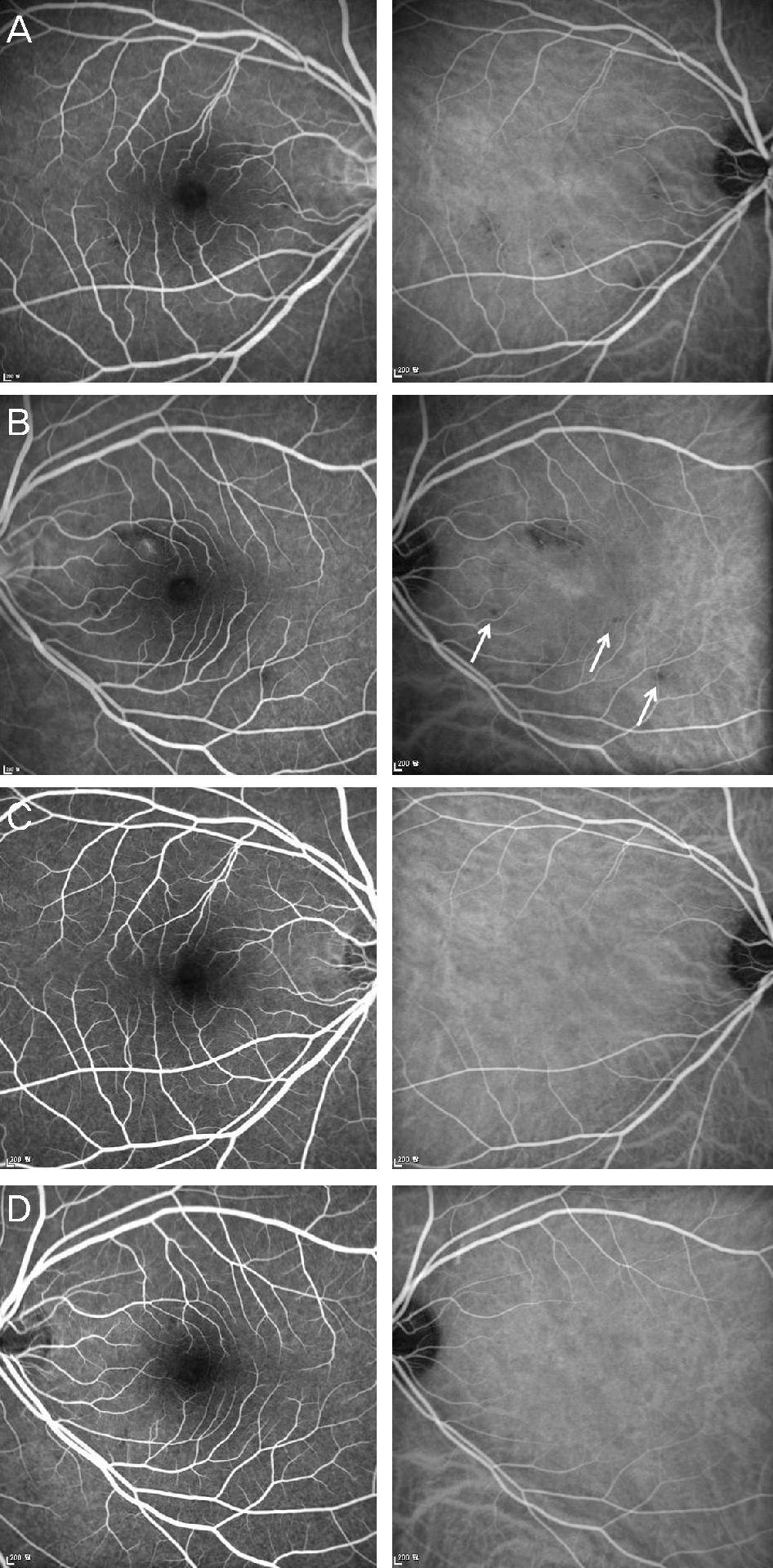
Figure 3.
(A, B) Optical coherence tomography showed irreg-ular density of outer neurosensory retina at the fovea in left eye. Focal subretinal fluid and retinal edema mainly affecting maculopapular bundle were observed in both eyes at patient’s first visit. (C, D) Six weeks after the first visit, macular thickening and subretinal fluid were resolved.
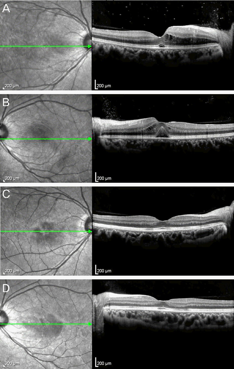
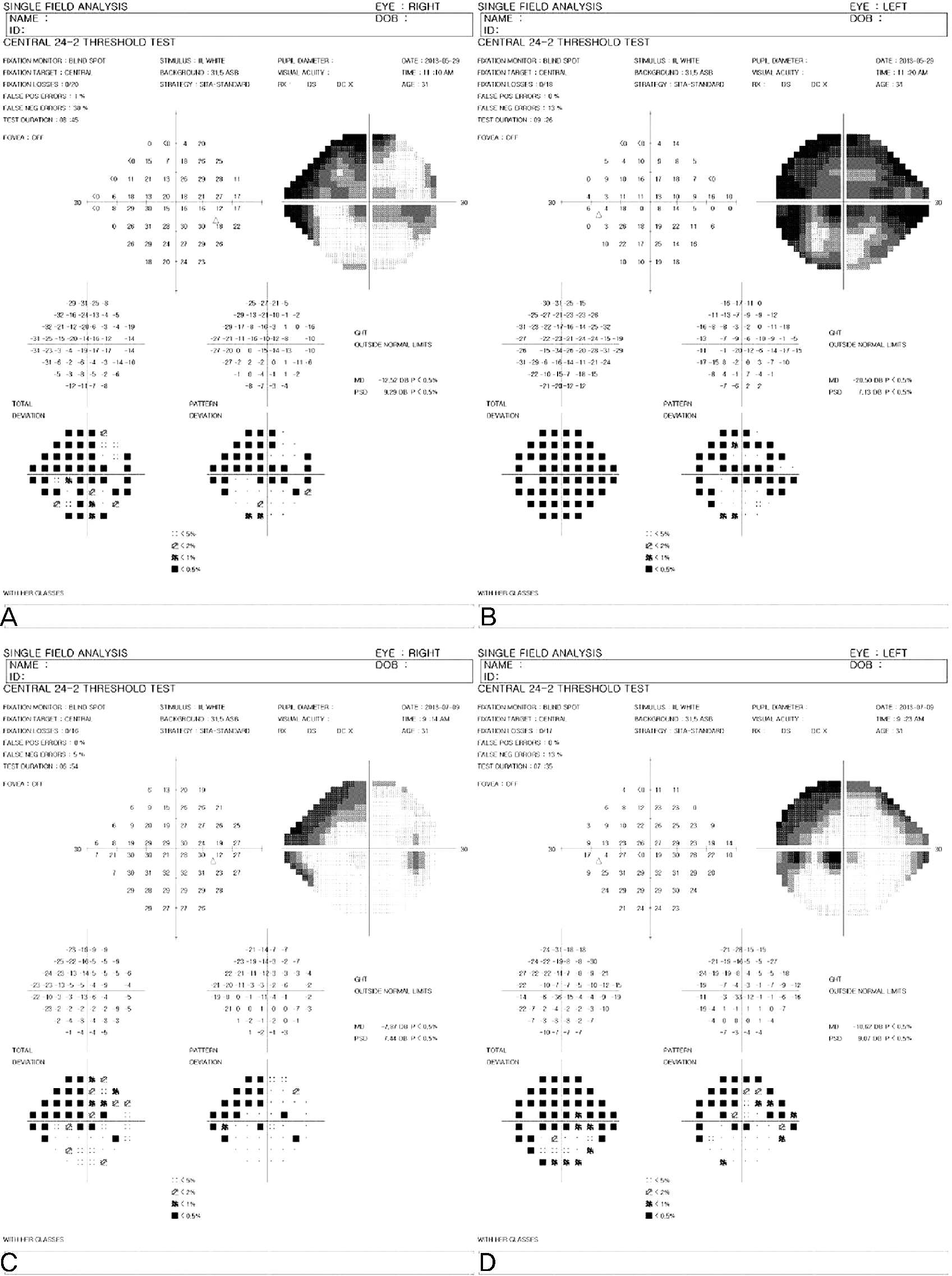
Figure 4.
(A, B) Humphrey visual fields show overall reduction of visual field and the increase of scotoma were observed in both eyes at patient’s first visit. (C, D) Six weeks after the first visit, peripheral visual field defects were improved in both eyes, but central scotoma remained in the left eye.
 XML Download