Abstract
Purpose
To report a case of bilateral Candida albicans endophthalmitis after robot-assisted laparoscopic radical prostatectomy (RALP) in a patient with prostate adenocarcinoma.
Case summary
A 61-year-old male patient had RALP for prostate adenocarcinoma. Four days after the surgery, the patient had a fever and pancytopenia. Two weeks after the surgery, the patient complained of decreased visual acuity in both eyes. Best-corrected visual acuity (BCVA) was counting fingers at 60 cm in the right eye and 20/63 in the left eye. The fundus examination showed vitreous opacity and yellow circumscribed chorioretinal lesions in both eyes. The blood and urine cultures were negative. An intravitreal voriconazole injection (100 µg/0.1 ml) was given in both eyes. However, the vitritis worsened, and BCVA decreased to counting fingers at 30 cm in the right eye and 60 cm in the left eye. Thus, combined phacoemulsification and 23-gauge microincisional vitrectomy surgery with a silicone oil injection was performed in both eyes at 1-week intervals. The vitrectomy sample culture was positive for Candida albicans in both eyes. Four months after the vitrectomy, BCVA was 20/100 in both eyes without subretinal lesions.
Figures and Tables
Figure 1
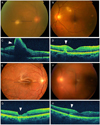
Fundus photographs and optical coherence tomography (OCT) of a patient with bilateral Candida albicans endogenous endophthalmitis. (A, B) Two weeks after robotic-assisted laparoscopic prostatectomy, the fundus examination showed yellow circumscribed subretinal lesions in the right macula, and in the superonasal and inferior to the left fovea. The black horizontal arrow in A and B shows the line scanned by OCT in C and D, respectively. (C, D) The OCT of the right eye shows a diffusely thickened retina. The retinal surface is highly elevated at the yellow subretinal lesion with whole retinal infiltration (arrowhead). The left eye shows a disruption in the retinal layers from the retinal pigment epithelium (RPE) to the inner nuclear layer (arrowhead). (E, F) Four months after 23-gauge microincisional vitrectomy surgery with silicone oil injection, the fundus examination shows resolution of the vitritis and subretinal infiltration. The fundus image of the left eye is slightly dimmed due to posterior capsular opacity. The black horizontal arrow in E and F shows the line scanned by OCT in G and H, respectively. (G, H) The OCT shows disruptions in the RPE and photoreceptor layers in both eyes.

References
1. Ficarra V, Cavalleri S, Novara G, et al. Evidence from robot-assisted laparoscopic radical prostatectomy: a systematic review. Eur Urol. 2007. 51:45–55. discussion 56.
2. Lingappan A, Wykoff CC, Albini TA, et al. Endogenous fungal endophthalmitis: causative organisms, management strategies, and visual acuity outcomes. Am J Ophthalmol. 2012. 153:162–166.
3. Toshikuni N, Ujike K, Yanagawa T, et al. Candida albicans endophthalmitis after extracorporeal shock wave lithotripsy in a patient with liver cirrhosis. Intern Med. 2006. 45:1327–1332.
4. Scheetz MH, McKoy JM, Parada JP, et al. Systematic review of piperacillin-induced neutropenia. Drug Saf. 2007. 30:295–306.
5. Breit SM, Hariprasad SM, Mieler WF, et al. Management of endogenous fungal endophthalmitis with voriconazole and caspofungin. Am J Ophthalmol. 2005. 139:135–140.
6. Sen P, Gopal L, Sen PR. Intravitreal voriconazole for drug-resistant fungal endophthalmitis: case series. Retina. 2006. 26:935–939.


 PDF
PDF ePub
ePub Citation
 Print
Print


 XML Download