Abstract
Purpose
To report the treatment results of mass excision and cryotherapy in a case of bilateral conjunctival amyloidosis.
Case summary
A 72-year-old man with conjunctival mass and foreign body sensations in both eyes visited our clinic. He was previously diagnosed with conjunctival lymphoma after conjunctival biopsy at another hospital. A yellow-pink colored mass was observed in the bulbar conjunctiva. A repeated conjunctival biopsy revealed the mass to be amyloid, consistent with the cervical lymph node biopsy results, and the diagnosis was changed to primary systemic amyloidosis. The patient was treated with melphalan and prednisolone; however, the ocular pain, symblepharon, and conjunctival mass progressed. A 360 degree conjunctivoperiotomy, mass excision, and repeated cryotherapy were performed in the more severely affected left eye. The patient was followed for one year, and there were no complications or progression of the conjunctival lesion.
References
1. Westermark P, Benson MD, Buxbaum JN, et al. Amyloid fibril protein nomenclature - 2002. Amyloid. 2002; 9:197–200.

2. Lee HM, Naor J, DeAngelis D, Rootman DS. Primary localized conjunctival amyloidosis presenting with recurrence of subconjunctival hemorrhage. Am J Ophthalmol. 2000; 129:245–7.

3. Moorman CM, McDonald B. Primary (localised non-familial) conjunctival amyloidosis: three case reports. Eye (Lond). 1997; 11:603–6.

4. Demirci H, Shields CL, Eagle RC Jr, Shields JA. Conjunctival amyloidosis: report of six cases and review of the literature. Surv Ophthalmol. 2006; 51:419–33.

5. Fraunfelder FW. Liquid nitrogen cryotherapy for conjunctival amyloidosis. Arch Ophthalmol. 2009; 127:645–8.

6. Rosenblatt M, Spitz GF, Friedman AH, Kazam ES. Localized conjunctival amyloidosis: case reports and review of literature. Ophthalmologica. 1986; 192:238–45.

7. Merlini G, Westermark P. The systemic amyloidoses: clearer understanding of the molecular mechanisms offers hope for more effective therapies. J Intern Med. 2004; 255:159–78.

9. Leibovitch I, Selva D, Goldberg RA, et al. Periocular and orbital amyloidosis: clinical characteristics, management, and outcome. Ophthalmology. 2006; 113:1657–64.
11. Eichler MD, Fraunfelder FT. Cryotherapy for conjunctival lym-phoid tumors. Am J Ophthalmol. 1994; 118:463–7.

12. Hazenberg BP, van Gameren II, Bijzet J, et al. Diagnostic and therapeutic approach of systemic amyloidosis. Neth J Med. 2004; 62:121–8.
13. Song BR, Kim YK, Yoo JH, Chu YC. Primary localized amyloidosis of bulbar conjunctiva and cornea. J Korean Ophthalmol Soc. 1993; 34:352–6.
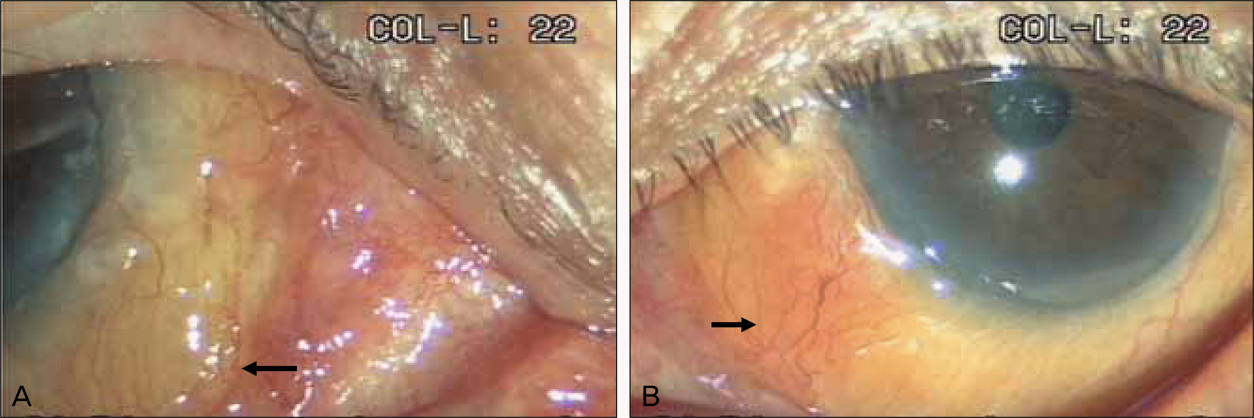
Figure 1.
A 4×3 mm sized yellow mass (A) (arrow) is seen in the inferomedial conjunctiva in the right eye. A 6×5 mm sized yel-low-pinkish mass (B) (arrow) is seen in the inferomedial conjunctiva in the left eye. The mass was larger and more severe in the left eye.


 PDF
PDF ePub
ePub Citation
 Print
Print


 XML Download