Abstract
Purpose
To identify prognostic factors associated with a favorable outcome after vitrectomy for patients with macular epiretinal membrane (ERM).
Methods
The authors retrospectively reviewed the records of 63 patients (64 eyes) with macular ERM, who were treated by vitrectomy between 2003 and 2008, and followed for more than 6 months.
Results
The mean follow-up period was 13.21 ± 9.11 months and the mean best corrected visual acuity after vitrectomy was log MAR 0.32 ± 0.34. Univariate analysis revealed the patients in the group with a postoperative log MAR of 0.3 or better had better preoperative visual acuity and shorter symptom duration; multivariate analysis revealed the same results. In 24 eyes, intraretinal structures which contained pseudoholes, intraretinal cysts, retinal folds and vitreoretinal traction were analyzed with Cirrus HD-OCT, however, there was no correlation with visual acuity after vitrectomy.
References
1. Poliner LS, Olk RJ, Grand MG, et al. Surgical management of premacular fibroplasia. Arch Ophthalmol. 1988; 106:761–4.

2. Wise GN. Clinical features of idiopathic preretinal macular fibrosis. Schoenberg Lecture. Am J Ophthalmol. 1975; 79:349–57.
3. de Bustros S, Thompson JT, Michels RG, et al. Vitrectomy for idiopathic epiretinal membranes causing macular pucker. Br J Ophthalmol. 1988; 72:692–5.

4. Pournaras CJ, Donati G, Brazitikos PD, et al. Macular epiretinal membranes. Semin Ophthalmol. 2000; 15:100–7.

5. Choi YK, Yoo JS, Kim MH. Result of surgery for epiretinal membrane and their recurrence. J Korean Ophthalmol Soc. 2000; 41:2357–62.
6. Rice TA, De Bustros S, Michels RG, et al. Prognostic factors in vitrectomy for epiretinal membranes of the macula. Ophthalmology. 1986; 93:602–10.

7. Pesin SR, Olk RJ, Grand MG, et al. Vitrectomy for premacular fibroplasia. Prognostic factors, long-term follow-up, and time course of visual improvement. Ophthalmology. 1991; 98:1109–14.
8. Wong JG, Sachdev N, Beaumont PE, Chang AA. Visual outcomes following vitrectomy and peeling of epiretinal membrane. Clin Experiment Ophthalmol. 2005; 33:373–8.

9. Kim HC, Kim HK, Lee HC. The effect of surgical management for macular epiretinal membranes. J Korean Ophthalmol Soc. 1995; 36:1713–20.
10. Lee HJ, Kim HC. The clinical outcome and prognostic factors of vitrectomy for macular epiretinal membranes. J Korean Ophthalmol Soc. 2003; 44:857–64.
11. Kawasaki R, Wang JJ, Mitchell P, et al. Racial difference in the prevalence of epiretinal membrane between caucasians and asains. Br J Ophthalmol. 2008; 92:1320–4.
12. Donati G, Kapetanios AD, Pournaras CJ. Complications of surgery for epiretinal membranes. Graefes Arch Clin Exp Ophthalmol. 1998; 236:739–46.

13. Ando A, Nishimura T, Uyama M. Surgical outome on combined procedures of lens extraction, intraocular lens implantation, and vitrectomy during removal of the epiretinal membrane. Ophthalmic Surg Lasers. 1998; 29:974–9.
14. Kim YC, Kim KS. The effect of internal limiting membrane peeling in treatment of idiopathic epiretinal membrane. J Korean Ophthalmol Soc. 2007; 48:1067–72.

15. Schadlu R, Tehrani S, Shah GK, Prasad AG. Long-term follow-up results of ILM peeling during vitrectomy surgery for premacular fibrosis. Retina. 2008; 28:853–7.

16. Kwok AKh, Lai TY, Yuen KS. Epiretinal membrane surgery with or without internal limiting membrane peeling. Clin Experiment Ophthalmol. 2005; 33:379–85.

17. Wilkins JR, Puliafito CA, Hee MR, et al. Characterization of epiretinal membranes using optical coherence tomography. Ophthalmology. 1996; 103:2142–51.

18. Mori K, Gehlbach PL, Sano A, et al. Comparison of epiretinal membranes of differing pathogenesis using optical coherence tomography. Retina. 2004; 24:57–62.

19. Massin P, Allouch C, Haouchine B, et al. Optical coherence tomography of idiopathic macular epiretinal membranes before and after surgery. Am J Ophthalmol. 2000; 130:732–9.

20. Kim CH, Kim JI, Cho HY, Kang SW. Correlation between preoperative OCT pattern and visual improvement in macular epiretinal membrane. J Korean Ophthalmol Soc. 2007; 48:75–82.
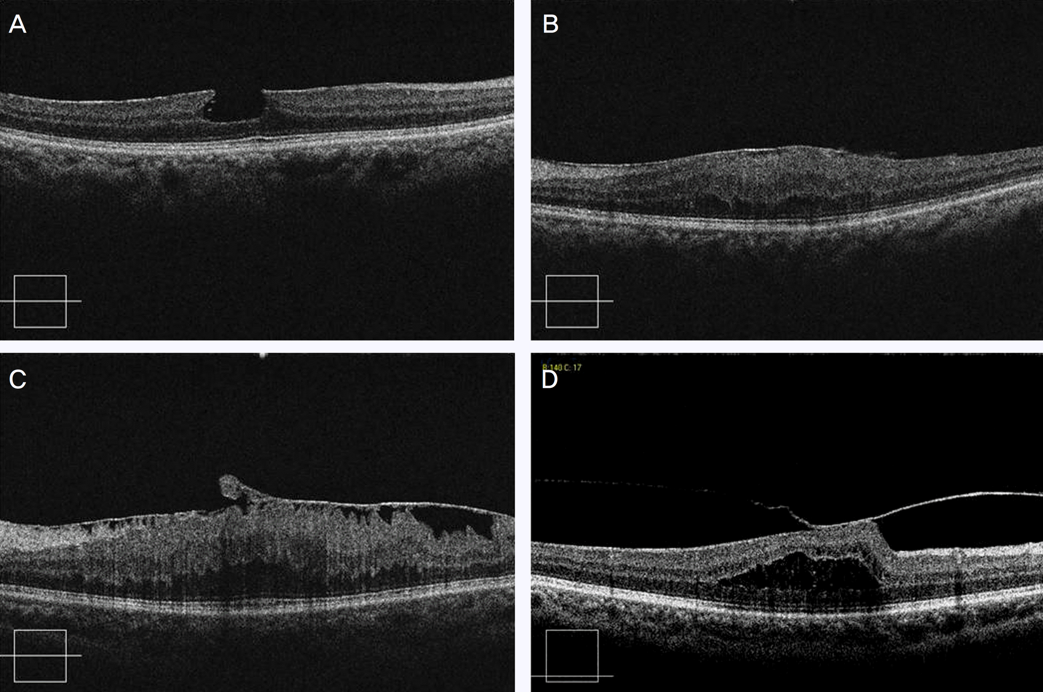
Figure 1.
The intraretinal structures of patients with macular ERM in Cirrus-HD OCT. (A) Pseudohole at fovea, (B) Diffuse retinal thickening of retina with globally adherent ERM, (C) Retinal fold with partially adherent ERM, (D) Vitreomacular traction with macular edema.

Table 1.
Postoperative visual outcome (log MAR)
| Visual Acuity change | Number of eyes (%) |
|---|---|
| Improved ≥ 2 lines | 46 (71.8) |
| Improved < 2 lines | 11 (17.2) |
| Unchanged | 5 (7.8) |
| Worsened | 2 (3.2) |
Table 2.
Results of univariate analysis (independent t-test)
| Factors |
Postoperative BCVA* (mean ± SD, log MAR) |
||
|---|---|---|---|
| > 0.3 | ≤ 0.3 | p-value† | |
| Preoperative visual acuity (log MAR) | 0.47 ± 0.32 | 0.89 ± 0.56 | 0.001 |
| Age (yr) | 66.58 ± 6.97 | 65.50 ± 8.96 | 0.588 |
| Symptom duration (mon) | 7.32 ± 6.53 | 14.28 ± 13.09 | 0.012 |
| Preoperative macular thickness (μ m) | 367.94 ± 143.64 | 388.56 ± 171.48 | 0.602 |
Table 3.
Results of univariate analysis (Chi-square)
| Factors |
Postoperative BCVA* (log MAR) |
||
|---|---|---|---|
| > 0.3 | ≤ 0.3 | p-value† | |
| Sex (M/F) | 10/24 | 7/23 | 0.777 |
| Etiology (primary/secondary) | 16/18 | 16/14 | 0.401 |
| Gas tamponade (yes/no) | 15/19 | 16/14 | 0.617 |
| ILM‡ peeling (yes/no) | 6/28 | 6/24 | 1.000 |
Table 4.
Results of multiple linear regression analysis
| Factors | p-value* |
|---|---|
| Age | 0.424 |
| Sex | 0.328 |
| Etiology | 0.831 |
| Preoperative visual acuity | 0.000 |
| Symptom duration | 0.000 |
| Gas tamponade | 0.781 |
| ILM† peeling | 0.305 |
| Preoperative macular thickness (μ m) | 0.979 |


 PDF
PDF ePub
ePub Citation
 Print
Print


 XML Download