Abstract
Purpose
To evaluate the therapeutic effect of bevacizumab in treating myopic choroidal neovascularization (CNV).
Methods
Medical records of the eyes that underwent intravitreal bevacizumab injection for myopic CNV and were followed up for more than one year were reviewed retrospectively. Best corrected visual acuity (BCVA), foveal thickness in optical coherence tomography and fluorescein angiography were investigated.
Results
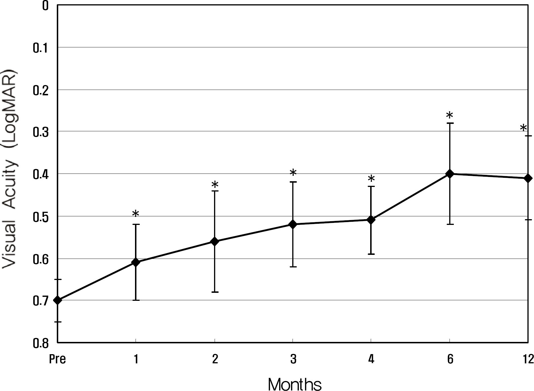
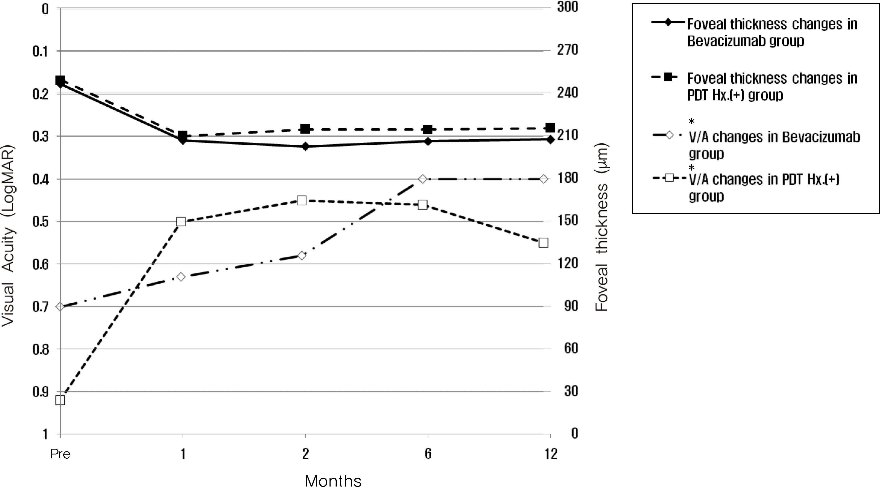
Twenty eyes of 18 patients were included in the present study. The average age was 41.5 years, average axial length was 28.5 mm, and average refractive error was −11.0 diopters. Visual acuity was maintained in all eyes, and 13 eyes improved by LogMAR 0.3 or more. Visual acuity improved significantly from 0.71 (0.2∼2.0) to 0.40 (0.1∼2.0, p =0.02) at six months and to 0.41 (0.1∼2.0, p =0.03) at one year. Central foveal thickness significantly decreased from 247.0 μ m to 207.5 μ m (p =0.03) at six months and to 208.5 μ m (p =0.04) at one year.
Go to : 
References
1. Chung EJ, Oh HS, Koh HJ, Lee SC, Kwon OW. Photodynamic Therapy in Practice: A Review of Experiences with Myopic CNV in Korean Patients. J Korean Ophthalmol Soc. 2005; 46:664–70.
2. Hotchkiss ML, Fine SL. Pathologic myopia and choroidal neovascularization. Am J Ophthalmol. 1981; 91:177–83.

3. Verteporfin in Photodynamic Therapy (VIP) Study Group. Photodynamic therapy of subfoveal choroidal neovascularization in pathologic myopia with verteporfin: 1year results of a randomized clinical trial-VIP report no. 1. Ophthalmology. 2001; 108:841–52.
4. Verteporfin in Photodynamic Therapy(VIP) Study Group. Photodynamic therapy of subfoveal choroidal neovascularization in pathologic myopia: 2-year results of a randomized clinical trial-VIP report no. 3. Ophthalmology. 2003; 110:667–73.
5. Kourlas H, Abrams P. Ranibizumab for the treatment of neovascular age-related macular degeneration: a review. Clinical Therapeutics. 2007; 29:1850–61.

6. Heier JS, Antoszyk AN, Pavan PR, et al. Ranibizumab for treatment of neovascular age-related macular degeneration: a phase I/II multicenter, controlled, multidose study. Ophthalmology. 2006; 113:633–4.
7. Steidl SM, Pruett RC. Macular complications associated with posterior staphyloma. Am J Ophthalmol. 1997; 123:181–7.

8. Brancato R, Trabucchi G, Introini U, et al. Indocyanine green angiography (ICGA) in pathologic myopia. Eur J Ophthalmol. 1996; 6:39–43.
9. Macular Photocoagulation Study Group. Laser photocoagulation of subfoveal Neovascular lesions in age related macular degeneration: results of a randomized clinical trial. Arch Ophthalmol. 1991; 109:1220–31.
10. Macular Photocoagulation Study Group. Subfoveal Neovascular lesions in age related macular degeneration: guidelines for evaluation and treatment in the macular photocoagulation study. Arch Ophthalmol. 1991; 109:1242–57.
11. Macular Photocoagulation Study Group. Laser photocoagulation for juxtafoveal choroidal neovascularization: five-year results from randomized clinical trials. Arch Ophthalmol. 1994; 112:500–9.
12. Macular Photocoagulation Study Group. Visual outcome after laser photocoagulation of subfoveal choroidal neovascularization secondary to age related macular degeneration. Arch Ophthalmol. 1994; 112:480–8.
13. Cohen SY, Laroche A, Leguen Y, et al. Etiology of choroidal neovascularization in young patients. Ophthalmology. 1996; 103:1241–5.

14. Bottoni F, Perego E, Airaghi P, et al. Surgical removal of subfoveal choroidal neovascular membranes in high myopia. Graefes Arch Clin Exp Ophthalmol. 1999; 237:573–82.

15. Thomas MA, Dickinson JD, Melberg NS, et al. Visual results after surgical removal of subfoveal choroidal neovascular membranes. Ophthalmology. 1994; 101:1384–96.

16. Ruiz-Moreno JM, De-la-Verga C. Surgical removal of subfoveal choroidal neovascularization in highly myopic patients. Br J Ophthalmol. 2001; 85:1041–3.
17. Pece A, Vadalà M, Isola V, Matranga D. Photodynamic therapy with Verteporfin for juxtafoveal choroidal neovascularization in pathologic myopia: A long-term follow-up study. Am J Ophthalmol. 2007; 143:449–54.

18. Kondo M, Ito Y, Miyata K, et al. Effect of axial length on laser spot size during photodynamic therapy: an experimental study in monkeys. Am J Ophthalmol. 2006; 141:214–5.

19. Yamamoto I, Rogers AH, Reichel E. Intravitreal bevacizumab (Avastin) as treatment for subfoveal choroidal neovascularization secondary to pathological myopia. Br J Ophthalmol. 2007; 91:157–60.
20. Sakaguchi H, Ikuno Y, Gomi F, et al. Intravitreal injection of bevacizumab for choroidal neovascularization associated with pathological myopia. Br J Ophthalmol. 2007; 91:161–5.
21. Song MH, Kim JY, Roh YJ. abdominal efficacy of intravitreal ranicizumab for myopic choroidal neovascularization. J Korean Ophthalmol Soc. 2009; 50:1027–34.
22. Wai-Man Chan, Timothy Y, David T, Lam DS. Intravitreal bevacizumab(Avastin) for Myopic Choroidal Neovascularization. Ophthalmology. 2007; 114:2190–6.
23. Fung AE, Lalwani GA, Rosenfeld PJ, et al. An optical coherence tomography-guided, variable dosing regimen with intravitreal ranivizumab (Lucentis) for neovascular age-related macular degeneration. Am J Ophthalmol. 2007; 143:679–80.
Go to : 
 | Figure 1.Graph shows changing in the mean best corrected visual acuity (logMAR) after intravitreal bevacizumab injection for choroidal neovascularization complicated with high myopia (* p<0.05). |
 | Figure 2.Number of eyes of which visual acuity improved, was stable, and decreased after intravitreal injection of bevacizumab for myopic choroidal neovascularization (* visual acuity). |
 | Figure 3.Graph shows changing in the mean central foveal thickness on optical coherence tomography after intravitreal bevacizumab injection for choroidal neovascularization complicated with high myopia (* p<0.05). |
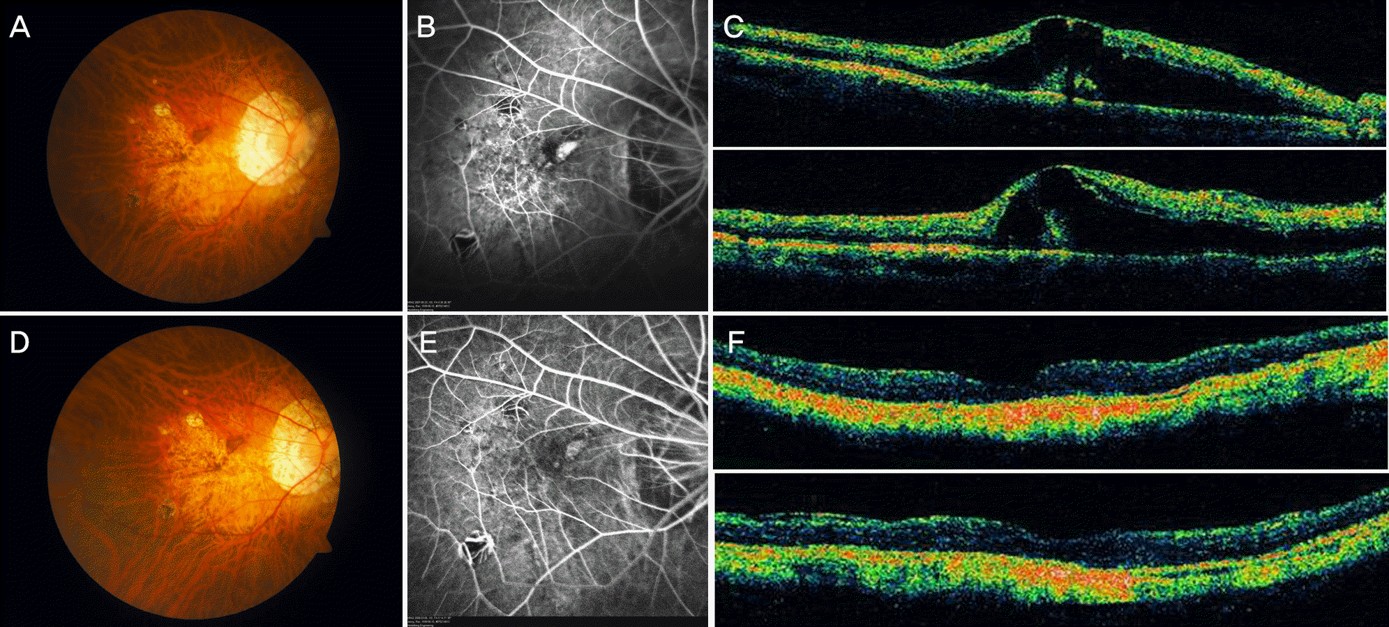
 | Figure 4.Bevacizumab was injected intravitreally two times at an interval of two months for myopic choroidal neovascularization (CNV) in the right eye of a 69-year-old woman. CNV is evident in a color photograph (A), fluorescein angiography (B) and optical coherent tomography (C). Visual acuity improved from 0.02 before injection to 0.1 at 6 months after the second injection, as well as findings in the various examinations (D, E, F). |
 | Figure 5.Graph shows difference in the mean best corrected visual acuity (logMAR) and the mean central foveal thickness on optical coherence tomography after intravitreal bevacizumab injection for choroidal neovascularization complicated with high myopia between the two groups. There was no significant difference between the two groups at one, two, three and twelve months after treatment (p<0.05; * visual acuity). |


 PDF
PDF ePub
ePub Citation
 Print
Print


 XML Download