

 PDF
PDF ePub
ePub Citation
 Print
Print


Abstract
Methods
Twenty subjects (40 eyes) underwent femtosecond LASIK and 20 subjects (40 eyes) underwent epi-LASIK for myopia with astigmatism. The results of each surgery were compared with regard to visual acuity, spherical equivalent, safety, efficacy, stability, predictability and high order aberration.
Results
Postoperative uncorrected visual acuities were 0.51 ± 0.11, 0.95 ± 0.08, and 0.97 ± 0.08 for epi-LASIK and 0.76 ±0.19, 0.97 ± 0.07, and 0.98 ± 0.06 for femtosecond LASIK at one week, one month, and two months after surgery, respectively. Femtosecond LASIK showed faster improvement in visual acuity. Postoperative spherical equivalents were −0.83 ± 0.24, −0.31 ± 0.19, and −0.27 ± 0.09 for epi-LASIK and −0.47 ± 0.21, −0.28 ± 0.15, and −0.25 ± 0.12 for femtosecond LASIK. Safety, efficacy, stability, and predictability showed no differences between the two groups. High order aberrations were increased significantly; however, no significant difference between the two groups was found.
References
1. Trokel SL, Srinivasan R, Braren B. Excimer laser surgery of the cornea. Am J Ophthalmol. 1983; 96:710–5.

2. Camellin M. Laser epithelial keratomileusis for myopia. J Refract Surg. 2003; 19:666–70.
3. Pallikaris IG, Katsanevaki VJ, Kalyvianaki MI, Naoumidi II. Advances in subepithelial excimer refractive surgery techniques: Epi-LASIK. Curr Opin Ophthalmol. 2003; 14:207–12.
4. Katsanevaki VJ, Kalyvianaki MI, Kavroulaki DS, Pallikaris IG. Epipolis laser in-situ keratomileusis: an evolving surface ablation procedure for refractive corrections. Curr Opin Ophthalmol. 2006; 17:389–93.
5. Trattler WB, Barnes SD. Current trends in advanced surface ablation. Curr Opin Ophthalmol. 2008; 19:330–4.
6. Netto MV, Mohan RR, Ambrósio R, et al. Wound healing in the cornea: a review of refractive surgery complications and new prospects for therapy. Cornea. 2005; 24:509–22.
7. Soong HK, Malta JB. Femtosecond lasers in ophthalmology. Am J Ophthalmol. 2009; 147:189–97. e2.
8. Kim JH, Lee D, Rhee KI. Flap thickness reproducibility in laser in situ keratomileusis with a femtosecond laser: optical coherence tomography measurement. J Cataract Refract Surg. 2008; 34:132–6.
9. Choi YS, Jung HJ, Lee KH. Comparison of clinical result of LASIK using between Femtosecond Laser and Microkeratome for correction of myopia. J Korean Ophthalmol Soc. 2007; 48:1041–7.
10. Durrie DS, Kezirian GM. Femtosecond laser versus mechanical keratome flaps in wavefront-guided laser in situ keratomileusis: prospective contralateral eye study. J Cataract Refract Surg. 2005; 31:120–6.
11. Patel SV, Maguire LJ, McLaren JW, et al. Femtosecond laser versus mechanical microkeratome for LASIK: a randomized controlled study. Ophthalmology. 2007; 114:1482–90.
12. Montes-Mico R, Rodriguez-Galietero A, Alio JL. Femtosecond laser versus mechanical keratome LASIK for myopia. Ophthalmology. 2007; 114:62–8.
13. Kim JH, Lee JE, Kim J-Y, Tchah HW. Early postoperative pain and visual outcomes following epipolis-laser in situ keratomileusis and photorefractive keratectomy. Korean J Ophthalmol. 2010; 24:143–7.
14. Teus MA, de Benito-Llopis L, Garcia-Gonzalez M. Comparison of visual results between laser-assisted subepithelial keratectomy and epipolis laser in situ keratomileusis to correct myopia and myopic astigmatism. Am J Ophthalmol. 2008; 146:357–62.
Figure 1.
Changes of uncorrected visual acuity at 1 week interval for postoperative 2 months after epi-LASIK and Femtosecond LASIK.* p value 0.05; t-test
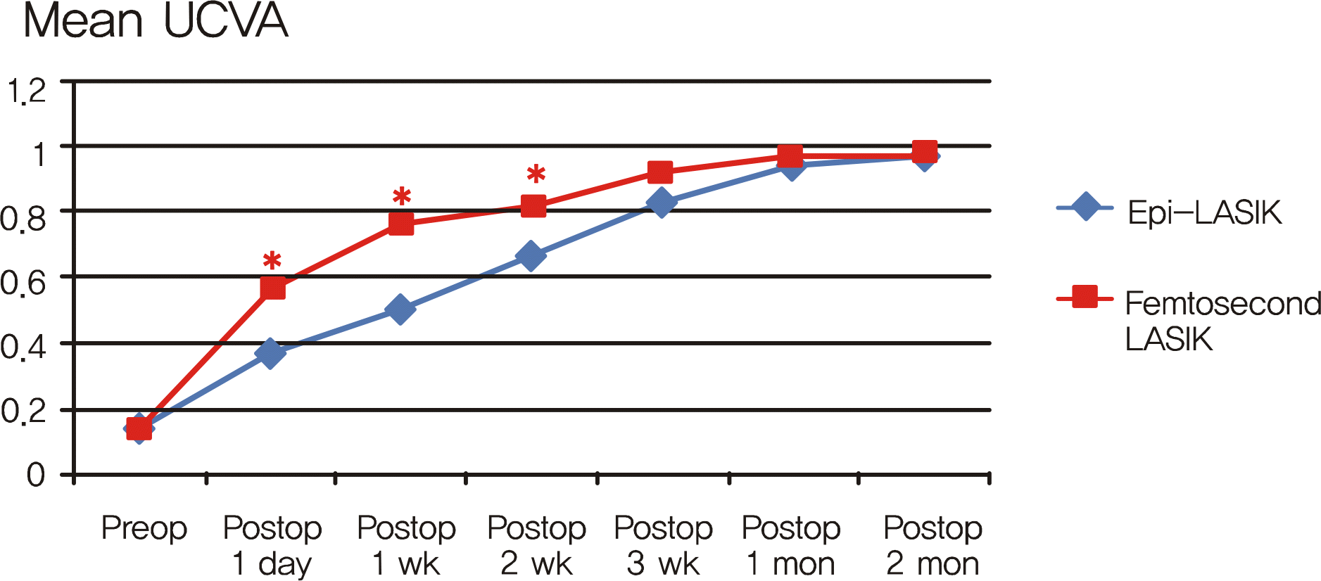
Figure 2.
Changes of best corrected visual acuity at 1 week interval for postoperative 2 months after epi-LASIK and Femtosecond LASIK.* p value <0.05; t-test
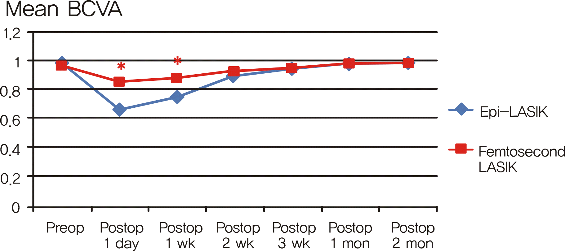
Table 1.
Baseline characteristics of eyes for epi-LASIK and Femtosecond LASIK
| | Epi-LASIK* | Femtosecond LASIK† | p value¶ |
|---|---|---|---|
| Age, mean (yr) | 24 ± 6.7 | 23 ± 5.7 | 0.829 |
| Sex (male/female) | 7/13 (20) | 6/14 (20) | |
| Preoperative UCVA‡ | 0.15 ± 0.12 | 0.14 ± 0.10 | 0.315 |
| Preoperative BSCVA§ | 0.98 ± 0.09 | 0.97 ± 0.13 | 0.256 |
| Sphere (D∏) | −5.05 ± 3.1 | −5.35 ± 2.8 | 0.391 |
| Cylinder (D) | −2.32 ± 1.8 | −2.55 ± 2.0 | 0.341 |
| SE# (D) | −5.87 ± 2.3 | −6.12 ± 2.7 | 0.298 |
| Pachymetry (µm) | 552 ± 61.8 | 548 ± 53.4 | 0.385 |
Table 2.
Mean uncorrected visual acuity after epi-LASIK and Femtosecond LASIK at postoperative 1 week and 1, 2 months
| | Epi-LASIK* | Femtosecond LASIK† | p value‡ |
|---|---|---|---|
| 1 week | 0.51 ± 0.14 | 0.76 ± 0.19 | < 0.01 |
| 1 month | 0.95 ± 0.08 | 0.97 ± 0.07 | 0.419 |
| 2 months | 0.97 ± 0.08 | 0.98 ± 0.06 | 0.336 |
Table 3.
Mean spherical equivalents after epi-LASIK and Femtosecond LASIK at postoperative 1 week and 1, 2 months
| | Epi-LASIK* | Femtosecond LASIK† | p value‡ |
|---|---|---|---|
| 1 wk | −0.83 ± 0.24 | −0.47 ± 0.21 | 0.007 |
| 1 mon | −0.31 ± 0.19 | −0.28 ± 0.15 | 0.237 |
| 2 mon | −0.27 ± 0.09 | −0.25 ± 0.12 | 0.342 |
Table 4.
Safety, efficacy, and predictability of epi-LASIK and Femtosecond LASIK at postoperative 2 months (in percentage)
| | Epi-LASIK* | Femtosecond LASIK† | p value∏ |
|---|---|---|---|
| Safety (BSCVA‡) | | | |
| Loss of 1 line | 6 | 4 | 0.386 |
| No loss | 84 | 83 | |
| Gain of 1 line | 10 | 13 | |
| Efficacy | | | |
| ≥ 20/20 | 82 | 90 | 0.183 |
| Predictability | | | |
| ± 0.50 D§ | 40 | 42 | 0.682 |
| ± 1.00 D | 84 | 89 | 0.329 |
| Stability | | | |
| Δ SE (SE2mo-SE1mo) | 0.04 | 0.03 | 0.728 |
Table 5.
Total HOA, coma, trefoil, spherical aberration of epi-LASIK and Femtosecond LASIK patients at pre- and postoperative 2 months (RMS, in microns)
| |
Epi-LASIK* |
Femtosecond LASIK† |
||||||
|---|---|---|---|---|---|---|---|---|
| Total HOA‡ | Coma | Trefoil | SA§ | Total HOA | Coma | Trefoil | SA | |
| Preop | 0.215 | 0.174 | 0.072 | 0.081 | 0.197 | 0.183 | 0.070 | 0.123 |
| Postop at 2 mo | 0.342 | 0.236 | 0.126 | 0.103 | 0.285 | 0.208 | 0.089 | 0.165 |
| Changes(%) | 159 | 136 | 175 | 127 | 145 | 114 | 127 | 134 |
| p value∏ | <0.01 | 0.025 | <0.01 | 0.034 | 0.017 | 0.038 | 0.012 | 0.045 |
 XML Download