

 PDF
PDF ePub
ePub Citation
 Print
Print


Abstract
Purpose
To compare retinal nerve fiber layer (RNFL) thickness obtained with Stratus optical coherence tomography (OCT) and Cirrus OCT.
Methods
Sixty-one normal eyes were evaluated with Stratus and Cirrus OCT on the same day, and the RNFL thicknesses measured by the two OCT machines were compared. The correlation between the two data sets was obtained using Pearson’s correlation coefficient. The correlation between RNFL thickness and the difference in data measured by the two OCT machines was then assessed.
References
1. Mikelberg FS, Yidegiligne HM, Shulzer M. Optic nerve axon count and axon diameter in patients with ocular hypertension and normal visual fields. Ophthalmology. 1995; 102:342–8.
2. Budenz DL, Michael A, Chang RT, et al. Sensitivity and specificity of the Stratus OCT for perimetric glaucoma. Ophthalmology. 2005; 112:3–9.
3. Huang D, Swanson EA, Lin CP, et al. Optical coherence tomography. Science. 1991; 254:1178–81.

4. Paunescu LA, Schuman JS, Price LL, et al. Reproducibility of nerve fiber thickness, macular thickness, and optic nerve head measurements using Stratus OCT. Invest Ophthalmol Vis Sci. 2004; 45:1716–24.
5. Koizumi H, Spaide RF, Fisher YL, et al. Three-dimensional evaluation of vitreomacular traction and epiretinal membrane using spectral-domain optical coherence tomography. Am J Ophthalmol. 2008; 145:509–17.
6. Leung CK, Cheung CY, Weinreb RN, et al. Comparison of macular thickness measurements between time domain and spectral domain optical coherence tomography. Invest Ophthalmol Vis Sci. 2008; 49:4893–7.
7. Forte R, Cennamo GL, Finelli ML, de Crecchio G. Comparison of time domain Stratus OCT and spectral domain SLO/OCT for assessment of macular thickness and volume. Eye. 2008; 1–8.
8. Kass MA, Heuer DK, Higginbotham EJ, et al. The ocular hypertension treatment study: a randomized trial determines that topical ocular hypotensive medication delays or prevents the onset of primary open-angle glaucoma. Arch Ophthalmol. 2002; 120:701–13.
9. Medeiros FA, Zangwill LM, Bowd C, Weinreb RN. Comparison of the GDx VCC scanning laser polarimeter, HRT II confocal scanning laser ophthalmoscope, and Stratus OCT optical coherence tomograph for the detection of glaucoma. Arch Ophthalmol. 2004; 122:827–37.
10. Zangwill LM, Bowd C, Berry CC, et al. Discriminating between normal and glaucomatous eyes using the Heidelberg retina tomograph, GDx nerve fiber analyzer, and optical coherence tomograph. Arch Ophthalmol. 2001; 119:985–93.
11. Colen TP, Tang NE, Mulder PG, Lemij HG. Sensitivity and specificity of new GDx parameters. J Glaucoma. 2004; 13:28–33.
12. El Beltagi TA, Bowd C, Boden C, et al. Retinal nerve fiber layer thickness measured with optical coherence tomography is related to visual function in glaucomatous eyes. Ophthalmology. 2003; 110:2185–91.
13. Ford BA, Artes PH, McCormick TA, et al. Comparison of data analysis tools for detection of glaucoma with the Heidelberg retina tomograph. Ophthalmology. 2003; 110:1145–50.
14. Bowd C, Weinreb RN, Williams JM, Zangwill LM. The retinal nerve fiber layer thickness in ocular hypertensive, normal, and glaucomatous eyes with optical coherence tomography. Arch Ophthalmol. 2000; 118:22–6.
15. Hoh ST, Greenfield DS, Mistlberger A, et al. Optical coherence tomography and scanning laser polarimetry in normal, ocular hypertensive, and glaucomatous eyes. Am J Ophthalmol. 2000; 129:129–35.
16. Medeiros FA, Zangwill LM, Bowd C, et al. Evaluation of retinal nerve fiber layer, optic nerve head, and macular thickness measurements for glaucoma detection using optical coherence tomography. Am J Ophthalmol. 2005; 139:44–55.
17. Sihota R, Sony P, Gupta V, et al. Diagnostic capability of optical coherence tomography in evaluating the degree of glaucomatous retinal nerve fiber damage. Invest Ophthalmol Vis Sci. 2006; 47:2006–10.
18. Hoffmann EM, Medeiros FA, Sample PA, et al. Relationship between patterns of visual field loss and retinal nerve fiber layer thickness measurements. Am J Ophthalmol. 2006; 141:463–71.
19. Jaffe GJ, Caprioli J. Optical coherence tomography to detect and manage retinal disease and glaucoma. Am J Ophthalmol. 2004; 137:156–69.
20. Wojtkowski M, Kowalczyk A, Leitgeb R, Fercher AF. Full range complex spectral optical coherence tomography technique in eye imaging. Opt Lett. 2002; 27:1415–7.
21. Legarreta JE, Gregori G, Punjabi OS, et al. Macular thickness measurements in normal eyes using spectral domain optical coherence tomography. Ophthalmic Surg Lasers Imaging. 2008; 39:S43–9.
22. Pons ME, Garcia-Valenzuela E. Redefining the limit of the outer retina in optical coherence tomography scans. Ophthalmology. 2005; 112:1079–85.
23. Srinivasan VJ, Monson BK, Wojtkowski M, et al. Characterization of outer retinal morphology with high-speed, ultrahigh-resolution optical coherence tomography. Invest Ophthalmol Vis Sci. 2008; 49:1571–9.
24. Chan A, Duker JS, Ishikawa H, et al. Quantification of photoreceptor layer thickness in normal eyes using optical coherence tomography. Retina. 2006; 26:655–60.
25. Jaffe GJ, Caprioli J. Optical coherence tomography to detect and manage retinal disease and glaucoma. Am J Ophthalmol. 2004; 137:156–69.
26. Tzamalis A, Kynigopoulos M, Schlote T, Haefliger I. Improved reproducibility of retinal nerve fiber layer thickness measurements with the repeat-scan protocol using the Stratus OCT in normal and glaucomatous eyes. Graefes Arch Clin Exp Ophthalmol. 2009; 247:245–52.
27. Polito A, Del Borrello M, Isola M, et al. Repeatability and reproducibility of fast macular thickness mapping with Stratus optical coherence tomography. Arch Ophthalmol. 2005; 123:1330–7.
28. Song YM, Uhm KB. Discrimination between normal and early stage of glaucomatous eyes using the Stratus optical coherence tomography. J Korean Ophthalmol Soc. 2007; 48:1675–85.
29. Hood DC, Raza AS, Kay KY, et al. A comparison of retinal nerve fiber layer (RNFL) thickness obtained with frequency and time domain optical coherence tomography (OCT). Opt Express. 2009; 17:3997–4003.
30. Hood DC, Fortune B, Arthur SN, et al. Blood vessel contributions to retinal nerve fiber layer thickness profiles measured with optical coherence tomography. J Glaucoma. 2008; 17:519–28.
31. Ghadiali Q, Hood DC, Lee C, et al. An analysis of normal variations in retinal nerve fiber layer thickness profiles measured with optical coherence tomography. J Glaucoma. 2008; 17:333–40.
Figure 1.
(A) Fundus photograph with white box indicating scanning area of Cirrus OCT, (B) Fundus photograph with the six radial line scanning pattern of Stratus OCT.
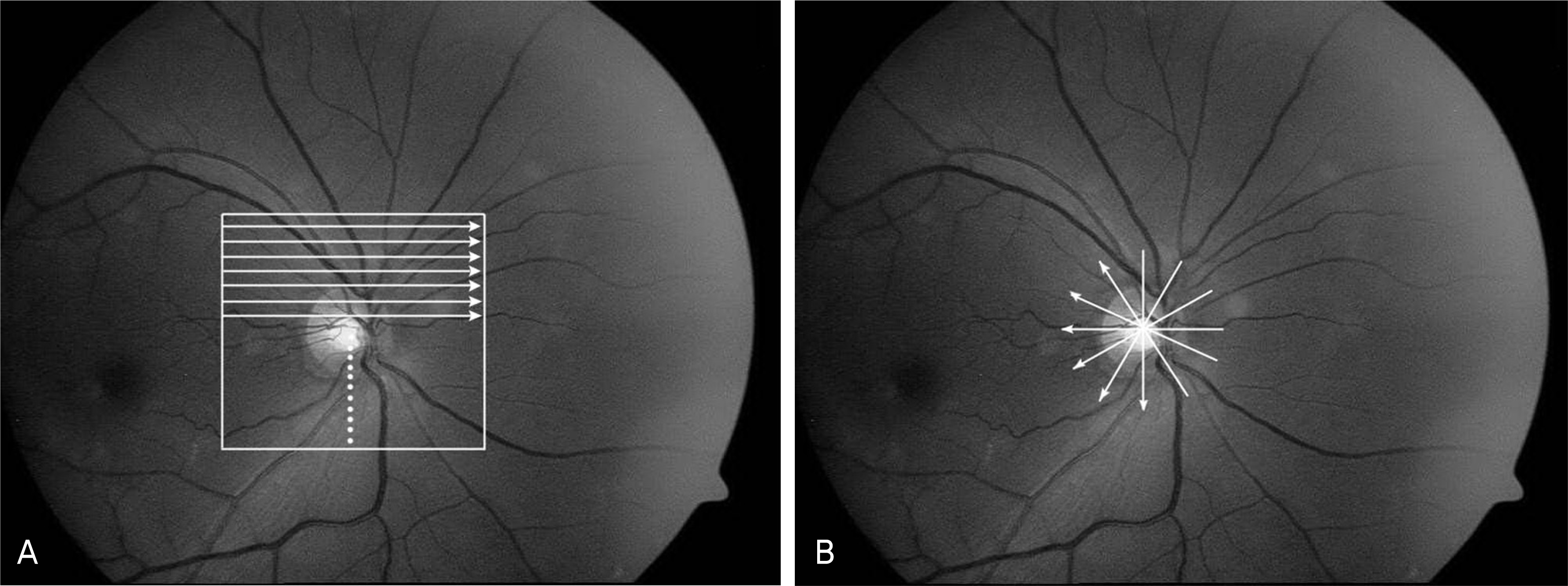
Figure 2.
Scatter plots of the Stratus RNFL thickness versus the Cirrus RNFL thickness. (A) Mean RNFL thickness, (B) Temporal RNFL thickness, (C) Superior RNFL thickness, (D) Nasal RNFL thickness, (E) Inferior RNFL thickness.
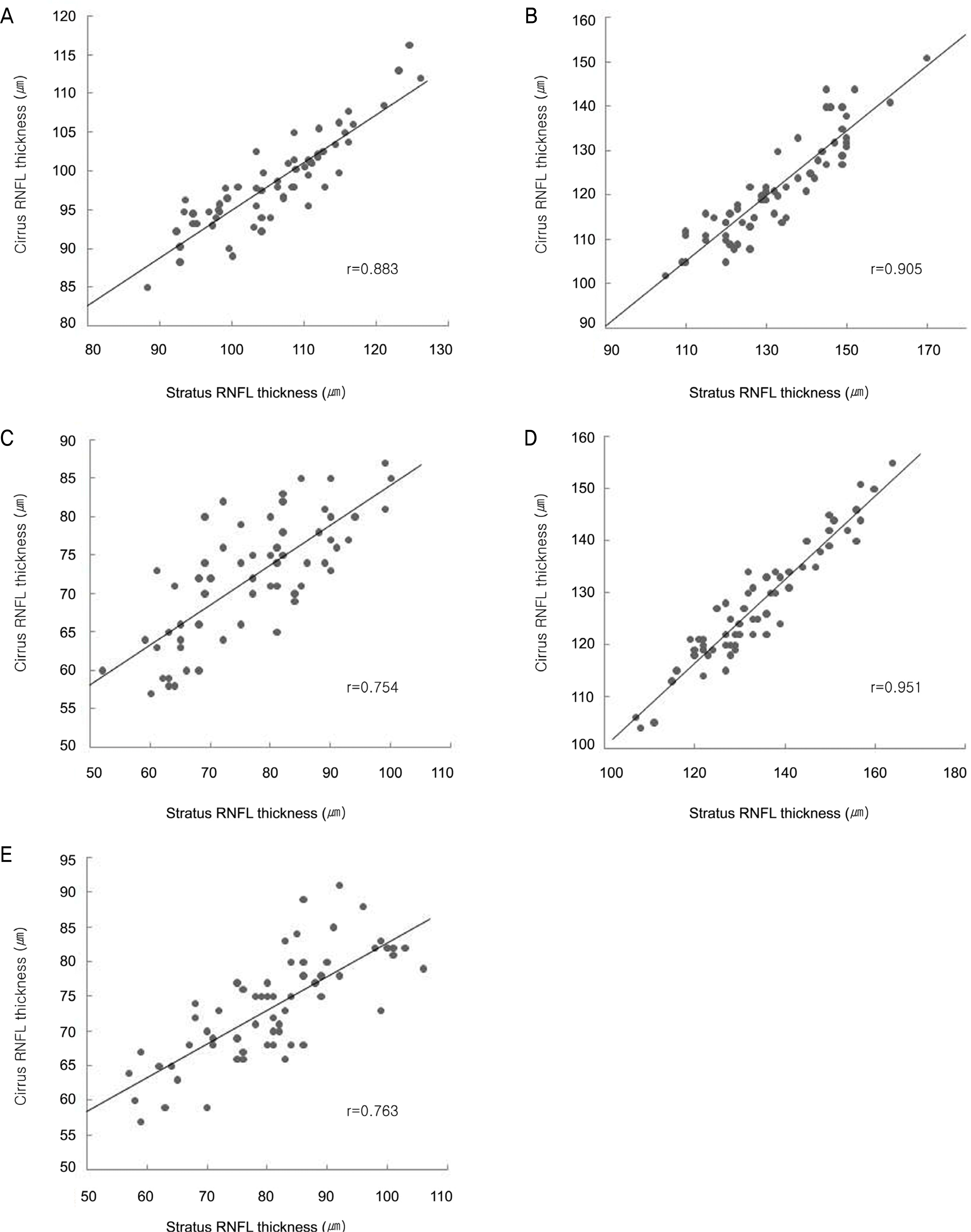
Figure 3.
Bland-Altman plots of Stratus minus Cirrus thickness differences versus the average RNFL. (A) Mean RNFL thickness, (B) Temporal RNFL thickness,(C) Superior RNFL thickness, (D) Nasal RNFL thickness,(E) Inferior RNFL thickness.
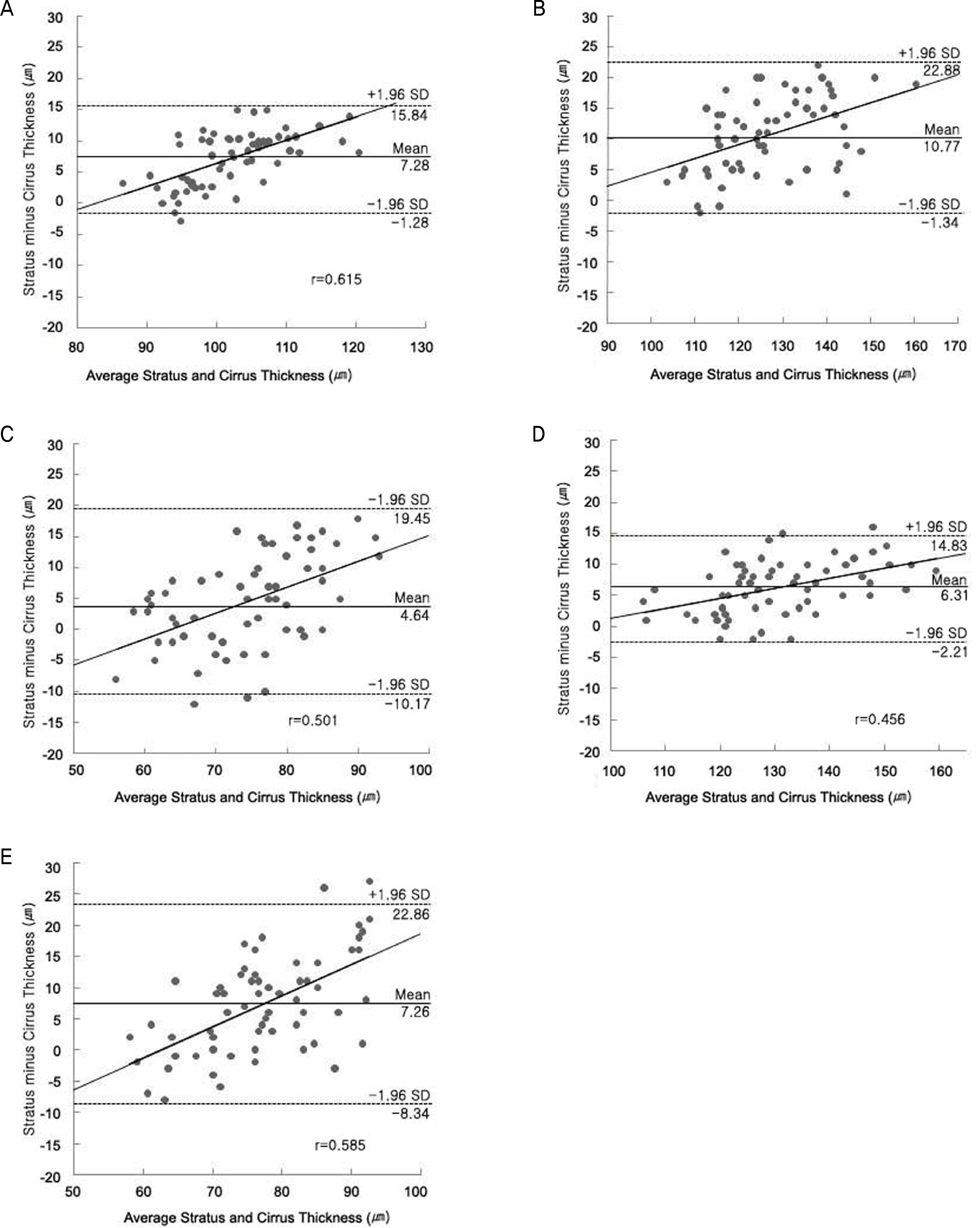
Figure 4.
(A) Stratus OCT and Cirrus OCT RNFL profiles.(B) Stratus OCT scan. (C) Cirrus OCT scan. Red dashed lines indicate two places where the two scans differ the most. And red arrows indicate shadow of retinal blood vessels.
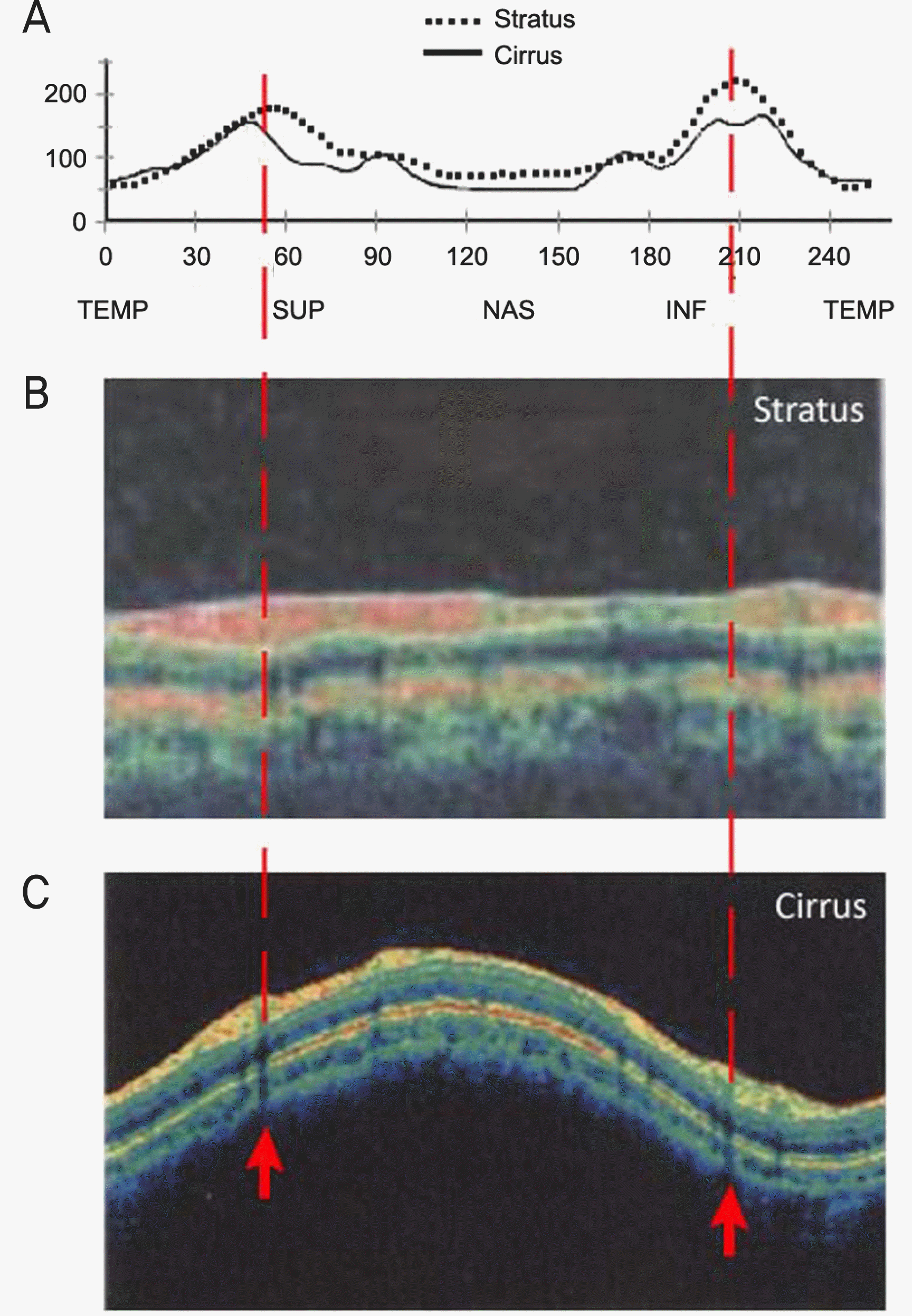
Table 1.
Basic characteristics of participants
Table 2.
Comparison of RNFL thickness scanned with Stratus and Cirrus OCT (n=61)
Table 3.
Comparison of Stratus and Cirrus OCT characteristics
 XML Download