

 PDF
PDF ePub
ePub Citation
 Print
Print


Abstract
Purpose
To evaluate differences between partial coherence laser interferometry (IOL-Master, Zeiss) and A-scan measurement of axial length and anterior chamber depth in silicone oil-filled eyes according to viscosity.
Methods
Using IOL-Master and A-scan, axial length and anterior chamber depth in silicone oil-filled eyes (n=54) and normal eyes (control, n=54) were measured and analyzed. In silicone oil-filled eyes, calculated axial lengths by A-scan using conversion factors, axial length multiplied by 0.71, and vitreous cavity multiplied by 0.64 (classic method) were compared with those calculated by IOL-Master. Anterior chamber depths were also analyzed., and axial lengths and anterior chamber depths were compared according to the viscosities of silicone oil for measurement by A-scan.
Results
Axial length and anterior chamber depth using IOL-Master were shorter than those using A-scan by 9.45±1.81 mm (p<0.05) and 0.11±1.29 mm, respectively. In normal eyes, axial length and anterior chamber depth using IOL-Master and A-scan were not significantly different. In silicone oil-filled eyes, axial length using IOL-Master and conversion factor was also not significantly different. At the highest silicone oil viscosity the difference in measured axial length was greatest (p<0.05) while the difference in anterior chamber depths was smallest.
References
1. Lucke K, Laqua H. Anatomic and functional success. Verlag. Silicone oil in the treatment of complicated retinal detachments: techniques, results and complications. Berlin, New York: Springer;1990. 1:chap. 2.
2. Moisseiev J, Bartov E, Cahane M, et al. Cataract extraction in eyes filled with silicone oil. Arch Ophthalmol. 1992; 110:1649–51.

3. Murray DC, Potamitis T, Good P, et al. Biometry of the silicone oil-filled eye. Eye. 1999; 13:913–24.
4. Ghoraba HH, El-Dorghamy AA, Atia AF, Ismail-Yassin Ael A. The problems of biometry in combined silicone oil removal and cataract extraction: a clinical trial. Retina. 2002; 22:589–96.
5. Choi JH, Roh GH. The reproducibility and accuracy of biometry parameter measurement from IOL Master. J Korean Ophthalmol Soc. 2004; 45:1665–73.
6. Chae JB, Park HR, Yoon YH. Axial length measurement in silicone oil-filled eyes using laser doppler interferometry. Retina. 2004; 24:655–7.
7. Nepp J, Krepler K, Jandrasits K, et al. Biometry and refractive outcome of eyes filled with silicone oil by standardized echography and partial coherence interferometry. Graefes Arch Clin Exp Ophthalmol. 2005; 243:967–72.
8. Santodomingo-Rubido J, Mallen EA, Wolffsohn JS. A new non-contact optical device for ocular biometry. Br J Ophthalmol. 2002; 86:458–62.
9. Takei K, Sekine Y, Okamoto F, Hommura S. Measurement of axial length of eyes with incomplete filling of silicone oil in the vitreous cavity using x ray computed tomography. Br J Ophthalmol. 2002; 86:47–50.
10. Larkin GB, Flaxel CJ, Leaver PK. Phacoemulsification and silicone oil removal through a single corneal incision. Ophthalmology. 1998; 105:2023–7.
11. Dietlein TS, Roessler G, Luke C, et al. Signal quality of biometry in silicone oil-filled eyes using partial coherence laser interferometry. J Cataract Refract Surg. 2005; 31:1006–10.
12. Lege BA, Haigis W. Laser interference biometry versus ultrasound biometry in certain clinical conditions. Graefes Arch Clin Exp Ophthalmol. 2004; 242:8–12.
13. Tehrani M, Krummenauer F, Blom E, Dick HB. Evaluation of the practicality of optical biometry and applanation ultrasound in 253 eyes. J Cataract Reflact Surg. 2003; 29:741–6.
14. Kim HJ, Kim HJ, Joo CK. Comparison of IOL Master, A-scan and Orbscan II for measurement of axial length and anterior chamber depth. J Korean Ophthalmol Soc. 2003; 44:1519–27.
Figure 1.
Axial length (AXL) and anterior chamber depth (ACD) without conversion in silicone oil-filled eyes. Axial lengths were significantly different between two methods (p<0.05), but anterior chamber depths were not (p>0.05).
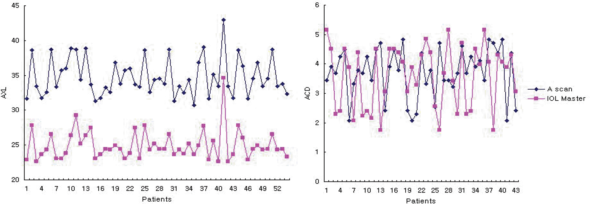
Figure 2.
Axial length (AXL) and anterior chamber depth (ACD) in contralateral eye. There were no significant differences between the two methods (p>0.05).
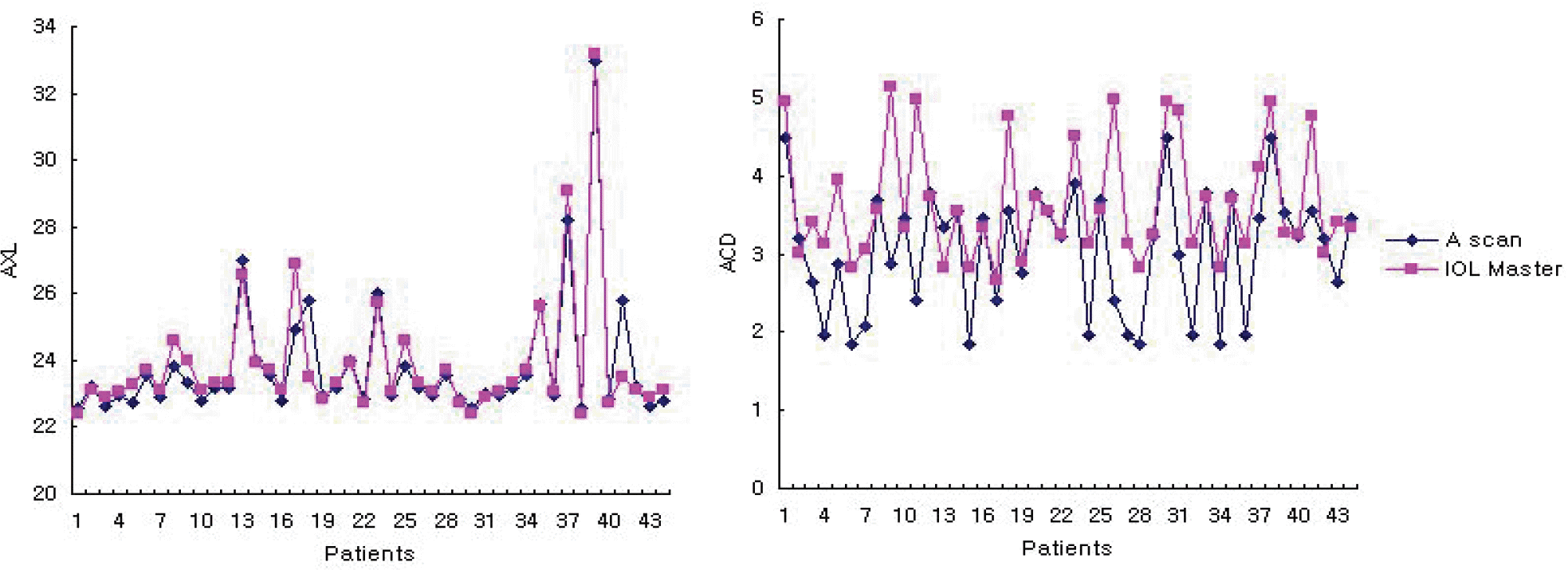
Figure 3.
Axial length by three methods in silicone oil-filled eye (A, B, C), by the two methods in contralateral eye (D, E). In silicone oil-filled eyes, axial lengths by IOL-master was not different to those by A scan with conversion factors (p>0.05). And in contralateral eyes, axial lengths were not different significantly between the two methods (p>0.05). A=IOL-Master; B=A-scan with conversion 0.64; C=A-scan with conversion 0.71; D=IOL- Master; E=A-scan.
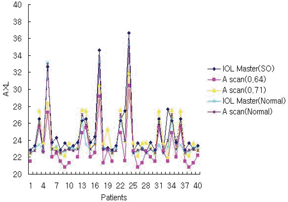
Table 1.
Difference and ratio of axial length, anterior chamber depth between A-scan and IOL-Master according to viscosity of silicone oil. As viscosity of silicone oil increases, difference of axial lengths was measured the largest (p<0.05, significantly different), anterior chamber depths the smallest. (average±standard deviation; AXL=axial length; ACD=anterior chamber depth)
 XML Download