

 PDF
PDF ePub
ePub Citation
 Print
Print


INTRODUCTION
The inferior alveolar nerve block (IANB) is one of the most common and important procedures in dentistry and has widespread applications in all fields of dentistry including oral surgery, endodontics, periodontics, and prosthodontics [1]. However, in practice, even with a standardized anesthetic technique, not all IANB procedures are successful. IANB failure rates have been reported as high as 29%-39% [234], and clinical studies have reported failure rates of 44%-81% in mandibular posterior teeth with irreversible pulpitis [567].
Some of the causes of IANB failure involve poor anesthetic techniques and anatomical variations such as differences in the ramus width and height, mandibular foramen position, musculature, and adipose tissue [38910]. Other potential explanations for IANB failure include a lowered pH of inflamed tissues, nerves with altered resting potentials, anesthetic-resistant sodium channels, and accessory innervations (mylohyoid nerve, cervical nerve, and auriculotemporal nerve) as well as the patient's anxiety level and factors related to the central core theory [6781112].
A conventional IANB involves the injection of local anesthetic fluid into the pterygomandibular space where the inferior alveolar nerve enters the mandibular foramen [10]. Therefore, the access to the mandibular foramen is considered to be the most important factor for the success of the IANB, and many studies have been performed to evaluate the exact location of the mandibular foramen for successful IANB [1314151617]. The location of the mandibular foramen is primarily located at the anteroposterior midpoint and two-thirds of the the length of the medial surface of the ramus, The position changes in relation to the occlusal plane and age. For example, the position in children is lower while the position in older edentulous patients is higher [1518].
The mandibular skeleton can be classified as retrognathic, prognathic, or normal [19]. In a three- dimensional (3-D) functional analysis of the mandible, Park et al. [20] found that the condyle and body length differed significantly among the three skeletal groups. This report revealed that the mean condyle length (distance from the mandibular foramen to the condyle tip) was significantly shorter in the retrognathic group and longer in the prognathic group when compared to that in the normal group. Conversely, the coronoid length (distance from the mandibular foramen to the coronoid tip) did not differ among the groups [20]. Since the position of the mandibular foramen, which is the reference point for an IANB, varies with the deviation of condylar length, and condylar length varies depending upon the mandibular growth pattern, it can be speculated that the mandibular characteristics are correlated with the success or failure of IANB. However, no previous research studies have reported on the contribution of skeletal characteristics to IANB success.
The purpose of this study was to examine the association between IANB failure and mandibular skeletal characteristics.
MATERIAL AND METHODS
This retrospective study was approved by the Institutional Review Board of the Yonsei University Dental Hospital (IRB number: 2-2011-0001). Between December 2008 and March 2010, 693 cases of lower third molar extraction from 575 patients were examined at the Department of Advanced General Dentistry of Yonsei University. Medical records for these cases were reviewed to obtain the demographic data for each subject. Of the 693 cases, 308 were in men and 385 in female, 347 were affecting left side and 346 the right side, and 118 patients had undergone a bilateral third-molar extraction. IANB anesthesia was performed by 24 residents. Using an aseptic technique, all patients received standard IANB injections using a total of 1.8 mL of 2% lidocaine with 1:100,000 epinephrine (Xylocaine, Astra-Zeneca LP, Dentsply, York, PA, USA). The solution was injected using self-aspirating syringes and 27-G long needles. After reaching the target area, aspiration was performed, and the solution was deposited. All surgical extraction procedures were performed by a single oral surgeon The success or failure of the IANB was evaluated by subjective, intraoperative symptoms especially pulpal pain during pulp cavity access for impacted tooth separation.
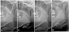
The mandibular skeletal characteristics were measured in duplicate by one dentist 7 days apart. The distance from the mandibular foramen to the condyle head (condylar distance) and that from the mandibular foramen to the tip of the coronoid process (coronoid distance) was measured using a dental picture archiving communication system. The condylar distance to coronoid distance (CC) ratio was then calculated (Fig. 1). Based on previous research, CC ratios of 1.07-1.22, < 1.07, and > 1.22 were classified as normal, retrognathic, and prognathic, respectively [2021]. The correlation between mandibular characteristics and the success of IANB was analyzed. A Pearson's chi-square test was used to analyze the correlation between the success or failure of the IANB and sex, treated side, and mandibular skeletal characteristics. The Statistical Package for the Social Sciences program (SPSS Science, Chicago, IL, USA) was used for all analyses and the level of statistical significance was set at P < 0.05.
RESULTS
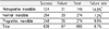
Of the 693 cases of IANB evaluated, 626 cases were successful and 67 cases failed; the overall failure rate was, thus, 9.67%. The age of the patients did not differ significantly between those who experienced successful anesthesia (26.6 ± 9.9 years, mean ± SD) and those who did not (26.6 ± 8.8 years; P = 0.997), and there was no significant difference in the IANB failure rate between men (34/308, 11.0%) and women (33/386, 8.6%; P = 0.302). In addition, although there was no statistically significant difference, the right side appeared to be more prone to IANB failure than the left side (Table 1).
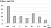
Of the 693 cases, 274 had a CC ratio in the range of 1.07-1.22 (i.e., a normal mandible), 145 had a CC ratio < 1.07 (i.e., a retrognathic mandible), and 274 had a CC ratio > 1.22 (i.e., a prognathic mandible). The IANB failure rates for the normal, retrognathic, and prognathic mandible groups were 7.3%, 14.5%, and 9.5%, respectively (Table 2), and the failure rate was highest in those with a CC ratio < 0.8 (Fig. 2). The chi-square test showed that the failure rate was significantly higher in the retrognathic group than in the normal group (P = 0.019). However, even though the failure rate of the prognathic group was found to be greater than that of normal group, there was no statistically significant difference between these two groups (P = 0.355). There was also no statistically significant difference between the failure rate of the retrognathic group and that of the prognathic group (P = 0.123).
DISCUSSION
The conventional IANB technique was first employed by William Halsted nearly a century ago [22]. This technique involves insertion of the needle into the pterygomandibular space by piercing the buccinator muscle. The injection point lies approximately 1 cm above the occlusal plane. With the syringe directed over the contralateral mandibular premolars, the puncture point should lie lateral to the pterygomandibular raphe and medial to the tendon insertion of the temporalis muscle on the anterior border of the ramus. This method delivers the anesthetic solution just above the mandibular foramen, which the inferior alveolar nerve enters, and distributes the anesthetic solution in the pterygomandibular space, but leaves other branches of the mandibular nerve, such as the lingual, buccal, and mylohyoid nerve, unaffected [23].
The mandibular foramen is an important anatomical landmark for the conventional IANB technique [1315]. According to Moss' functional matrix theory, the mandibular foramen is considered the main reference point for expressing mandibular growth [2024]. The mandible has six functional growth units (the symphysis, body, dentoalveolar, angle, coronoid, and condyle) each of which is affected by the surrounding functional matrix. In addition, the overall mandibular growth is the sum of the growth of each independent unit [242526]. Moss also mentioned that the balance of growth between the condylar and coronoid units contributes most to normal or abnormal growth. However, others contend that the milestone structures associated with mandibular growth are the mandibular and mental foramina, which are the first points of mandibular ossification and are located at the junction of each functional unit [26].
Park et al. analyzed and compared the functional unit growth patterns for normal, prognathic, and retrognathic mandibles using reference points that included the mandibular foramen. The authors used a linear analysis of 3-D-reconstructed computed tomography data and found that the condyle length differed significantly among the three groups, but the coronoid length did not. The results showed that the condylar units exhibits a more profound and consistent effect on mandibular growth than the body unit, and that the relative CC ratio is longest in the mandibular prognathism group and shortest in the mandibular retrognathism group. Their study also found that the relative CC ratio gradually increased in the order of the retrognathic, normal, and prognathic mandible groups (0.92, 1.08, and 1.16, respectively) [20]. Furthermore, Chang and Lee [21] studied the linear CC ratio in normal, adult Korean faces using a panoramic view and reported that the total mean CC ratio was 1.145 ± 0.079.
Thus, since condylar length varies depending upon the mandibular growth pattern, and the location of the mandibular foramen varies according to the condylar length, we speculated that the skeletal characteristics of the mandible could be related to the success or failure of IANB. In our study, we classified each mandible as normal, retrognathic, or prognathic based on the normal CC ratio range in Chang and Lee's study[21]. The IANB failure rate was 7.3% in the normal mandible group but was higher in both the retrognathic and prognathic group when compared to the normal group. The highest failure rate was found in the retrognathic mandible group and, within this group, the IANB failure rate was highest in cases with a CC ratio < 0.8. However, the failure rate among those with a more severe prognathic mandible, those with a CC ratio > 1.4, was lower than for those with a normal mandible.
In our study, the increase of IANB failure in those with a retrognathic mandible may be related to the position of the mandibular foramen. If the injection point lies approximately 1 cm above the occlusal plane, the anesthetic agents are likely to be infused inferior to the mandibular foramen since the position of the mandibular foramen in retrognathic mandibles is higher than in normal mandibles as a result of the short condylar length. In addition, Epars et al. reported that the vertical distance between the occlusal plate and the mandibular foramen was correlated to the patient's age and the vertical facial type. The distance between the foramen and the occlusal plane was significantly positively correlated to the patient's age, and negatively correlated with the vertical facial morphology. Furthermore, the foramen-occlusal plane distance increases more in short-face individuals than in long-face individuals [1417]. The study by Epars et al. showed that the IANB failure rate increases in patients with a short facial height because of the high position of the mandibular foramen. However, their study did not include skeletal characteristics.
IANB failure may also be related to the ramal inclination and divergence. If the ramal divergence is large, it is easy to access the mandibular foramen with an anesthetic needle. However, if the ramal divergence is small, the anesthetic needle can only access the front of the mandibular foramen and the possibility of IANB failure increases. For example, the analysis by Park et al. found that the ramal flaring angle (the angle between the condyle-coronoid plane and the angular plane) of a retrognathic mandible is larger than that of both normal and prognathic mandibles [20]. In another study using 3-dimensional computed tomography, the lateral ramal inclination (the angle of the gonion-condyle process to the midsagittal plane) was small in retrognathic patients [27]. Because both a larger ramal flaring angle and a smaller lateral ramal inclination indicate that mandible divergence is small, the risk of IANB failure may be larger in retrognathic mandibles.
In addition, the mouth opening of a retrognathic patient is small because the condyle is short. When the mouth opening is not adequate, the inferior alveolar nerve, which descends from above, is relaxed and away from the medial wall of the ramus. Consequently, the distance from the target area leads to inadequate anesthesia [2].
Therefore, to increase the success rate of IANB in patients with retrognathic mandibles, the anesthetic solution should be injected at a higher point. Other anesthetic methods, such as the Gow-Gates and Akinosi techniques, which are not correlated with the location of the mandibular foramen, could also be used.
However, in a prognathic mandible, the anesthetic agents are likely to be infused superior to the mandibular foramen since it is positioned lower than in the normal mandible as a result of the long condylar length. In addition, the mouth opening is larger in those with a more severe prognathic mandible, which facilitates access to the anatomical structures and, thus, is likely to reduce the failure rate. For example, Keros et al. found that the position of the mandibular foramen was lower in patients with successful anesthesia than in patients with unsuccessful anesthesia [28] supporting this hypothesis.
Our study has some limitations. First, the IANB was performed by 24 different residents, not by one practitioner. This does not meet the requirements of a control study; however, it can be interpreted as randomizing the procedure to be performed by many different general dentists, which would increase the study strength. Second, our study did not examine the position of the mandibular foramen. Therefore, further studies are needed to assess the correlation between mandibular skeletal characteristics and the vertical position of the mandibular foramen.
The results of the present study suggest that dental professionals may be able to predict the difficulty of an IANB before implementation by calculating the CC ratio of an individual patient. Taking into consideration the skeletal characteristics of patients may also increase the rates of successful and safe IANBs.

 XML Download