

 PDF
PDF ePub
ePub Citation
 Print
Print


INTRODUCTION
Pilocytic astrocytomas are the most common glial cell neoplasms in children and the most common pediatric cerebellar tumor [1]. Generally, they originate from the cerebellum, brainstem, and optic nerve [12]. However, lobar tumors have been reported rarely [1]. Most pilocytic astrocytomas are benign, and anaplastic or malignant transformation is uncommon unless there is a history of radiation exposure [3].
Meningiomas are benign tumors originating from arachnoid cap cells [2]. They exhibit homogeneous enhancing masses on magnetic resonance imaging (MRI). They have typical image findings known as a dural tail sign [4]. It is a homogenously enhanced thickening of the dural margin with peripheral tapering [45]. Sunburst patterns (radial divergence of feeder arterial branches) on angiography and bony erosion on computed tomography (CT) scans are included in typical imaging findings of meningiomas [5]. The incidence of cystic meningiomas varies from 1.6% to 10% of all meningiomas, and the commonest location is the cerebral convexity, particularly the frontal and parietal regions [6].
In this article, we describe a case of a supratentorial pilocytic astrocytoma with early anaplastic transformation, strongly suspected to be a cystic meningioma in radiographic findings. This case study was approved by the Ethics Committee of the Pusan National University Hospital.
CASE REPORT
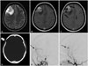
A 28-year-old female visited our institute because of headache for a month. The consciousness level was alert and motor grades of the extremities were normal. However, she had repeated vomiting for several days. This symptom was likely due to increased intracranial pressure, and therefore brain imaging was performed. The brain CT scan showed abnormal low density on the right frontal lobe and bony thinning on the right frontal bone (Fig. 1D). Brain MRIs revealed cystic and enhancing mass on the right frontal lobe (Fig. 1A and B). Dural enhancement suspected to be the dural tail sign was identified (Fig. 1C). Conventional angiography was performed and sunburst appearance through the right middle meningeal artery was confirmed (Fig. 1E and F). These radiographic findings suggested a diagnosis of a cystic meningioma.
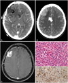
A surgical excision was performed. Intraoperatively, the dura was intact and there was no abnormal findings at the craniotomy eroded bone site. The mass had a well-defined arachnoid plane. Also, there was no dural attachment or adhesions (Fig. 2A). A biopsy was taken and sent to the pathologist for intraoperative diagnosis, and the result of the frozen biopsy was a glioma. After cyst fenestration, the mass was totally removed. The final pathology findings described a proliferation of piloid glial cell with Rosenthal fibers and eosinophilic granular bodies (Fig. 2B and C). Biphasic pattern was also identified. The Ki-67 was 1%. However, some scattered mitotic figures were identified. A maximum of 3 mitoses per 10 high power field showed that it was difficult to diagnose the patient as an anaplastic astrocytoma. Also, slightly increased cellularity was also shown, but there was no necrotic tissue. Therefore, the histologic findings confirmed a diagnosis of pilocytic astrocytoma with transformation to a more cellular and mitotically active lesion.
Postoperatively, enhanced CT scans revealed that the tumor was totally removed. The symptoms improved and there were no abnormal neurologic signs. Additional treatment such as radiation was not performed because pilocytic astrocytoma are known to be benign. There were no specific symptoms during follow-up. However, CT scans after nine months for routine follow-up showed a mass-like lesion. An MRI with gadolinium was performed, and the tumor recurrence was identified (Fig. 3). The recurrent mass was removed surgically. Pathologic diagnosis was an anaplastic astrocytoma (Fig. 3D). Postoperative radiation therapy of 60 Gy was performed. After radiation, there was no tumor recurrence for 3 years.
DISCUSSION
The MRI findings of this mass showed a well enhancing lesion which included cystic portions, and the lesion seemed to communicate with the dura. These findings coincided with a dural tail sign. Classically, the MRI findings of a cystic meningioma have been described as an extra-axial lesion with enhancement of solid portion and presence of a dural tail [67]. This findings were very similar to this mass. Therefore, we believed that this mass was a cystic meningioma. Furthermore, there were other image findings that correlated with a meningioma, such as thinning of the involved skull on CT scans, and sunburst sign on angiography.
Typical radiographic findings of pilocytic astrocytomas are well known. Typical MRI findings of pilocytic astrocytomas are usually an enhancing mass with a non-enhancing cystic component [18]. Also, in general pilocytic astrocytomas are well-circumscribed and round [2]. These findings are similar to those of cystic meningiomas [6]. However, there is little information about the incidence and biologic behavior of supratentorial pilocytic astrocytomas [9]. There are also few reports related to radiographic findings of these tumors. Chourmouzi et al. [8] reviewed radiologic findings for all types of pilocytic astrocytomas, including supratentorial pilocytic astrocytomas. They classified supratentorial pilocytic astrocytomas into three categories: chiasmatic/hypothalamic, optic and hemispheric area. According to this classification criteria, this case belonged to the hemispheric area. The most characteristic imaging finding of the hemispheric area was same as the typical infratentorial pilocytic astrocytoma, similar to a cystic meningioma [8]. Therefore, preoperative prediction was difficult.
Several supratentorial pilocytic astrocytomas mimicking other intracranial tumors have been reported. Skipworth et al. [1] reported three suprasellar pilocytic astrocytomas mimicking a craniopharyngioma. A review of the literature revealed only one other case of supratentorial pilocytic astrocytoma mimicking a meningioma on preoperative imaging. Hong et al. [2] reported a case of pilocytic astrocytoma mimicking a clinoidal meningioma. On preoperative MRI, there was an extra-axial mass in the right clinoid region with homogenous enhancement and a dural tail sign [2]. They concluded that this was a right clinoidal meningioma radiologically. However, the final pathologic diagnosis was pilocytic astrocytoma.
Pilocytic astrocytomas are known to have a clinically benign course after total surgical resection, and there are many reports that report supratentorial pilocytic astrocytomas also have relative good outcomes [9]. On the other hand, this case was an early transformed to anaplastic astrocytoma after nine postoperative months. Only two cases of supratentorial pilocytic astrocytomas with spontaneous anaplastic or malignant change have been reported [1011]. The period of anaplastic change was about 3 years [11]. We repeatedly reviewed the pathologic findings and consulted other institutes due to the unusual behaviors of this case. Nevertheless, no error was found in the pathologic diagnosis process.
Anaplastic transformation of pilocytic astrocytomas are described in the World Health Organization classification (WHO blue book) of tumors of the central nervous system [12]. Unlike glioblastomas, the prognosis of this tumor is not known to be uniformly grim [1213]. Therefore, the WHO blue book recommend that the diagnosis of a pilocytic astrocytoma with anaplasia is preferable, although grading criteria and nomenclature are yet to be defined [12]. Pilocytic astrocytomas with anaplasia are known to be associated with radiation therapy, however, they have also occurred in patients without radiation [313]. In our case, there was no previous irradiation.
In conclusion, we describe a rare case of supratentorial pilocytic astrocytoma with early anaplastic transformation with similar radiologic findings of convexity meningiomas. Although there are many reports that supratentorial pilocytic astrocytomas are clinically benign, there is still a lack of information regarding accurate prognosis. Therefore, short-term and regular follow-up imaging is needed after surgical resection of supratentorial pilocytic astrocytomas. Also, a well-enhancing mass with a dural tail sign is not always representative of meningiomas. So, physicians must consider a supratentorial pilocytic astrocytoma in their differential diagnosis of a well-enhancing mass with a dural tail sign on the frontal and parietal convexity areas.

 XML Download