

 PDF
PDF ePub
ePub Citation
 Print
Print


Seong Kee Shin, Ki Chun Kim , Youngju Roh, Jongkyu Kim, *
, Youngju Roh, Jongkyu Kim, *
Abstract
Symptomatic neuromas after amputation can be troublesome to treat and make it difficult to properly fit a brace. Surgical management is required when conservative management such as prosthetic socket modification or local injections fail. However, small cutaneous nerves adhere to adjacent soft tissue and they are difficult to locate. The authors suggest that ultrasonography guided tattoo localization using a charcoal suspension is useful to find a visually indistinguishable neuroma.
Figures and Tables
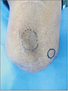
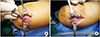
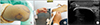
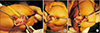
Figure 2
A cutaneous nerve on lateral side of the amputee end was dissected (A) and embedded into muscle layer (B).
References
1. Nashold BS Jr, Goldner JL, Mullen JB, Bright DS. Long-term pain control by direct peripheral-nerve stimulation. J Bone Joint Surg Am. 1982; 64:1–10.

2. Kim WH, Kim HJ, Kim SH, Jung JH, Park HY, Lee J, et al. Ultrasound-guided dual-localization for axillary nodes before and after neoadjuvant chemotherapy with clip and activated charcoal in breast cancer patients: a feasibility study. BMC Cancer. 2019; 19:859. DOI: 10.1186/s12885-019-6095-1.
3. Helvie MA, Ikeda DM, Adler DD. Localization and needle aspiration of breast lesions: complications in 370 cases. AJR Am J Roentgenol. 1991; 157:711–714. DOI: 10.2214/ajr.157.4.1892023.
4. Czarnecki DJ, Feider HK, Splittgerber GF. Toluidine blue dye as a breast blocalization marker. AJR Am J Roentgenol. 1989; 153:261–263. DOI: 10.2214/ajr.153.2.261.
5. Saarela AO, Kiviniemi HO, Rissanen TJ. Preoperative methylene blue staining of galactographically suspicious breast lesions. Int Surg. 1997; 82:403–405.
6. Sewak S, Graham P, Nankervis J. Tattoo allergy in patients receiving adjuvant radiotherapy for breast cancer. Australas Radiol. 1999; 43:558–561. DOI: 10.1046/j.1440-1673.1999.00733.x.
7. Soran A, Kanbour-Shakir A, Bas O, Bonaventura M. A tattoo pigmented node and breast cancer. Bratisl Lek Listy. 2014; 115:311–312. DOI: 10.4149/BLL_2014_063.
8. Lager DJ, O'Connor JC, Robinson RA, Brown RC, Urdaneta LF. Factitious microcalcifications in breast biopsy material: laboratory-induced error by use of tattoo powder for specimen mammography. J Surg Oncol. 1989; 40:281–282. DOI: 10.1002/jso.2930400415.
9. Vilska J. [Indications and contra-indications of activated charcoal in a poison control center. The viewpoint of the Poison Control Center of Finland]. J Toxicol Clin Exp. 1989; 9:295–298. French.
10. Sperry K. Tattoos and tattooing. Part i: history and methodology. Am J Forensic Med Pathol. 1991; 12:313–319. DOI: 10.1097/00000433-199112000-00008.
 XML Download