

 PDF
PDF ePub
ePub Citation
 Print
Print


INTRODUCTION
Silicone-gel-filled breast implants are widely used for breast augmentation and breast reconstruction following mastectomy (1). Postoperative conditions such as hematoma, infection, capsular contracture, silicone granuloma formation, and implant rupture have been reported, and rupture is one of the most common complications of silicone implant augmentation (2). The gold standard for diagnosing and evaluating implant rupture is magnetic resonance imaging (MRI) (3). Numerous publications have outlined MRI features of implant rupture (23456).
We present the case of a patient who experienced silicone breast implant rupture 10 years after implantation. MRI of the breast showed intra- and extracapsular ruptures of the implant and mass suggesting silicone granuloma. Unlike most cases of silicone implant ruptures, which exhibit collapse of the implant shell and decreased breast size, MRI showed swelling of the breast and maintenance of implant volume rather than collapse.
CASE REPORT
A 50-year-old woman presented with a history of gradual enlargement of her right breast that had persisted for about one month. She reported mild pain but no skin changes or fever. The results of all laboratory tests were within normal ranges. She had undergone augmentation mammoplasty with silicone implants 10 years prior to presentation. A physician examined her breasts and decided to perform breast MRI to evaluate possible implant complications.
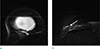
On breast MRI, bilateral implants were seen in subpectoral locations, and the right breast showed asymmetric enlargement. On T2-weighted images (T2WI), silicone gel with high signal intensity was seen both inside and outside the implant shell, representing intracapsular rupture and gel leakage (Fig. 1b). Although it was difficult to evaluate the fibrous capsule, silicone gel with high signal intensity was seen within the pectoralis minor and major muscles, superior and inferior to the enlarged breast, respectively, and possibly represented extracapsular rupture. In addition, a 5.0 × 1.6 cm lesion with an irregular shape was seen in the posterior portion of the right breast outside the implant shell. The lesion showed iso-signal intensity compared with the silicone inside the implant shell along with small foci of subtle high signal intensity on a T1-weighted image (T1WI) (Fig. 1a).
The small foci showed subtle high signal intensity on T1WI with mild enhancement on gadolinium-enhanced subtraction images and was considered indicative of silicone granuloma (Fig. 1c). Different fluid-fluid levels were noted inside and outside the implant shell, and nondependent portions of low signal intensity on T2WI were thought to reflect recent or old hemorrhages. There was no evidence of implant shell collapse; rather, the volume of the implant was maintained and even slightly increased compared with the intact implant on the contralateral side (Figs. 1, 2).
During surgery, the surgeons observed a rupture in the silicone implant shell and a silicone granuloma. The implant, fibrous capsule, and granuloma were removed via the inframammary folds. Upon histopathologic examination, chronic granulomatous inflammation was discovered in the removed mass lesion and diagnosed as granuloma. A fluid analysis was also performed to detect infection, but there was no bacterial growth in culture.
DISCUSSION
Silicone-implant ruptures can be classified as intracapsular or extracapsular, depending on the location of the rupture. Intracapsular rupture is defined as rupture of the implant shell elastomer with silicone leakage that does not macroscopically extend beyond the fibrous capsule that normally forms around silicone implants. Most implant ruptures (80–90%) are intracapsular. On MRI, the well-known ‘linguine sign,’ which represents multiple curvilinear low-signal-intensity lines within the high-signal-intensity silicone gel, is considered the hallmark of intracapsular rupture. These curvilinear lines represent the collapsed implant shell floating within the silicone gel. Minor ruptures of recently manufactured silicone-gel implants with multilayer shells, a barrier layer, and thick silicone gel show as the ‘keyhole sign’ or ‘noose sign’ on MRI, which represents free silicone within a radial fold without collapse of the implant shell. Extracapsular silicone-implant rupture is less common and is defined as rupture of both the implant shell and the fibrous capsule, with silicone leakage that extends beyond the fibrous capsule into surrounding tissues. On MRI, focal areas of iso- to low signal intensity on T1 fat-suppressed images and high signal intensity on water-suppressed T2WI represent free silicone (4). When free silicone gel incites intense fibrotic reactions in the surrounding tissue, silicone granulomas (also called siliconomas) are formed. Silicone granuloma shows as nearly iso- or slightly hypointense to fat on water-suppressed fast spin-echo T2WI and may increase much as breast carcinomas do (25). On ultrasonography, silicone granuloma may show complex cystic lesions or a ‘snowstorm appearance’, depending on the amount of extravagated silicone gel and the extent of the fibrous and foreign-body reactions (1). Siliconomas in axillary, supraclavicular, or internal mammary lymph nodes may mimic the metastatic lymphadenopathy of breast cancer (7).
There are many studies reporting intra- or extracapsular ruptures of the silicone bag and silicone granuloma (23456). Our case was unusual, and this is the first report of breast enlargement after silicone-implant rupture. We detected fluid-fluid levels of various signal intensities both inside and outside the implant elastomer. The implant volume was maintained instead of collapsing, as is usually observed. As noted above, collapse of the implant shell is a natural result of silicone-implant rupture. We hypothesized that a small rupture and chronic leakage of silicone gel can result in silicone granuloma formation and acute inflammation or hemorrhage. Consequently, it can cause swelling of the breast and implant. Additionally, the high osmotic gradient across the hydrogel implant may cause swelling of the implant and resistance against implant collapse (8).
In conclusion, we report a rare case of intra- and extracapsular silicone breast implant rupture that resulted in fluid-fluid levels of various signal intensities, silicone granulomas, and unexpected enlargement of the breast.
 XML Download