

 PDF
PDF ePub
ePub Citation
 Print
Print


Abstract
Purpose
This study was designed to optimize the flip angle (FA) and scan timing of the hepatobiliary phase (HBP) using the 3D T1-weighted, gradient-echo (GRE) imaging with controlled aliasing in parallel imaging results in higher acceleration (CAIPIRINHA) technique on gadoxetic acid-enhanced 3T liver MR imaging.
Materials and Methods
Sixty-two patients who underwent gadoxetic acid-enhanced 3T liver MR imaging were included in this study. Four 3D T1-weighted GRE imaging studies using the CAIPIRINHA technique and FAs of 9° and 13° were acquired during HBP at 15 and 20 min after intravenous injection of gadoxetic acid. Two abdominal radiologists, who were blinded to the FA and the timing of image acquisition, assessed the sharpness of liver edge, hepatic vessel clarity, lesion conspicuity, artifact severity, and overall image quality using a five-point scale. Quantitative analysis was performed by another radiologist to estimate the relative liver enhancement (RLE) and the signal-to-noise ratio (SNR). Statistical analyses were performed using the Wilcoxon signed rank test and one-way analysis of variance.
Results
The scores of the HBP with an FA of 13° during the same delayed time were significantly higher than those of the HBP with an FA of 9° in all the assessment items (P < 0.01). In terms of the delay time, images at the same FA obtained with a 20-min-HBP showed better quality than those obtained with a 15-min-HBP. There was no significant difference in qualitative scores between the 20-min-HBP and the 15-min-HBP images in the non-liver cirrhosis (LC) group except for the hepatic vessel clarity score with 9° FA. In the quantitative analysis, a statistically significant difference was found in the degree of RLE in the four HBP images (P = 0.012). However, in the subgroup analysis, no significant difference in RLE was found in the four HBP images in either the LC or the non-LC groups. The SNR did not differ significantly in the four HBP images. In the subgroup analysis, 20-min-HBP imaging with a 13° FA showed the highest SNR value in the LC-group, whereas 15-min-HBP imaging with a 13° FA showed the best value of SNR in the non-LC group.
Conclusion
The use of a moderately high FA improves the image quality and lesion conspicuity on 3D, T1-weighted GRE imaging using the CAIPIRINHA technique on gadoxetic acid, 3T liver MR imaging. In patients with normal liver function, the 15-min-HBP with a 13° FA represents a feasible option without a significant decrease in image quality.
References
1. Kim BS, Angthong W, Jeon YH, Semelka RC. Body MR imaging: fast, efficient, and comprehensive. Radiol Clin North Am. 2014; 52:623–636.
2. Semelka RC, Martin DR, Balci NC. Magnetic resonance imaging of the liver: how I do it. J Gastroenterol Hepatol. 2006; 21:632–637.

3. Semelka RC, Helmberger TK. Contrast agents for MR imaging of the liver. Radiology. 2001; 218:27–38.
4. Rofsky NM, Lee VS, Laub G, et al. Abdominal MR imaging with a volumetric interpolated breathhold examination. Radiology. 1999; 212:876–884.
5. Yu MH, Lee JM, Yoon JH, Kiefer B, Han JK, Choi BI. Clinical application of controlled aliasing in parallel imaging results in a higher acceleration (CAIPIRINHA)volumetric interpolated breathhold (VIBE) sequence for gadoxetic acid-enhanced liver MR imaging. J Magn Reson Imaging. 2013; 38:1020–1026.
6. Kim BS, Lee KR, Goh MJ. New imaging strategies using a motion-resistant liver sequence in uncooperative patients. Biomed Res Int. 2014; 2014:142658.
7. Wright KL, Harrell MW, Jesberger JA, et al. Clinical evaluation of CAIPIRINHA: comparison against a GRAPPA standard. J Magn Reson Imaging. 2014; 39:189–194.
8. Park YS, Lee CH, Kim IS, et al. Usefulness of controlled aliasing in parallel imaging results in higher acceleration in gadoxetic acid-enhanced liver magnetic resonance imaging to clarify the hepatic arterial phase. Invest Radiol. 2014; 49:183–188.
9. Huppertz A, Balzer T, Blakeborough A, et al. Improved detection of focal liver lesions at MR imaging: multicenter comparison of gadoxetic acid-enhanced MR images with intraoperative findings. Radiology. 2004; 230:266–275.
10. Seale MK, Catalano OA, Saini S, Hahn PF, Sahani DV. Hepatobiliary-specific MR contrast agents: role in imaging the liver and biliary tree. Radiographics. 2009; 29:1725–1748.
11. Goodwin MD, Dobson JE, Sirlin CB, Lim BG, Stella DL. Diagnostic challenges and pitfalls in MR imaging with hepatocyte-specific contrast agents. Radiographics. 2011; 31:1547–1568.
12. Sofue K, Tsurusaki M, Tokue H, Arai Y, Sugimura K. Gd-EOB-DTPA-enhanced 3.0 T MR imaging: quantitative and qualitative comparison of hepatocyte-phase images obtained 10 min and 20 min after injection for the detection of liver metastases from colorectal carcinoma. Eur Radiol. 2011; 21:2336–2343.
13. Motosugi U, Ichikawa T, Tominaga L, et al. Delay before the hepatocyte phase of Gd-EOB-DTPA-enhanced MR imaging: is it possible to shorten the examination time? Eur Radiol. 2009; 19:2623–2629.
14. Haradome H, Grazioli L, Al manea K, et al. Gadoxetic acid disodium-enhanced hepatocyte phase MRI: can increasing the flip angle improve focal liver lesion detection? J Magn Reson Imaging. 2012; 35:132–139.
15. Bashir MR, Husarik DB, Ziemlewicz TJ, Gupta RT, Boll DT, Merkle EM. Liver MRI in the hepatocyte phase with gadolinium-EOB-DTPA: does increasing the flip angle improve conspicuity and detection rate of hypointense lesions? J Magn Reson Imaging. 2012; 35:611–616.
16. Bashir MR, Merkle EM. Improved liver lesion conspicuity by increasing the flip angle during hepatocyte phase MR imaging. Eur Radiol. 2011; 21:291–294.
17. Cho ES, Yu JS, Park AY, Woo S, Kim JH, Chung JJ. Feasibility of 5-minute delayed transition phase imaging with 30 degrees flip angle in gadoxetic acid-enhanced 3D gradient-echo MRI of liver, compared with 20-minute delayed hepatocyte phase MRI with standard 10 degrees flip angle. AJR Am J Roentgenol. 2015; 204:69–75.
18. Chang KJ, Kamel IR, Macura KJ, Bluemke DA. 3.0-T MR imaging of the abdomen: comparison with 1.5 T. Radiographics. 2008; 28:1983–1998.
19. Erturk SM, Alberich-Bayarri A, Herrmann KA, Marti-Bonmati L, Ros PR. Use of 3.0-T MR imaging for evaluation of the abdomen. Radiographics. 2009; 29:1547–1563.
20. Kim S, Mussi TC, Lee LJ, Mausner EV, Cho KC, Rosenkrantz AB. Effect of flip angle for optimization of image quality of gadoxetate disodium-enhanced biliary imaging at 1.5 T. AJR Am J Roentgenol. 2013; 200:90–96.
21. Bashir MR, Breault SR, Braun R, Do RK, Nelson RC, Reeder SB. Optimal timing and diagnostic adequacy of hepatocyte phase imaging with gadoxetate-enhanced liver MRI. Acad Radiol. 2014; 21:726–732.
22. van Kessel CS, Veldhuis WB, van den Bosch MA, van Leeuwen MS. MR liver imaging with Gd-EOB-DTPA: a delay time of 10 minutes is sufficient for lesion characterisation. Eur Radiol. 2012; 22:2153–2160.
23. Kim SH, Kim SH, Lee J, et al. Gadoxetic acid-enhanced MRI versus triple-phase MDCT for the preoperative detection of hepatocellular carcinoma. AJR Am J Roentgenol. 2009; 192:1675–1681.
24. Kim HY, Choi JY, Park CH, et al. Clinical factors predictive of insufficient liver enhancement on the hepatocyte-phase of Gd-EOB-DTPA-enhanced magnetic resonance imaging in patients with liver cirrhosis. J Gastroenterol. 2013; 48:1180–1187.
25. Kanki A, Tamada T, Higaki A, et al. Hepatic parenchymal enhancement at Gd-EOB-DTPA-enhanced MR imaging: correlation with morphological grading of severity in cirrhosis and chronic hepatitis. Magn Reson Imaging. 2012; 30:356–360.
Fig. 1.
Graphs (signal intensity and contrast-to-noise ratio [CNR] to flip angle) of water, fat, and gadoxetic acid obtained from an in-vitro phantom. Changing the flip angle affects the signal intensity of water, fat, and gadoxetic acid (a, b). The shape of a water curve shows a specific flip angle at 6 degrees, which produces maximum signal intensity (a). The signal intensity of water converges with fat at 14 degrees (a, b). The CNR between gadoxetic acid and water is increased by raising the flip angle 12.4% per degree (b).
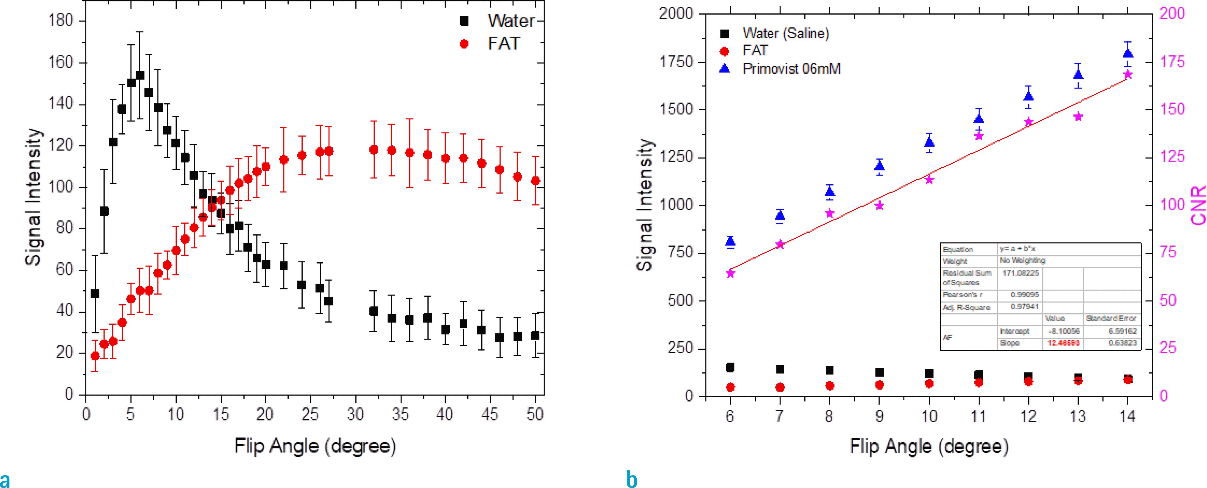
Fig. 2.
MR images of a 44-year-old woman with a history of breast cancer and normal liver function. Four hepatobiliary phase (HBP), gadoxetic acid-enhanced 3D-gradient echo (GRE) images using the CAIPIRINHA technique obtained at 20 min (a) and 15 min (b) after contrast injection with a 13° flip angle (FA) and at 20 min (c) and 15 min (d) after contrast injection with 9° FA. Moderately high flip angle HBP images (a, b) showed the best delineation of intrahepatic vessels, compared with standard flip angle HBP images (c, d). At moderately high flip angle HBP images (a, b), intrahepatic vessels (arrow) were well defined with low signal intensity. The background liver showed high signal intensity due to enhanced, normal parenchyma, resulting in excellent conspicuity. The relative liver enhancement was highest with transverse, T1-weighted 3D-GRE images obtained 15 min after contrast injection with a 13° FA (b), followed by T1-weighted, 3D-GRE images obtained 20 min after contrast injection with a 13° FA (a), T1-weighted, 3D-GRE images obtained 20 min after contrast injection with a 9° FA (c), and T1-weighted, 3D-GRE images obtained 15 min after contrast injection with a 9° FA (d).
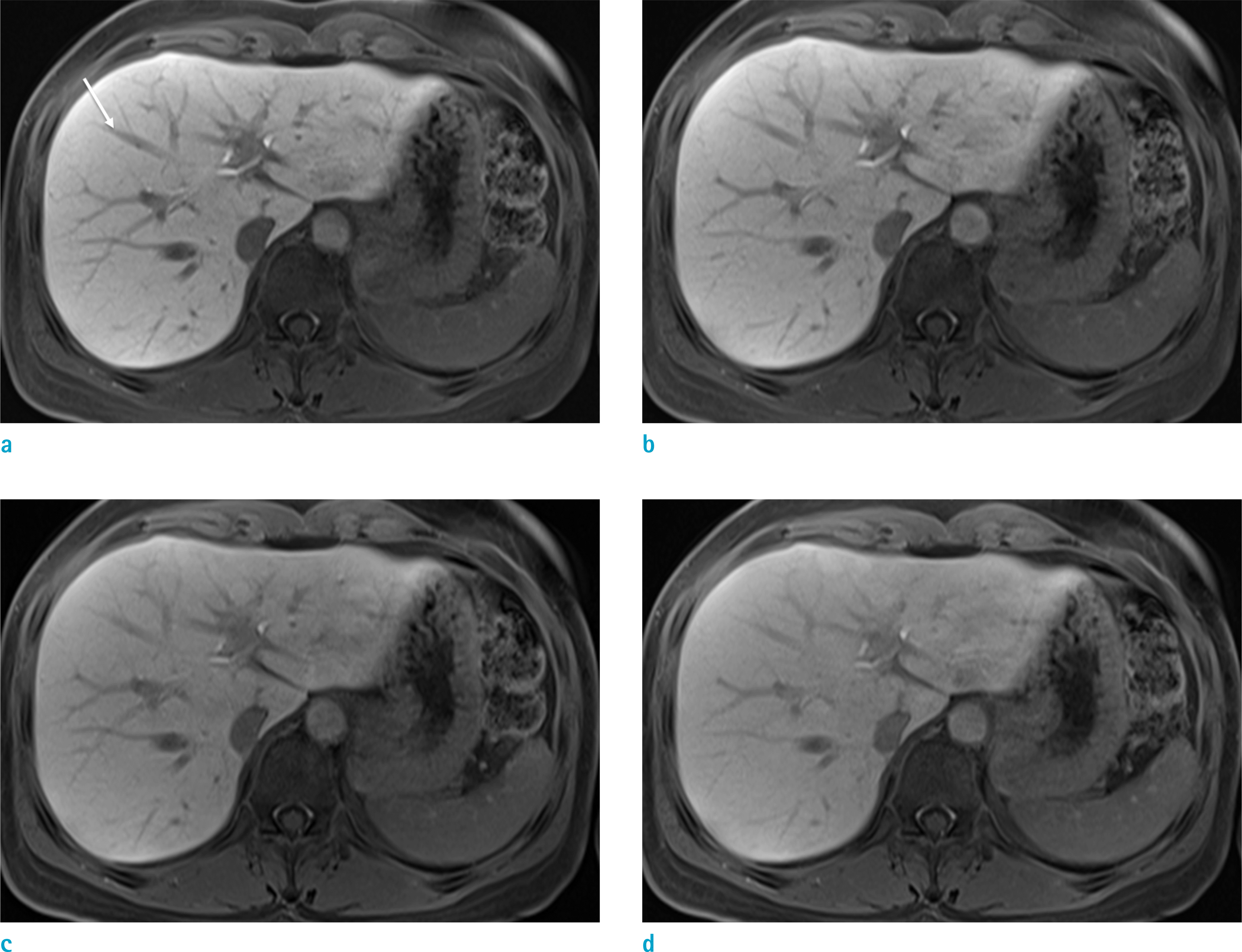
Fig. 3.
MR images of a 70-year-old man with liver cirrhosis (Child-Pugh class: A) show hepatocellular carcinoma (arrow). Four hepatobiliary phase (HBP), gadoxetic acid-enhanced, 3D-gradient echo (GRE) images using the CAIPIRINHA technique obtained at 20 min (a) and 15 min (b) after contrast injection with 13° flip angle (FA) and at 20 min (c) and 15 min (d) after contrast injection with 9° FA. Moderately high flip angle HBP images (a, b) demonstrate clearly defined hepatocellular carcinoma. The fine intrahepatic vasculatures (short arrow) are well defined in moderately high flip angle HBP images (a, b).
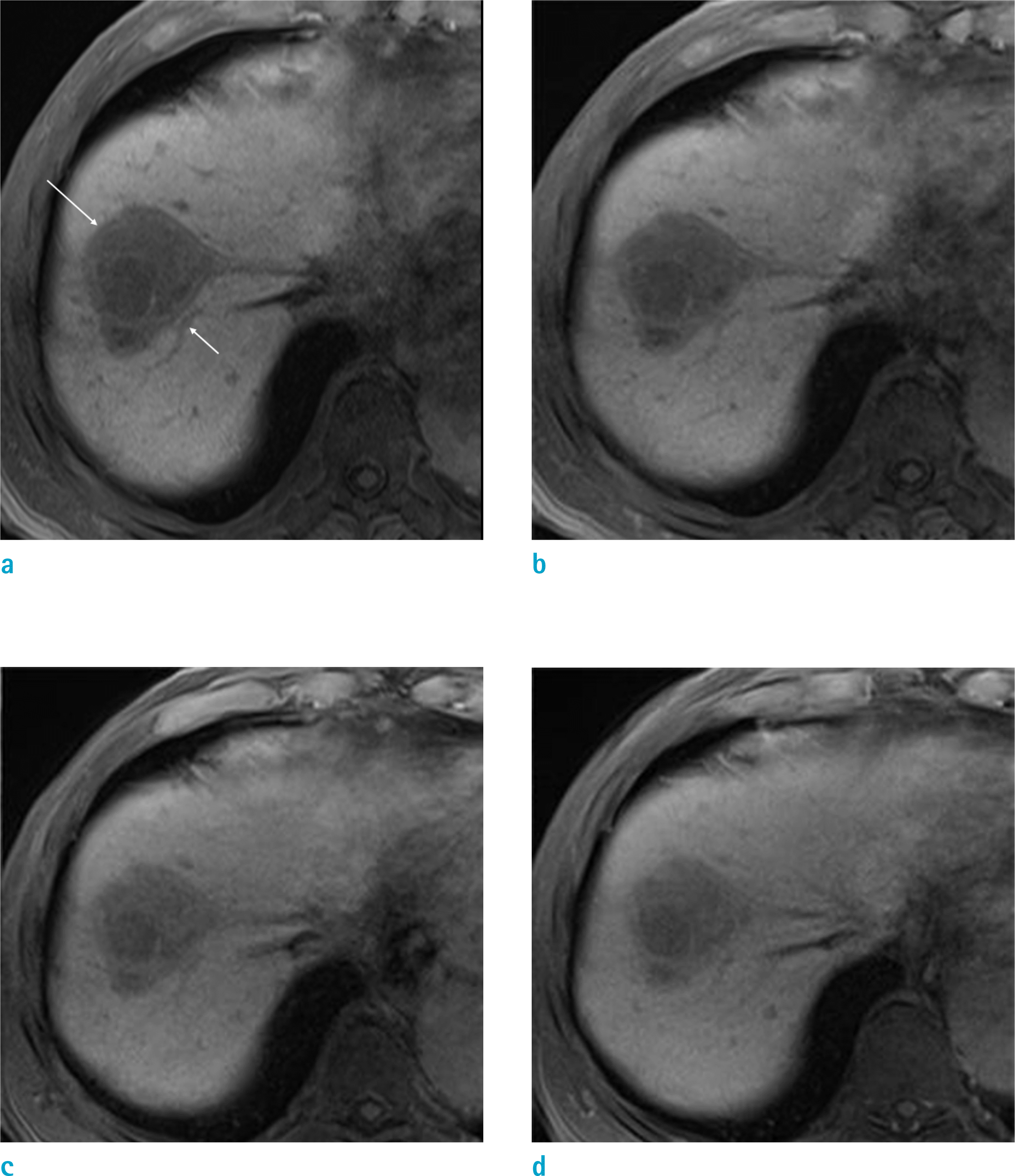
Table 1.
Results of the Qualitative Analysis: Comparison of the Hepatobiliary Phases at Different Flip Angles and Delayed Times
| Quality parameter | Flip angle and delayed time | P value | ||||||||
|---|---|---|---|---|---|---|---|---|---|---|
| Same flip angle | Same delayed time | Different flip angle and delayed time | ||||||||
| 20 min 13° | 15 min 13° | 20 min 9° | 15 min 9° | 20 min 13° vs. 15 min 13° | 20 min 9° vs. 15 min 9° | 20 min 13° vs. 20 min 9° | 15 min 13° vs. 15 min 9° | 15 min 13° vs. 20 min 9° | 20 min 13° vs. 13 min 9° | |
| Liver edge sharpness | ||||||||||
| Total | 4.77 | 4.69 | 4.63 | 4.54 | 0.083 | 0.047* | 0.001* | 0.001* | 0.288 | 0.000* |
| LC | 4.76 | 4.64 | 4.55 | 4.47 | 0.021* | 0.084 | 0.011* | 0.001* | 0.185 | 0.000* |
| Non-LC | 4.78 | 4.76 | 4.76 | 4.64 | 0.854 | 0.236 | 0.034* | 0.564 | 1.000 | 0.272 |
| Hepatic vessel clarity | ||||||||||
| Total | 4.50 | 4.33 | 4.07 | 3.88 | 0.001* | 0.004* | 0.000* | 0.000* | 0.001* | 0.000* |
| LC | 4.43 | 4.24 | 3.93 | 3.80 | 0.003* | 0.04* | 0.000* | 0.000* | 0.005* | 0.000* |
| Non-LC | 4.62 | 4.46 | 4.28 | 4.00 | 0.143 | 0.04* | 0.000* | 0.002* | 0.103* | 0.001* |
| Lesion conspicuity | ||||||||||
| Total | 4.73 | 4.58 | 4.26 | 4.13 | 0.003* | 0.008* | 0.000* | 0.000* | 0.000* | 0.000* |
| LC | 4.64 | 4.42 | 4.08 | 3.96 | 0.005* | 0.034* | 0.000* | 0.000* | 0.003* | 0.000* |
| Non-LC | 4.92 | 4.89 | 4.62 | 4.46 | 0.317 | 0.102 | 0.009* | 0.025* | 0.02* | 0.01* |
| Artifact severity | ||||||||||
| Total | 4.48 | 4.40 | 4.33 | 4.25 | 0.223 | 0.194 | 0.005* | 0.001* | 0.119 | 0.002* |
| LC | 4.42 | 4.32 | 4.22 | 4.21 | 0.257 | 0.655 | 0.06 | 0.004* | 0.057 | 0.005* |
| Non-LC | 4.56 | 4.48 | 4.50 | 4.32 | 0.518 | 0.244 | 0.023* | 0.083 | 0.891 | 0.098 |
| Overall image quality | ||||||||||
| Total | 4.60 | 4.48 | 4.31 | 4.23 | 0.041* | 0.073 | 0.000* | 0.000* | 0.001* | 0.000* |
| LC | 4.55 | 4.42 | 4.18 | 4.13 | 0.029* | 0.157 | 0.000* | 0.000* | 0.001* | 0.000* |
| Non-LC | 4.66 | 4.58 | 4.50 | 4.38 | 0.51 | 0.236 | 0.004* | 0.011 | 0.219 | 0.024* |
Table 2.
Results of Quantitative Analysis: Comparison of the Hepatobiliary Phase at Different Flip Angles and Times
| Parameter | Flip angle and delayed time | P value | |||
|---|---|---|---|---|---|
| 20 min 13° | 15 min 13° | 20 min 9° | 15 min 9° | ||
| Relative liver enhancement | |||||
| Total | 0.9141 ± 0.3751 | 0.8819 ± 0.3454 | 0.7683 ± 0.3006 | 0.7546 ± 0.2782 | 0.012* |
| LC | 0.9123 ± 0.3577 | 0.8760 ± 0.3307 | 0.7639 ± 0.9258 | 0.7515 ± 0.2800 | 0.073 |
| Non-LC | 0.9168 ± 0.4070 | 0.8906 ± 0.3728 | 0.7748 ± 0.3140 | 0.7593 ± 0.2812 | 0.272 |
| SNR of the liver parenchyma | |||||
| Total | 251.06 ± 78.62 | 245.51 ± 72.69 | 239.76 ± 28.28 | 239.72 ± 60.89 | 0.757 |
| LC | 245.84 ± 86.75 | 232.49 ± 73.48 | 232.41 ± 56.00 | 236.84 ± 67.71 | 0.836 |
| Non-LC | 258.79 ± 65.71 | 264.78 ± 68.42 | 250.62 ± 61.01 | 243.98 ± 50.15 | 0.652 |
 XML Download