

 PDF
PDF ePub
ePub Citation
 Print
Print


INTRODUCTION
Nodular hidradenoma is a benign skin tumor with sweat gland origin. It mostly occurs in adults, but a few cases of nodular hidradenoma in children have been reported. It grows slowly, over a long period of time, and without symptoms [1]. At present, complete excision with tumor free margins is thought to be a standard treatment for hidradenoma, because malignant transformation may occur [2]. Herein, we present a case of nodular hidradenoma in a 29-month-old girl on her left axilla.
Case Report
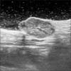
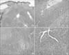
A 29-month-old girl was referred to our department with a one-year history of tender, asymptomatic, multiple verruciform nodule under the left axillary area (Fig. 1). Its initial size was about 0.5 cm in diameter, and has slowly grown over a year to 2 cm in diameter. Ultrasonography revealed a well-defined ovoid hypoechoic mass at the subcutaneous layer, with prominent internal vascularity, and several small internal cystic areas (Fig. 2). Excisional operation was done, and histopathologic examination showed a well-demarcated lobulated dermal nodule, with solid and cystic areas. The lesion was centrally located in the dermis, with no connection to the overlying epidermis. Both solid and cystic areas consisted of polygonal cells, with abundant eosinophilic to pale cytoplasm. Occasional ductal differentiation lined by cuboidal epithelium was observed. No nuclear atypia, necrosis or abnormal mitosis was found (Fig. 3). The histological findings were consistent with the diagnosis of nodular hidradenoma. During 3 years of the follow up period, the tumor didn't recur.
DISCUSSION
Nodular hidradenoma, which is an uncommon benign skin tumor of the sweat gland, has several designations, including eccrine acrospiroma, and clear cell hidradenoma. The age presentation varies, but mainly occurs in adults, with a peak incidence in the seventh decade [1]. Women are more affected than men, and head and upper extremities are more frequently involved than lower extremities [3]. Few cases of hidradenoma that occurred in children in their 1st decade have been reported [4]. It usually presents as a solitary, tender nodule or papule, with a size of 1 to 2 cm in diameter. It is usually asymptomatic, grows slowly over many years, and has a high locoregional reccurence rate [4].
Pathologically, hidradenomas are circumscribed, noncapsulated, multilobular masses that lie in the dermis, with no connection to the overlying epidermis [5]. They are mainly solid, but may show cystic areas containing mucinous material. Ductal differentiations are often present. They are composed of multiple lobulated masses of epithelial cells and tubular lumina of variable size and number [56]. Malignant transformations, known as hidranocarcinoma, are rare, and generally develops de novo but can evolve from a preexisting benign hidradenoma in extremely rare case [67]. After the malignant transformations, it shows slowly growing and no obvious changing for several years. Metastases may occur first in regional lymph nodes and the most common hematogenic metastases appear in lung. In widespread disease, chemotherapy and radiotherapy have proven ineffective [7]. The definitive treatments for both hidradenoma and hidradenocarcinoma is complete excision [567].
In summary, this case confirms that nodular hidradenoma can develop in a child as young as 29 months of age, which, to our knowledge, is the youngest yet to be reported. Once the diagnosis is established, long-term follow-up after complete excision is recommended, for the possibility of malignant transformation.

 XML Download