

 PDF
PDF ePub
ePub Citation
 Print
Print


If the surgery of cancer accompanying the removal or lymph nodes resulted in the swelling progressed in extremities, it is called obstructive secondary lymphedema. In the initial stage, bandage or drug therapy could be used to treat the lymphedema. However, after some time, patients with lymphedema feel heavier and painful while suffering from aesthetic reasons.
Treatment of the secondary lymphedema at the lower extremities is largely divided into two methods: removal of lymphatic tissue and increase of lymphatic perfusion. Increasing lymphatic perfusion include a lympho-lymphatic bypass, vascularized lymph node transfer, and various types of lympho-venous shunt operations1. Among them, the lymphaticovenous Anastomosis is known as the treatment of effectiveness and minimal invasiveness. However, it requires supermicrosurgical technique to perform anastomosis of vessels with 0.5 mm2.
The author is going to perform the Lymphaticovenous Aana stomosis to the patient with obstructive secondary lymphedema at her left extremity and to see the satisfactory results.
CASE REPORT
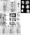
She is a 59-year-old patient who underwent the hysterectomy to treat uterine bleeding six years ago. During the progress of healing, the pain recurred two years ago; therefore, she was diagnosed with metastatic carcinoma. In the department of Obstetrics of this hospital, she underwent the Radical cuff resection, Bilateral Pelvic Lymphadenectomy, Periarterial Lymphadenectomy and total omentectomy. After the surgeries, the swelling was progressing and got worse in last six months and the patient visited the hospital (Fig. 1A). The result of lymphoscintigraphy scan showed the dermal backflow at left lower extremity but not the left inguinal lymph node activity (Fig. 1B). The result of computed tomography scan showed the subcutaneous edema at the left extremity and fluid collection at deep fascia (Fig. 1C).
Local anesthesia was used. Firstly, two lymph vessels were end-to-side anastomosed with one superficial vein at ankle. After that, the lymph vessels at the surrounding of knee and inguinal was end-to-side anastomosed with the superficial vein (Fig. 1D). The leg circumference was measured at the following level: 20 cm above the knee joint, knee joint, and 20 cm below the knee joint.
The patient felt the decrease of leg circumference after three months of surgery. There were wrinkles observed at the ankle and knee joint. At 6 months and 1 year, the patient's left leg was 43.3% and 13.5% larger than her right leg, respectively (a 32.8% reduction in volume differential), and the pain and heavy feeling were lessened. Also, there were wrinkles observed near the ankle and knee, therefore, the patient was very satisfied (Fig. 1E).
DISCUSSION
Patients with lymphedema are classified into primary lymphedema and secondary lymphedema. One of secondary lymphedema is the secondary obstructive lymphedema that occurs after tumor removal and lymphatic duct where cancer is spread. The treatment of secondary obstructive lymphedema is lymphaticovenous anastomosis, which has effectiveness and minimal invasiveness234. For successful Lymphaticovenous Anastomosis, it is important to find the availability of functional lymphatic vessels and to connect with veins. Generally, at the initial stage of lymph edema, there is high availability of functional lymphatic vessels, which will turn into a good result5. Also, the suitable vein in compatible size should be in proper location, which will reduce the backflow at the anastomosed part. Therefore, the veins less than 0.8 mm is advantageous than larger one6. Lymphaticovenous Anastomosis includes end-to-end, end-to-side anastomosis7 and Intravascular stenting method7 as well.
Generally, an end-to-end anastomosis may be an equally effective method that is technically simpler to perform and arguably more hemodynamically efficient, and an end-to-side anastomosis is indicated when the distal flow of the recipient vessel must be preserved or when a significant vessel size discrepancy exists. It is reported that the size and pressure of vein are bigger in lower than upper extremities, therefore, end-to-side anastomosis at lower extremities are often performed3. The end-to-side anastomosis was performed at all four parts in this case.
Comparing Lymphaticovenous anastomosis with vein anastomosis at the amputated distal fingers, it is found that they have the same issues; the size of veins to be anastomosed is small in a size of 1 mm and the vein is hard to find. Meanwhile, the lymphaticovenous anastomosis is rather tension-free than anastomosis of veins at amputated distal fingers. Also, the spasm of the vein was not an issue. The end-to-end anastomosis is usually used for the anastomosis of amputated distal finger since the size of veins is similar, while side-to-end anastomosis was used for four cases of the lymphaticovenous anastomosis since the superficial veins were bigger in size.
If the features of lymphaticovenous anastomosis are understood, we could expect a good result from patients with secondary obstructive lymphedema.
 XML Download