

 PDF
PDF Citation
 Print
Print


INTRODUCTION
Warfarin is a vitamin K antagonist that exerts an anticoagulant effect by reducing the levels of vitamin K-dependent coagulation factors II, VII, IX, and X [1]. It is widely used to prevent thrombotic complications in prosthetic heart valves. Warfarin treatment also reduces the levels of vitamin K-dependent anticoagulation factors, such as proteins C and S [2].
Close monitoring of warfarin anticoagulation therapy is imperative to prevent bleeding or thrombotic complications. The most widely used monitoring parameter is the international normalized ratio (INR) of prothrombin time (PT), which is measured using traditional coagulation analyzers or point-of-care testing (POCT) coagulometers [1]. As POCT coagulometers use capillary whole blood from a fingertip, they do not require blood collection and provide quick results [3]. Hence, the use of POCT by individual patients as well as in hospital settings is increasing.
Despite the practicality and speed of POCT, studies have reported considerable discrepancy between INR values provided by POCT and those from conventional laboratory tests, especially in high INR ranges, indicating that POCT results need to be confirmed by conventional laboratory tests [4-6]. The cause of the INR discrepancy between POCT and conventional laboratory tests is currently unclear.
Recent advances in coagulation measurement have led to the introduction of the thrombin generation assay (TGA) in coagulation laboratories [7, 8]. The TGA measures the total amount of thrombin generated in plasma following stimulation with a tissue factor, termed endogenous thrombin potential (ETP). It simultaneously measures the initiation time of thrombin generation (lag time), highest thrombin generation (peak thrombin), and time to peak thrombin [9]. TGA parameters have been suggested as potential markers of bleeding or thrombotic disorders [8]. We previously reported that the TGA well represented the anticoagulation status of patients receiving warfarin therapy [2].
We compared the INR of POCT (CoaguChek XS Plus; Roche Diagnostics, Mannheim, Germany) with that of a conventional laboratory test (ACL TOP 750; Instrumentation Laboratory SpA, Milan, Italy) in patients who received warfarin anticoagulation therapy after cardiac surgery. Coagulation and anticoagulation factor tests and TGAs were performed to clarify which test is more reliable for assessing changes in coagulation factors induced by warfarin.
MATERIALS AND METHODS
Study population
In total, 404 samples from 128 patients who received warfarin anticoagulation therapy after cardiac surgery between August 2020 and June 2021 in Seoul National University Hospital, Seoul, Korea, were collected prospectively. All patients were given an appointment to draw venous blood after obtaining informed consent from them to take part in the trial. The study was approved by the Institutional Review Board of Seoul National University Hospital (IRB No. 2006-231-1138). Paired POCT and conventional laboratory INR tests were performed. Four samples from two patients were excluded because the POCT INR was >8.0 (i.e., out of range), leaving 400 samples from 126 patients for analysis. The INR tests were repeated up to five times per patient during hospitalization or at each outpatient visit.
Demographic and clinical data were obtained from medical records. The study population comprised 59 men and 67 women, and their mean age was 63.2±10.9 years. The patients were taking warfarin to prevent or treat clinical conditions, such as mechanical valve group (N=98), bioprosthetic valve (N=16), atrial fibrillation (N=10), and other conditions (N=2) (Supplemental Data Table S1). The patients’ INR values ranged from 1.0 to <8.0.
Tests
INR levels in the fingertip capillary blood from the patients were measured using CoaguChek XS Plus. Simultaneously, venous blood samples were collected into a tube containing 3.2% buffered sodium citrate. The tubes were transferred to a conventional laboratory and centrifuged at 1,550×g for 15 minutes. The plasma obtained after centrifugation was used to measure the INR by a conventional laboratory test using a standard coagulation analyzer ACL TOP 750.
All coagulation factor tests were performed using the ACL TOP 750 analyzer. The coagulation factors were measured by a PT-based clotting test using HemosIL RecombiPlasTin reagent (ISI 1.0) for factors II, V, VII, and X (Instrumentation Laboratory, Lexington, MA, USA) and by an activated partial thromboplastin-based clotting test using SynthASil reagent for factors VIII, XI, XI, and XII (Instrumentation Laboratory SpA). Fibrinogen was measured using the Fibrinogen-C XL kit (Instrumentation Laboratory SpA). Proteins C and S were also tested using the ACL TOP 750 analyzer.
Thrombin generation was measured as previously described [8]. Briefly, 20 μL of reagent containing tissue factor at a final concentration of 1 or 5 pmol/L, as well as phospholipids or thrombin calibrators, was distributed in each well of 96-well plates, and 80 μL of test plasma was added. After the addition of 20 μL of fluorogenic substrate in HEPES buffer containing CaCl2, fluorescence was measured using a Fluoroskan Ascent fluorometer (Thermo Labsystems, Helsinki, Finland), and thrombin generation curves were calculated using the Thrombinoscope software (Thrombinoscope, Maastricht, the Netherlands). The curves were analyzed using parameters that describe the initiation, propagation, and termination phases of thrombin generation, including lag time, peak thrombin, time to peak, and ETP.
Statistical analysis
CoaguChek XS Plus and ACL TOP 750 INR values were compared using Pearson’s correlation coefficient, Passing–Bablok regression analysis, and Bland–Altman plots. The agreement between the INR values from the two tests according to three ranges of clinical decision making on anticoagulant dosing was analyzed based on agreement and Cohen’s kappa values. The Mann–Whitney U test was used to compare the groups. The correlations between INR values and coagulation or anticoagulation factors were assessed using Pearson’s correlation coefficient. Contributions of coagulation or anticoagulation factors to the INR discrepancy were analyzed using logistic regression and multiple linear regression analyses.
Statistical analyses were performed using SPSS 23 for Windows (IBM Corp., Armonk, NY, USA) and MedCalc Statistical Software (version 17.2, MedCalc Software BV, Ostend, Belgium). P<0.05 was considered statistically significant.
RESULTS
Comparison of CoaguChek XS Plus and ACL TOP 750 INR values
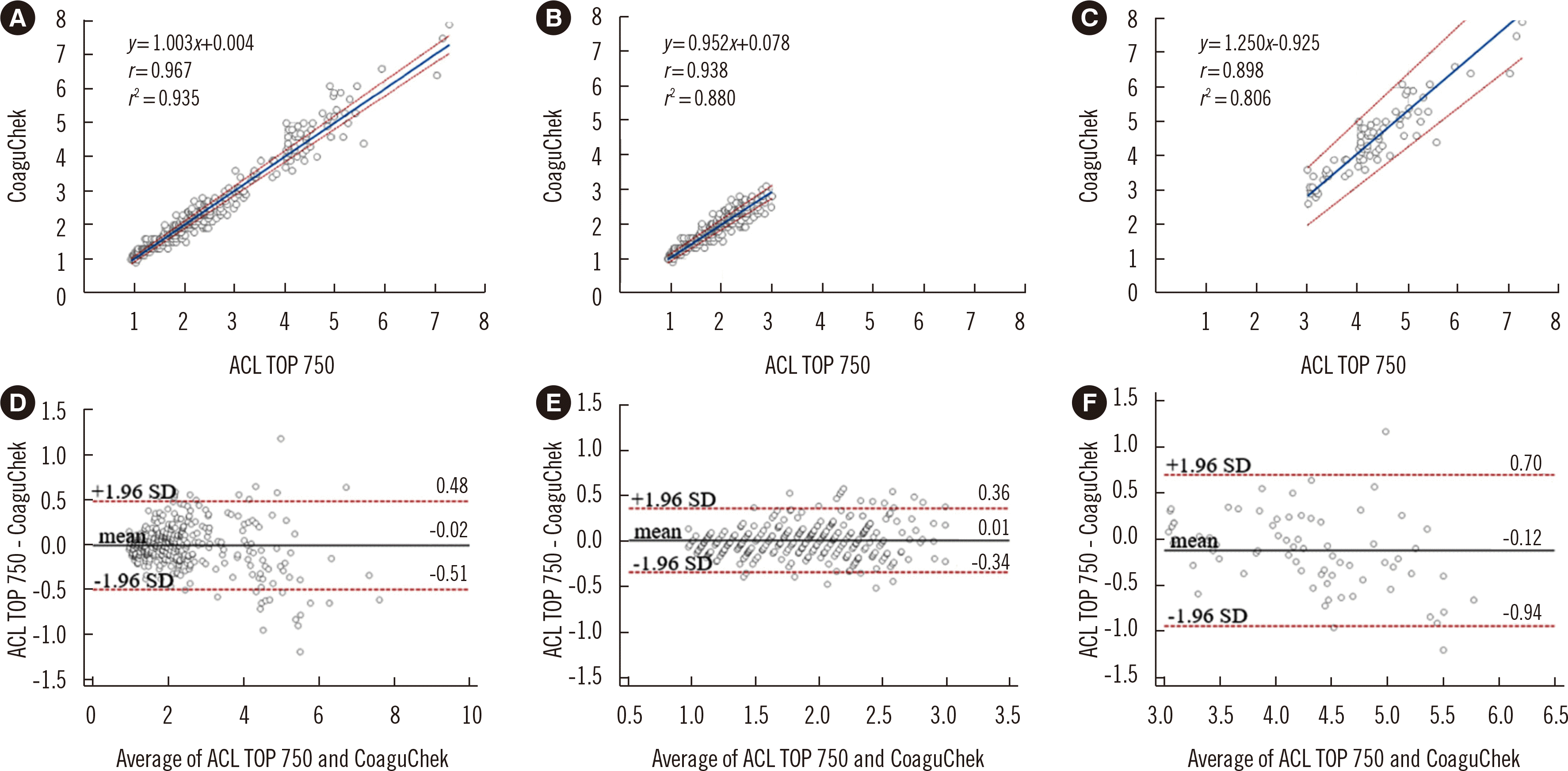
In the entire INR range, the correlation between CoaguChek XS Plus and ACL TOP 750 INR values was excellent, with no significant deviation from linearity and a Pearson’s correlation coefficient (r) of 0.967 (95% confidence interval [CI], 0.960–0.973; P<0.001) (Fig. 1A). The correlation coefficient was 0.938 at INR values ≤3.0 and 0.898 at INR values >3.0. The spread between the CI limits was larger at INR values >3.0 than at INR values ≤3.0 (Fig. 1B and C).
The mean difference between CoaguChek XS Plus and ACL TOP 750 INR values in the entire INR range was –0.02 INR (Fig. 1D). The ACL TOP 750 INR differed from the CoaguChek XS Plus INR by –0.51 to 0.48 INR with 95% limits of agreement. The mean difference in the INR was 0.01 (±1.96 SD, –0.34 to 0.36) at INR values ≤3 and –0.12 (±1.96 SD, –0.94 to 0.70) at INR values >3 (Fig. 1E, F). At INR values >3, the absolute mean difference in the INR tended to increase, and the interval, which represents the sum of the absolute values of the upper and lower 95% limits of agreement, was greater than at INR values ≤3.
Agreement between CoaguChek XS Plus and ACL TOP 750 INR values in terms of the range for clinical decision making on anticoagulant dosing
The agreement between CoaguChek XS Plus and ACL TOP 750 INR values was analyzed according to three INR ranges (subtherapeutic, therapeutic, and supratherapeutic) for clinical decision making on anticoagulant dosing (Table 1). The overall agreement between the two tests was 90.5% (362/400). The kappa index was 0.882 (95% CI, 0.845–0.919).
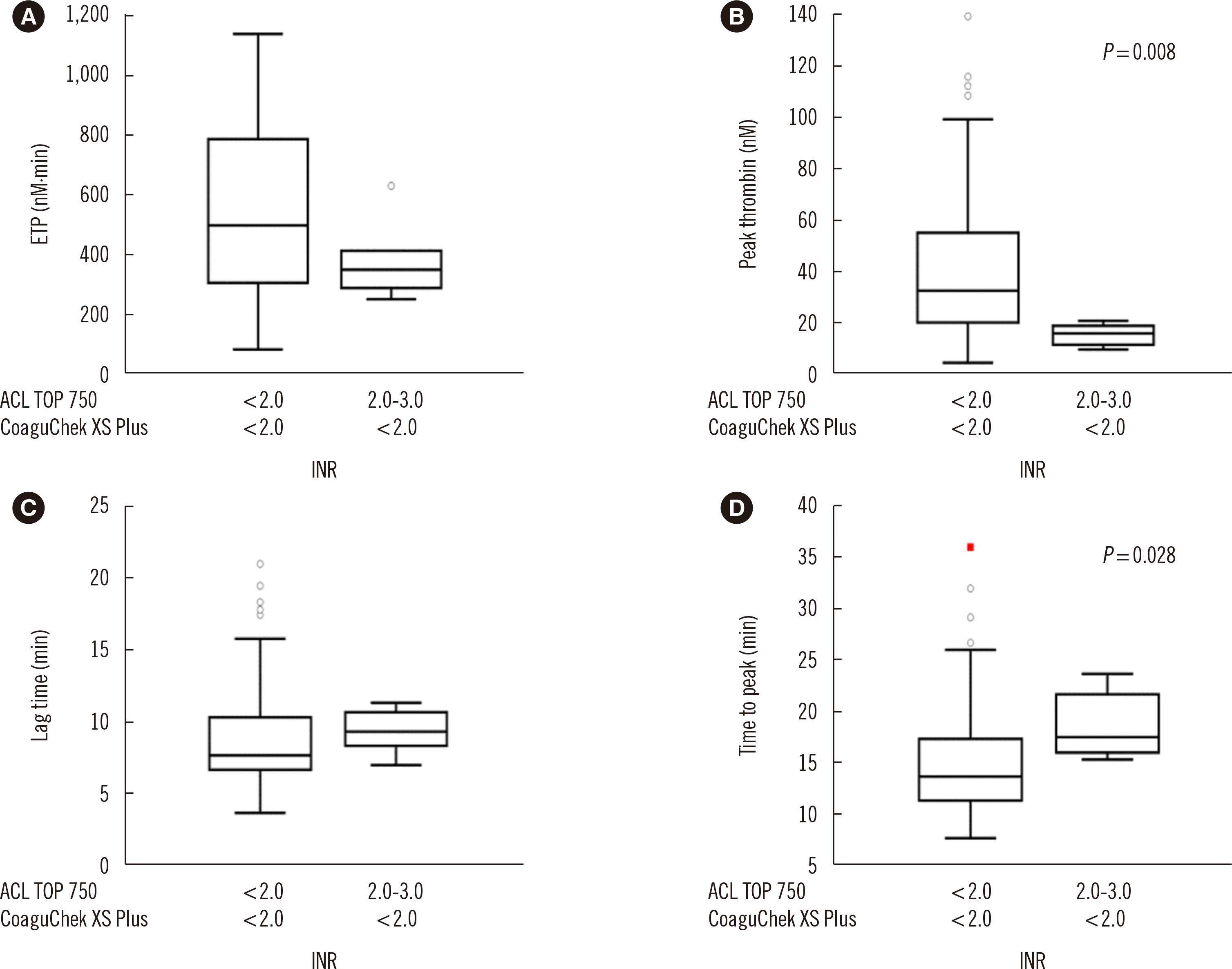
To determine which test is more reliable when discrepancy between the two tests occurs, the TGA was performed in samples that showed values in the subtherapeutic INR range (<2.0) in both tests (accord group, N=169) and those that showed a value in the subtherapeutic range (<2.0) by CoaguChek XS Plus, but in the therapeutic range (2.0≤ INR ≤3.0) by ACL TOP 750 (discrepancy group 1, N=12) (Fig. 2). The ETP was lower, albeit not significantly, in the accord group than in discrepancy group 1 (Fig. 2A). Peak thrombin was significantly lower, and the time to peak was significantly higher in discrepancy group 1 (Fig. 2B, D). The lag time was higher, albeit not significantly, in discrepancy group 1 (Fig. 2C). These data indicated that ACL TOP 750 INR values were better reflected by the degree of thrombin formation (low values of ETP and peak thrombin and high values of lag time and time to peak) than CoaguChek XS Plus INR values. In addition, TGA values were compared between the accord group and another discrepancy group, which included samples showing a value in the therapeutic range (2.0≤ INR ≤3.0) by CoaguChek XS Plus, but in the subtherapeutic range (<2.0) by ACL TOP 750 (discrepancy group 2, N=16) (Supplemental Data Fig. S1). Although the tendency was similar to that in discrepancy group 1, the difference in TGA values between the accord group and discrepancy group 2 was not significant.
Correlations between INR and coagulation and anticoagulation factors
Both CoaguChek XS Plus and ACL TOP 750 INR values were significantly correlated with fibrinogen and vitamin K-dependent coagulation factors (II, VII, IX, and X) and anticoagulation factors (proteins C and S) (Supplemental Data Table S2). The correlation coefficient values were higher for ACL TOP 750 INR values than for CoaguChek XS Plus INR values. Among all coagulation and anticoagulation factors, factor II exhibited the strongest correlation with both CoaguChek XS Plus and ACL TOP 750 INR values.
The contributions of coagulation and anticoagulation factors to INR values were determined using multiple linear regression analysis (Supplemental Data Table S3). Extrinsic coagulation factors (VIII, IX, XI, and XII) were excluded from the analysis because they do not contribute to PT test results. The coefficient of determination (modified R2) was higher for ACL TOP 750 INR values (0.622) than for CoaguChek XS Plus INR values (0.578), indicating that 62.2% of the variance in ACL TOP 750 INR values and 57.8% of that in CoaguChek XS Plus INR values was explained by variance in coagulation and anticoagulation factors. Factors II and V and proteins C and S had significant standardized regression coefficients (β) to both INR values of both tests.
Factors contributing to the significant INR discrepancy between the two tests
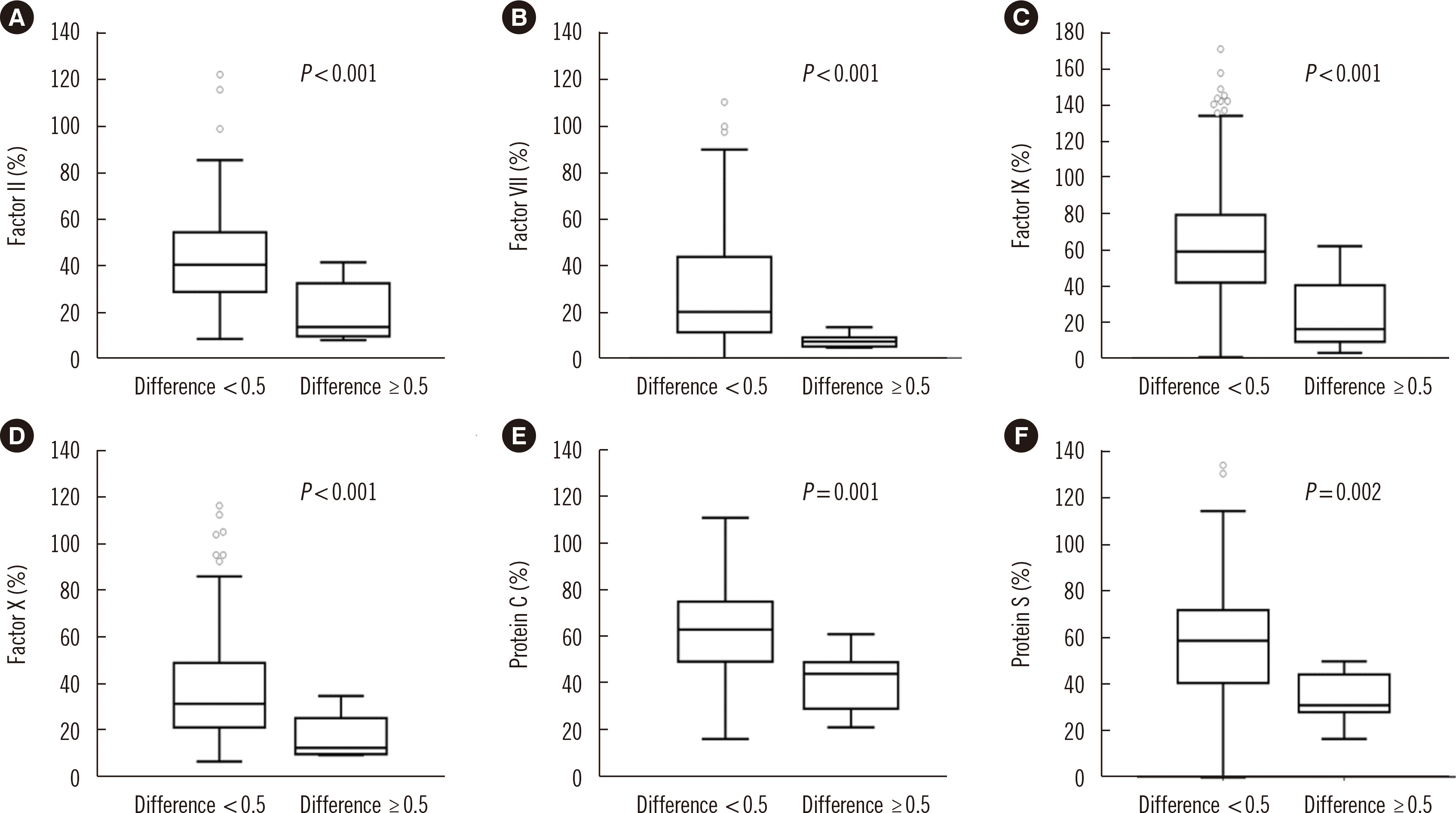
We defined the discrepancy in INR as significant when the absolute INR differences between the CoaguChek XS Plus and ACL TOP 750 results were ≥0.5. Among the 400 tests, 27 (6.7%) tests showed significant INR differences (Supplemental Data Table S4). At INR values ≥3.0, 21 (29.2%) tests showed significant differences.
To determine the factors contributing to the significant INR discrepancy (≥0.5), logistic regression analysis was performed (Table 2), which revealed that vitamin K-dependent coagulation and anticoagulation factors significantly contributed to the significant INR discrepancy. The odds ratios were <1. The median values of coagulation and anticoagulation factors were significantly lower in the group with an INR difference <0.5 than in the group with an INR difference ≥0.5 (Fig. 3).
DISCUSSION
Since the reporting of satisfactory results of a comparison between POCT INR values and conventional laboratory test INR values, POCT monitoring is widely accepted as a reliable option for monitoring warfarin therapy [10]. However, there have been significant discrepancies between POCT and conventional laboratory INR values, particularly in high INR ranges [4-6]. In agreement herewith, our results showed that the correlation and agreement between CoaguChek XS Plus and ACL TOP 750 INR values were excellent in the overall INR range, but at high INR ranges (INR >3), the mean difference in INR values increased.
As the TGA uses physiological tissue factor concentrations to trigger the coagulation cascade and evaluates the total amount of thrombin generated in plasma, studies have recommended this test for monitoring warfarin therapy [8]. Although there is no gold-standard method to measure the true anticoagulation status in patients treated with warfarin, we used the TGA to investigate which INR values are more reliable with respect to dosing decision for the anticoagulation therapy. The ETP and peak thrombin levels were lower, and the corresponding lag time and time to peak were higher in samples with 2.0≤ INR ≤3.0 by ACL TOP 750 and in samples with INR <2.0 by CoaguChek XS Plus than in samples with INR <2.0 by ACL TOP 750, indicating that INR values measured by ACL TOP 750 (2.0≤ INR ≤3.0) better represent low levels of thrombin generation than those measured by CoaguChek XS Plus (INR<2.0). In other words, conventional laboratory test INR values are more reliable than POCT INR values for predicting plasma thrombin generation induced by warfarin.
Studies have shown considerable discrepancy between POCT and conventional laboratory test INR values at high INR ranges, but its cause had not been established [4-6]. We performed coagulation and anticoagulation factor tests to determine the possible causes. Vitamin K-dependent coagulation and anticoagulation factors were significantly correlated with INR values. Of note, the correlation coefficients were higher for ACL TOP 750 than for CoaguChek XS Plus, suggesting that conventional laboratory test INR values better reflect the decreases in vitamin K-dependent coagulation factors. In multiple linear regression analysis, the coagulation and anticoagulation factors better explained the variance in conventional laboratory test INR values than that in POCT INR values. These results support that conventional laboratory test INR values better reflect warfarin-induced changes in coagulation and anticoagulation factor levels than POCT INR values.
We demonstrated that vitamin K-dependent coagulation (II, VII, IX, and X) and anticoagulation (proteins C and S) factors contributed to the significant INR discrepancy (≥0.5). The odds ratios were <1, indicating that as the levels of these factors decreased, significant INR differences were likely to occur. We consider the decreases in the vitamin K-dependent coagulation and anticoagulation factors to be a plausible cause of the discrepancy between POCT and conventional laboratory test INR values.
In comparison with conventional laboratory tests, POCT has several advantages: it saves time and money by reducing the time to results and requires a lower amount of blood, which is especially important for patients with life-threatening illnesses and outpatients with chronic illnesses who are treated at home [11]. However, the precision and accuracy of POCT are lower than those of conventional laboratory tests [12]. According to INR monitoring guidelines for POCT [13], a measurement that yields an INR value between 4 and 8 should be repeated using POCT to ensure that the increased INR value is not due to poor sample quality or analytical error. If the repeat value is >8.0 or differs by more than 0.5 units from the initial value, a conventional laboratory test INR measurement is recommended. As our results showed that conventional laboratory test INR values are more reliable for predicting thrombin generation as measured by the TGA and better reflect the changes in coagulation and anticoagulation factors than POCT when there is a significant difference between conventional laboratory test and POCT INR values, a confirmatory conventional laboratory test is required, which is in line with previous guidelines [13].
This study had a few limitations. First, we analyzed INR values using only one type of POCT device. Therefore, the comparison results and possible causes of discrepancies identified in our study should be considered with caution when applied to other POCT devices. Second, we focused on INR values and could not investigate the clinical outcomes, including bleeding and thrombosis complications, because none of our patients experienced any such complications. Clinical outcome results may need to be included in future studies. Third, some samples analyzed in our study were collected from patients who were treated with warfarin within one month after cardiac surgery. Warfarin exerts an anticoagulant effect by reducing the levels of vitamin K-dependent coagulation factors, which have relatively short half-lives; therefore, at least one month is required for warfarin levels to attain therapeutic equilibria [14]. Nevertheless, we demonstrated a significant discrepancy between POCT and conventional laboratory test INR values at high INR ranges.
In summary, INR values measured by POCT (CoaguChek XS Plus) were compared with those measured using a conventional laboratory analyzer (ACL TOP 750) in patients receiving warfarin therapy after cardiac surgery. The CoaguChek XS Plus and ACL TOP 750 INR values were consistent in the therapeutic INR range, but there was a tendency of increasing difference in the supratherapeutic INR range. The conventional laboratory test better reflected the decreases in thrombin formation and coagulation factors induced by warfarin than POCT. The possible causes of the INR difference between the two tests were decreases in the levels of vitamin K-dependent coagulation and anticoagulation factors. As conventional laboratory test INR values are more reliable than POCT INR values, a confirmatory conventional laboratory test is required at high POCT INR values. Future study using a high number of test samples and surveying clinical outcomes is required to determine the cutoff level for the high INR range to confirm POCT INR values.
 XML Download