Abstract
Purpose
To compare clinical outcomes of Descemet's stripping automated endothelial keratoplasty (DSAEK) between different graft insertion methods
Methods
The clinical records of 32 eyes of 30 DSAEK patients were retrospectively analyzed. Patients were divided into 2 groups according to graft insertion method. Group A: Taco-folding, group B: Tan-endoglide. The best corrected visual acuities (BCVA), intraocular pressures, astigmatism, endothelial cell count, central corneal thickness and complications were evaluated pre and post-operatively.
Results
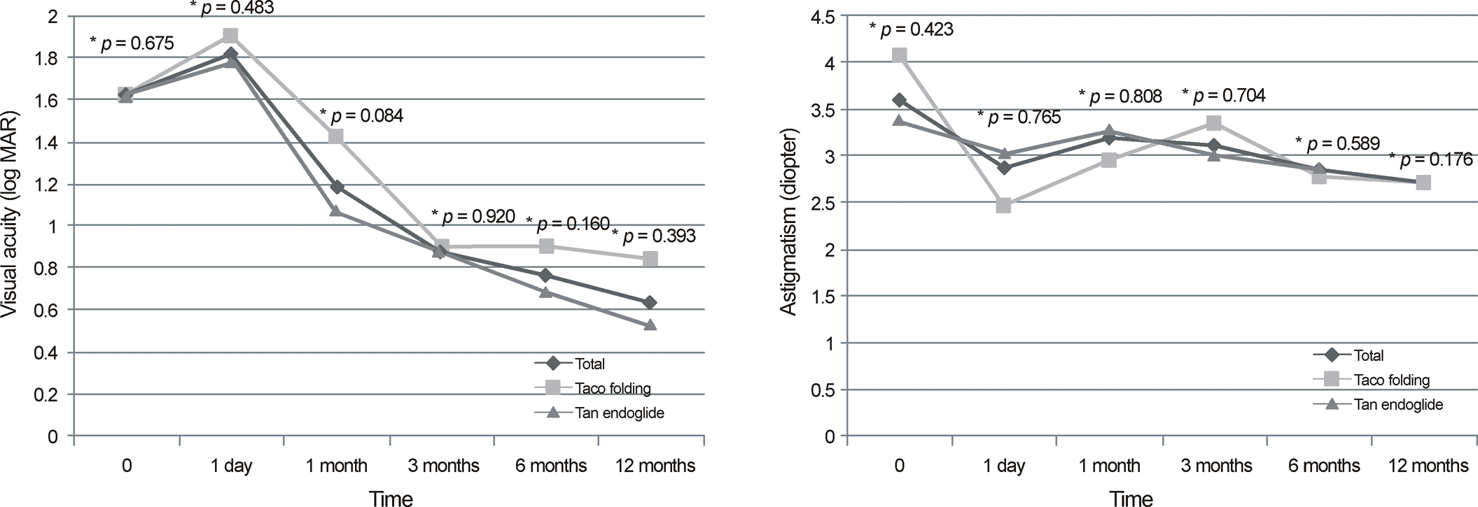
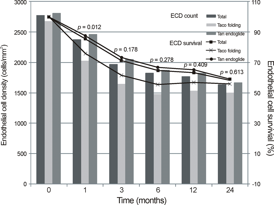
The average follow-up period was 19 months (range 1-67). Postoperative log MAR visual acuity had significantly improved both from 1.63 (log MAR) to 0.69 and 0.53 at 12 months in each group (p = 0.035 p = 0.000). Mean endothelial cell survival of each group at 1 month postoperative were 75.8% (range 62.7-88.6) and 87.7% (range 70.2-97.9), respectively (p = 0.012). The differences of BCVA improvement and endothelial cell survival between the groups at 12 months were not significant (p = 0.393, p = 0.544).
References
1. Melles GR, Eggink FA, Lander F, et al. A surgical technique for posterior lamellar keratoplasty. Cornea. 1998; 17:618–26.

2. Terry MA, Ousley PJ. Replacing the endothelium without corneal surface incisions or sutures: the first United States clinical series using the deep lamellar endothelial keratoplasty procedure. Ophthalmology. 2003; 110:755–64. discussion 764.
3. Price FW Jr, Price MO. Descemet's stripping with endothelial ker- atoplasty in 200 eyes: Early challenges and techniques to enhance donor adherence. J Cataract Refract Surg. 2006; 32:411–8.
5. Cheng YY, Pels E, Nuijts RM. Femtosecond-laser-assisted Descemet's stripping endothelial keratoplasty. J Cataract Refract Surg. 2007; 33:152–5.

6. Busin M, Bhatt PR, Scorcia V. A modified technique for Descemet membrane stripping automated endothelial keratoplasty to mini- mize endothelial cell loss. Arch Ophthalmol. 2008; 126:1133–7.
7. Khor WB, Mehta JS, Tan DT. Descemet stripping automated endo- thelial keratoplasty with a graft insertion device: surgical technique and early clinical results. Am J Ophthalmol. 2011; 151:223–32.e2.
8. Price FW Jr, Price MO. Descemet's stripping with endothelial ker- atoplasty in 50 eyes: a refractive neutral corneal transplant. J Refract Surg. 2005; 21:339–45.
9. Seo WM, Kim HK. Early result of femtosecond laser assisted de- scemet's membrane stripping endothelial keratoplasty. J Korean Ophthalmol Soc. 2008; 49:40–7.
10. Durrie DS, Kezirian GM. Femtosecond laser versus mechanical keratome flaps in wavefront-guided laser in situ keratomileusis: prospective contralateral eye study. J Cataract Refract Surg. 2005; 31:120–6.
11. Lee JS, Park YG, Yoon KC. Long-term results of Descemet's strip- ping automated endothelial keratoplasty in Korea. J Korean Ophthalmol Soc. 2010; 51:1431–7.
12. Bahar I, Kaiserman I, McAllum P, et al. Comparison of posterior lamellar keratoplasty techniques to penetrating keratoplasty. Ophthalmology. 2008; 115:1525–33.

13. Mearza AA, Qureshi MA, Rostron CK. Experience and 12-month- results of descemet-stripping endothelial keratoplasty (DSEK) with a small-incision technique. Cornea. 2007; 26:297–83.

14. Koenig SB, Covert DJ, Dupps WJ Jr, Meisler DM. Visual acuity, refractive error, and endothelial cell density six months after Descemet stripping and automated endothelial keratoplasty (DSAEK). Cornea. 2007; 26:670–4.

15. Terry MA, Chen ES, Shamie N, et al. Endothelial cell loss after Descemet's stripping endothelial keratoplasty in a large pro- spective series. Ophthalmology. 2008; 115:488–96.
16. Price MO, Price FW Jr. Endothelial cell loss after descemet strip- ping with endothelial keratoplasty influencing factors and 2-year trend. Ophthalmology. 2008; 115:857–65.
17. Terry MA. Endothelial keratoplasty: a comparison of complication rates and endothelial survivial between precut tissue and surgeon-cut tissue by a single DSAEK surgeon. Trans Am Ophthalmol Soc. 2009; 107:184–91.
18. Yamazoe K, Yamazoe K, Shinozaki N, Shimazaki J. Influence of the precutting and overseas transportation of corneal grafts for Descemet stripping automated endothelial keratoplasty on donor endothelial cell loss. Cornea. 2012; 1–4.

19. Balidis M, Konidaris VE, Ioannidis G, Boboridis K. Descemet's stripping endothelial automated keratoplasty using Tan Endoglide endothelium insertion system. Transplant Proc. 2012; 44:2759–64.

20. Yokogawa H, Kobayashi A, Sugiyama K. Clinical evaluation of a new donor graft inserter for Descemet's stripping automated endo- thelial keratoplasty. Ophthalmic Surg Lasers Imaging. 2012; 43:50–6.
21. Moon BG, Kim JH, Lee JE, et al. Long-term clinical outcomes of femtosecond LASER-assisted Descemet's stripping endothelial keratoplasty. J Korean Ophthalmol Soc. 2011; 52:679–89.

22. Cheng YY, Schouten JS, Tahzib NG, et al. Efficacy and safety of femtosecond laser-assisted corneal endothelial keratoplasty: a randomized multicenter clinical trial. Transplantation. 2009; 88:1294–302.

23. Khor WB, Teo KY, Mehta JS, Tan DT. Descemet stripping auto- mated endothelial keratoplasty in complex eyes: results with a do- nor insertion device. Cornea. 2013; 32:1063–8.
Figure 1.
Post-operative visual acuity and astigmatism changes of of Descemet's stripping automated endothelial keratoplasty (DSAEK) patients by insertion method. *Mann-Whitney U test between Taco-folding group and Tan-endoglide group.

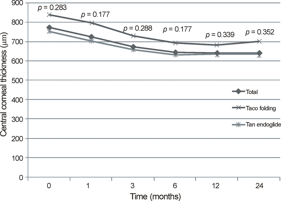
Figure 2.
Central corneal thickness of Descemet's stripping automated endothelial keratoplasty (DSAEK) patients by donor insertion method. *Mann-Whitney U test between Taco-folding group and Tan-endoglide group.

Figure 3.
Mean endothelial cell density and endothelial cell survival of Descemet's stripping automated endothelial keratoplasty (DSAEK) patients by insertion method. *Mann-Whitney U test between Taco-folding group and Tan-endoglide group.

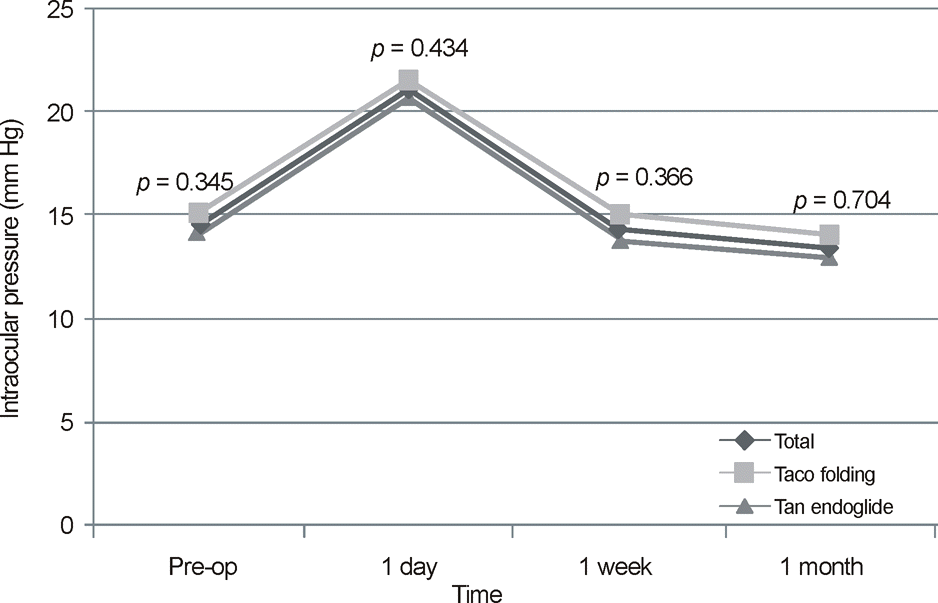
Figure 4.
Early post-operative intraocular pressure changes of of Descemet's stripping automated endothelial keratoplasty (DSAEK) patients by insertion method. *Mann-Whitney U test between Taco-folding group and Tan-endoglide group.

Table 1.
Demographic and Operative Details of Descemet Stripping Automated Endothelial Keratoplasty (DSAEK) Patients.
Table 2.
Pre and post-operative clinical data of Descemet Stripping Automated Endothelial Keratoplasty (DSAEK) Patients
|
BCVA (log MAR) mean (range) |
IOP (mm Hg) mean (range) |
Astigmatism (D) mean (range) |
|||||||||||||
|---|---|---|---|---|---|---|---|---|---|---|---|---|---|---|---|
| Time | Preop | 3 months | 6 months | 12 months | p-value* | Preop | 3 months | 6 months | 12 months | p-value† | Preop | 3 months | 6 months | 12 months | p-value* |
| Taco-folding | 1.63 | 0.91 | 0.78 | 0.69 | 0.035 | 15.2 | 14.1 | 13.7 | 14.6 | 0.134 | 4.10 | 3.35 | 2.78 | 2.71 | 0.028 |
| (n = 10) | (0.7-3.0) | (0.3-1.7) | (0.3-1.4) | (0.3-1.1) | (7-19) | (11-18) | (10-16) | (8-19) | (1.25-6.25) | (1-5.25) | (1.25-4.25) | (1.5-4.25) | |||
| (n = 8) | (n = 7) | (n = 7) | (n = 8) | (n = 7) | (n = 7) | (n = 7) | (n = 8) | (n = 7) | (n = 7) | ||||||
| Tan-endoglide | 1.63 | 0.89 | 0.69 | 0.53 | 0.000 | 14.3 | 13.1 | 13.5 | 13.3 | 0.876 | 3.40 | 3.03 | 2.88 | 2.71 | 0.030 |
| (n = 22) | (0.4-3.0) | (0.2-2.0) | (0.1-1.7) | (0.1-1.0) | (6-24) | (5-22) | (6-23) | (8-19) | (0.75-6.25) | (0.5-6.5) | (0.75-6.25) | (0.5-6.25) | |||
| (n = 21) | (n = 20) | (n = 13) | (n = 21) | (n = 20) | (n = 13) | (n = 18) | (n = 21) | (n = 20) | (n = 13) | ||||||
| p-value‡ | 0.675 | 0.920 | 0.160 | 0.393 | 0.345 | 0.704 | 0.589 | 0.176 | 0.423 | 0.704 | 0.589 | 0.176 | |||
| All eyes | 1.63 | 0.90 | 0.80 | 0.72 | 0.000 | 14.6 | 13.4 | 13.6 | 13.7 | 0.453 | 3.60 | 3.11 | 2.84 | 2.71 | 0.004 |
| (n = 32) | (0.4-3.0) | (0.2-2.0) | (0.1-2.0) | (0.2-1.7) | (6-24) | (5-23) | (6-23) | (8-19) | (0.75-6.25) | (0.5-6.5) | (0.75-6.25) | (0.5-6.25) | |||
| (n = 29) | (n = 27) | (n = 20) | (n = 29) | (n = 27) | (n = 20) | (n = 25) | (n = 29) | (n = 27) | (n = 20) | ||||||
Table 3.
Postoperative complications and survival rate of Descemet's stripping automated endothelial keratoplasty (DSAEK) patients


 PDF
PDF ePub
ePub Citation
 Print
Print


 XML Download