

 PDF
PDF ePub
ePub Citation
 Print
Print


Abstract
Purpose
To evaluate the visual prognosis and time course of the foveal reattachment according to operation methods and pre-operative factors using optical coherence tomography (OCT) after successful reattachment surgery in cases of macula-off rhegmatogenous retinal detachment.
Methods
From Jan 2006 to Aug 2008, 47 patients who underwent retinal reattachment surgery for macula-off retinal detachment who were followed up for more than 12 months were enrolled in this study. Thirty patients underwent vitrectomy, and 17 patients underwent a scleral buckle procedure. Ophthalmological examinations, including best-corrected visual acuity, slit-lamp examination, fundus examination, and OCT, were performed before surgery and at 1, 1.5, 3, 6, and 12 months after surgery.
References
1. Machemer R. The importance of fluid absorption, traction, abdominal currents, and chorioretinal scars in the therapy of abdominal retinal detachments. XLI Edward Jackson memorial lecture. Am J Ophthalmol. 1984; 98:681–93.
2. Burton TC. Preoperative factors influencing anatomic success rates following retinal detachment surgery. Trans Sect Ophthalmol Am Acad Ophthalmol Otolaryngol. 1977; 83:499–505.
3. Tani P, Robertson DM, Langworthy A. Prognosis for central abdominal and anatomic reattachment in rhegmatogenous retinal abdominal with macular detached. Am J Ophthalmol. 1981; 92:611–20.
4. Escoffery RF, Olk RJ, Grand MG, Boniuk I. Vitrectomy without scleral buckling for primary rhegmatogenous retinal detachment. Am J Ophthalmol. 1985; 99:275–81.

5. Mcperson AR, O'Malley RE, Butner RW, Beltangady SS. Visual acuity after surgery for retinal detachment with macular involvement. Ann Ophthalmol. 1982; 14:639–45.
6. Park JM, Shim HS, Bae JH. A clinical study on rhegmatogenous retinal detachment. J Korean Ophthalmol Soc. 1993; 34:1154–61.
7. Sharma T, Challa JK, Ravishankar KV, Murugesan R. Scleral buckling for retinal detachment. Predictors for anatomic failure. Retina. 1994; 14:338–43.
8. Kaufman PL. Prognosis of primary rhegmatogenous retinal detachment. 2. Accounting for and predicting final visual acuity in surgically reattached cases. Acta Ophthalmol. 1976; 54:61–74.
9. Chang SD, Kim IT. abdominal visual recovery after scleral abdominal procedure of rhegmatogenous retinal detachment involving the macula. Korean J Ophthalmol. 2000; 14:20–6.
10. Barr CC. The histopathology of successful retinal reattachment. Retina. 1990; 10:189–94.
11. Machemer R. Experimental retinal detachment in the owl monkey. IV. The reattached retina. Am J Ophthalmol. 1968; 66:1075–91.
12. Aaberg TM, Machmer R. Correlation of naturally occurring abdominals with long-term retinal detachment in the owl monkey. Am J Ophthalmol. 1970; 69:640–50.
13. Puliafito CA, Hee MR, Schuman JS, et al. Optical coherence abdominal of ocular disease. Thorofare NJ: Slack;1996. p. 3–15.
14. Wolfensberger TJ, Gonvers M. Optical coherence tomography in the evaluation of incomplete visual acuity recovery after mac-ula-off retinal detachments. Graefes Arch Clin Exp Ophthalmol. 2002; 240:85–9.
15. Kaga T, Fonseca RA, Dantas MA, et al. Optical coherence abdominal of bleb-like subretinal lesions after retinal reattachment surgery. Am J Ophthalmol. 2001; 132:120–1.
16. Benson SE, Schlottmann PG, Bunce C, et al. Optical coherence tomography analysis of the macular after scleral buckle surgery for retinal detachment. Ophthalmology. 2007; 114:108–12.
17. Theodossiadis PG, Georgalas IG, Emefietzoglou J, et al. Optical coherence tomography findings in the macula after treatment of rhegmatogenous retinal detachments with spared macula preoperatively. Retina. 2003; 23:69–75.
18. Diddie KR, Ernest JT. Uveal blood flow after 360 degrees constriction in the rabbit. Arch Ophthalmol. 1980; 98:729–30.
19. Yoshida A, Feke GT, Green GJ, et al. Retinal circulatory changes after scleral buckling procedures. Am J Ophthalmol. 1983; 95:182–8.
20. Kang SW, Kim JH, Shin WJ, Kim JI. Subretinal fluid bleb after successful scleral buckling and cryotherapy for retinal detachment. Am J Ophthalmol. 2008; 146:205–10.
21. Hagimura N, Iida T, Suto K, Kishi S. Persistent foveal retinal abdominal after successful rhegmatogenous retinal detachment surgery. Am J Ophthalmol. 2002; 133:516–20.
22. Wolfensberger TJ. Foveal reattachment after macula-off retinal detachment occurs faster after vitrectomy than after buckle surgery. Ophthalmology. 2004; 111:1340–3.
23. Benson SE, Schlottmann PG, Bunce C, et al. Optical coherence tomography analysis of the macula after vitrectomy surgery for retinal detachment. Ophthalmology. 2006; 113:1179–83.
24. Ku M, Sohn HJ, Lee DY, Nam DH. Foveal reattachment after scleral buckling vs vitrectomy for macula-off retinal detachment. J Korean Ophthalmol Soc. 2009; 50:399–404.
25. Kang JH, Yoon HS. Foveal retinal detachment diagnosed by abdominal coherence tomography after successful retinal detachment surgery. J Korean Ophthalmol Soc. 2005; 46:1637–41.
26. Na KS, Kim CG. The usefulness of optical coherence tomography in macula-off retinal detachment. J Korean Ophthalmol Soc. 2006; 47:1597–604.
27. Lincoff H, Kreissig I. Optical coherence tomography of abdominal displacement of optic disc pit maculopathy. Br J Ophthalmol. 1998; 82:367–72.
28. Bird AC. Pathogenesis of serous detachment of the retina and abdominal epithelium. Ryan SJ, editor. Retina. 3rd ed.St. Louis: Mosby;2001. 2:chap. 59.
29. Auh SJ, Kang SW. Delayed absorption of subretinal fluid after scleral buckling procedure for rhegmatogeous retinal detachment. J Korean Ophthalmol Soc. 2000; 41:1177–86.
30. Benson WE, Nantawan P, Morse PH. Characteristics and abdominal of retinal detachments with demarcation lines. Am J Ophthalmol. 1977; 84:641–4.
31. Wallyn RH, Hilton GF. Subretinal fibrois in retinal detachment. Arch Ophthalmol. 1979; 97:2128–9.
32. Park CS, Chang WH, Cha SC, Park YH. Surgical results of scleral buckling in retinal detachments with subretinal traction band. J Korean Ophthalmol Soc. 2004; 45:1092–8.
33. Chung SJ, Chung SK, Chung SM. Chemical analysis of subretinal fluid in rhegmatogenous retinal detachment. J Korean Ophthalmol Soc. 1992; 33:605–8.
34. Lee SJ, Huh K. Drainge vs. nondrainage of subretinal fluid in scleral buckling procedure. J Korean Ophthalmol Soc. 1998; 39:2082–7.
35. Baba T, Hirose A, Moriyama M, Mochizuki M. Tomographic abdominal and visual recovery of acute macula-off rhegmatogenous abdominall detachment. Graefes Arch Clin Exp Ophthalmol. 2004; 242:576–81.
36. Lecleire-Collet A, Muraine M, Menard JF, Brasseur G. Predictive visual outcome after macula-off retinal detachment surgery using optical coherence tomography. Retina. 2005; 25:44–53.
37. Seo JH, Woo SJ, Park KH, et al. Influence of persistent abdominal fluid on visual outcome after successful scleral buckle abdominal for macula-off retinal detachment. Am J Ophthalmol. 2008; 145:915–22.
Figure 1.
Scatterplot illustrating the relation between postoperative best-corrected visual acuity (BCVA, LogMAR) and height of foveal detachment (μm) in optical coherence tomography (OCT).
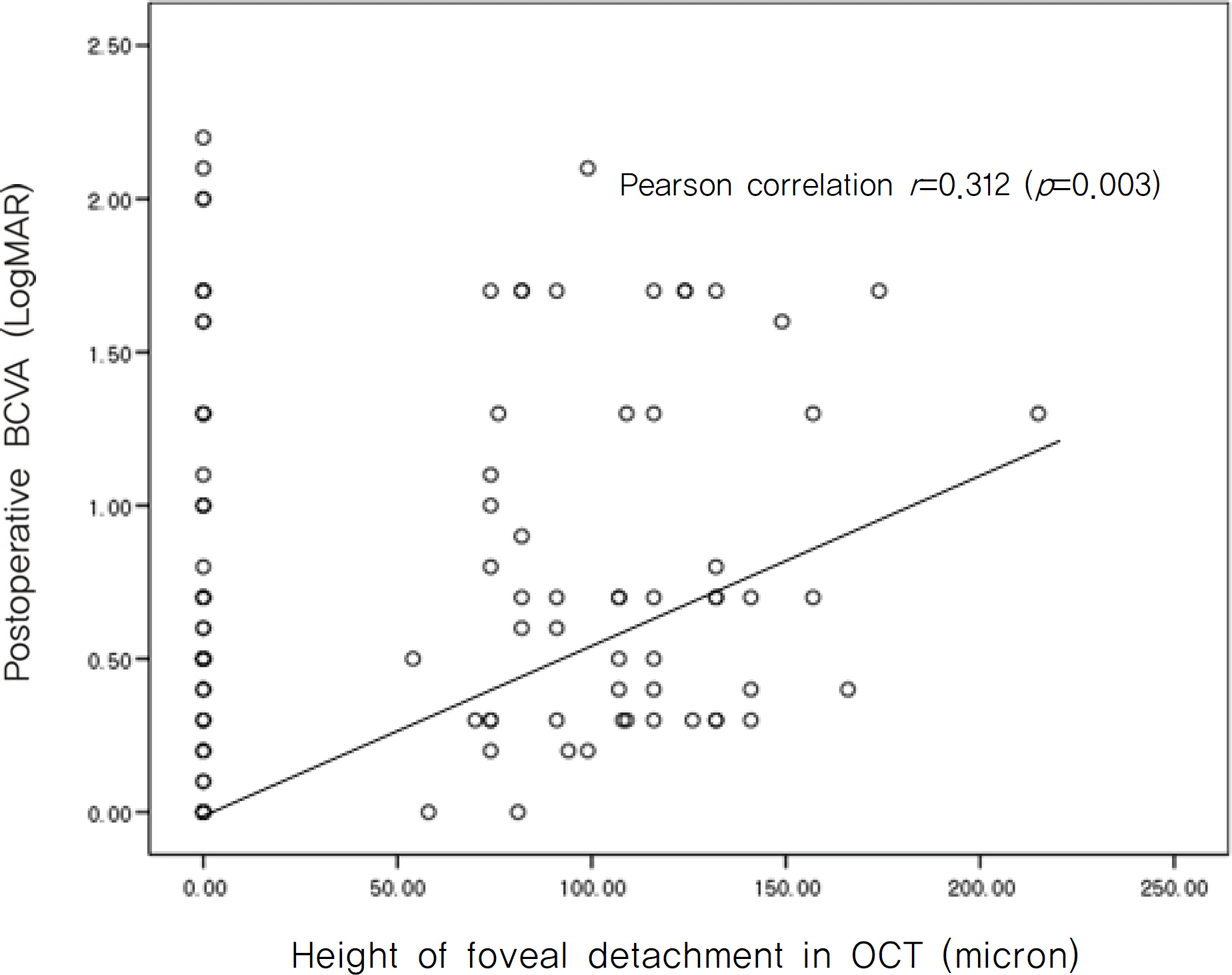
Figure 2.
Scatterplot illustrating the relation between improvement of best-corrected visual acuity (BCVA, LogMAR) and foveal reattachment time (days) by optical coherence tomography (OCT) after buckling procedure.
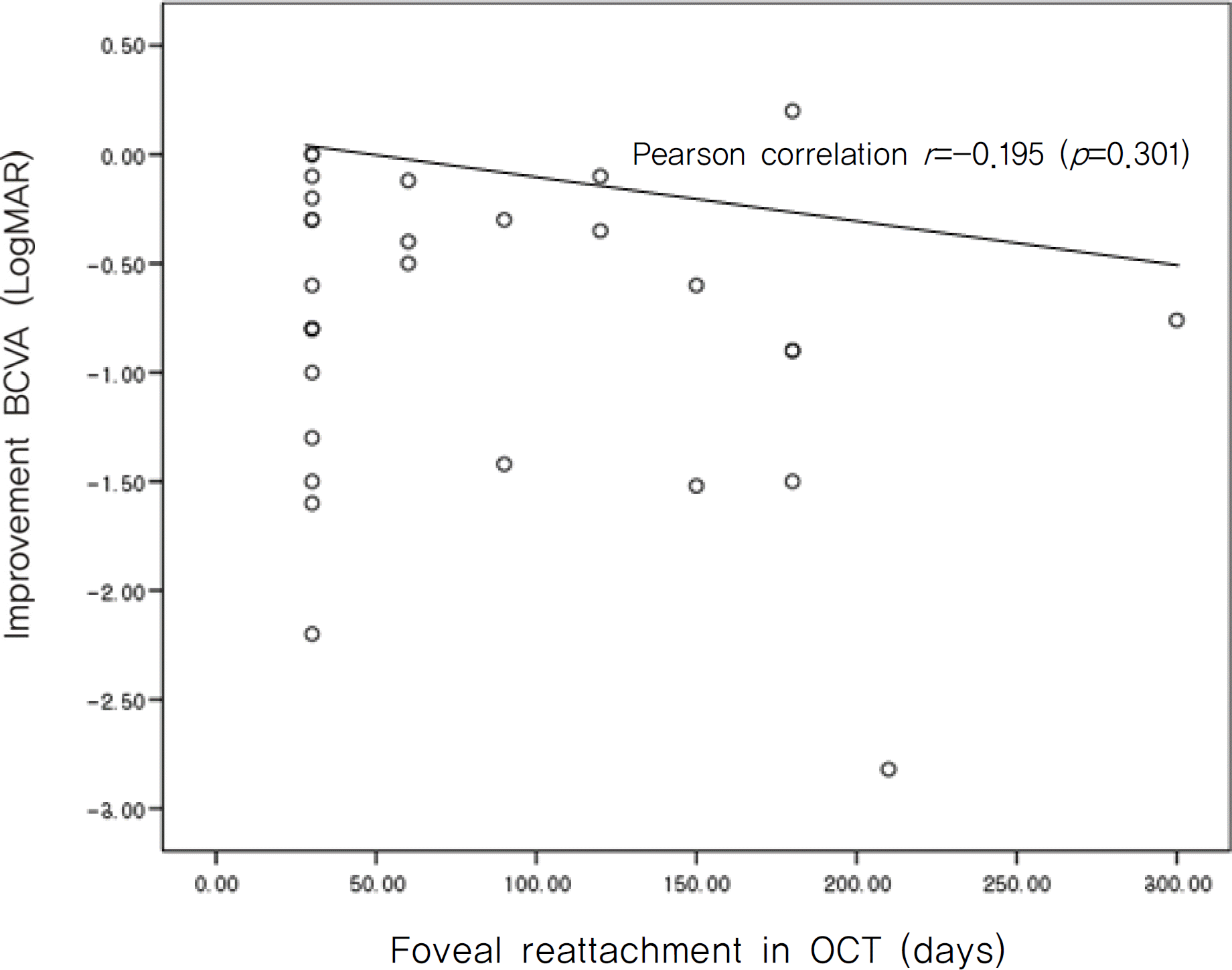
Table 1.
Clinical characteristics of patients (I)
| Variables | Data | ||
|---|---|---|---|
| Age (years) | 35.6±18.9 (10–73) | ||
| Male/Female (No. of eyes) | 28/19 | ||
| Right/Left eye (No. of eyes) | 22/25 | ||
| Duration of symptom (days) | 42.31±117.5 (3–730) | ||
| Extent of retinal detachment (hours) | 5.49±1.99 (2∼12) | ||
| Phakia/Pseudophakia (No. of eyes) | 40/7 | ||
| Preoperative BCVA* (LogMAR) | 1.27±0.79 (0.3–2.6) | ||
| Operation methods | No. of eyes (%) | ||
| Encircling buckle | with subretinal fluid drainage | 7 (14.9%) | |
| without subretinal fluid drainage | C3F8 gas† (+) | 1 (2.1%) | |
| C3F8gas (−) | 3 (6.4%) | ||
| Segmental buckle | with subretinal fluid drainage | C3F8 gas (+) | 3 (6.4%) |
| C3F8 gas (−) | 7 (14.9%) | ||
| without subretinal fluid drainage | C3F8 gas (+) | 1 (2.1%) | |
| C3F8 gas (−) | 8 (17.0%) | ||
| Vitrectomy with fluid gas exchange | 17 (36.2%) |
Table 2.
Clinical characteristics of patients (II)
| Variables | Results | P-value* |
|---|---|---|
| Preoperative BCVA† (LogMAR) | 1.27±0.79 | |
| Postoperative BCVA (LogMAR) | ||
| 1 month | 1.01±0.59 | 0.034 |
| 3 months | 0.528±0.39 | <0.0001 |
| 6 months | 0.363±0.38 | 0.004 |
| 12 months | 0.369±0.38 | 0.02 |
| Height of foveal detachment (μm) | ||
| 1 month | 37.73±56.93 (0∼157) | |
| 3 months | 19.74± 42.2 (0∼132) | <0.0001 |
| 6 months | 3.68±17.84 (0∼99) | 0.018 |
| Timing of foveal reattachment by OCT‡ ( | days) 68.29.14±63.7 (4∼210) |
Table 3.
Comparison between vitrectomy and scleral buckling procedure
| Vitrectomy (n=17) | Scleral Buckle (n=30) | P-value | |
|---|---|---|---|
| Age (years) | 50.71±12.36 | 27.07±16.62 | <0.0001* |
| Extent of retinal detachment (hours) | 5.17±1.77 | 5.67±2.12 | 0.557* |
| Duration of Symptom (days) | 12.06±21.48 (1–90) | 59.47±144.17 (3–730) | 0.007* |
| Lens status (n, %) | |||
| Phakia | 11 (64.7%) | 29 (96.7%) | 0.006† |
| Pseudophakia | 6 (35.3%) | 1 (3.3%) | 0.006† |
| Preoperative BCVA‡ (LogMAR) | 1.62±0.76 | 1.08±0.74 | 0.019* |
| Postoperative BCVA (LogMAR) | |||
| 1 month | 1.27±0.70 | 0.96±0.51 | 0.122* |
| 3 months | 0.59±0.36 | 0.54±0.43 | 0.588* |
| 6 months | 0.43±0.38 | 0.44±0.52 | 0.624* |
| 12 months | 0.399±0.44 | 0.317±0.22 | 0.670* |
| Improvement of BCVA (LogMAR) | –1.36±0.68 | –0.78±0.7 | 0.005* |
| Timing of foveal reattachment by OCT§ (days) | 37.06±16.87 (1–90) | 86±73.37 (4–300) | 0.013* |
| Foveal reattachment by OCT in less than 6 weeks (n, %) | 17 (100%) | 15 (50%) | <0.0001† |
Table 4.
Comparison between encircling and segmental scleral buckle
| Encircling buckle (n=11) | Segmental buckle (n=19) | P-value | |
|---|---|---|---|
| Age (years) | 17.90±7.34 | 32.37±18.28 | 0.012* |
| Extent of retinal detachment (hours) | 6.36±2.62 | 5.26±1.73 | 0.25* |
| Duration of symptom (days) | 123.54±228.03 (3–730) | 22.37±25.28 (4–75) | 0.35* |
| Preoperative BCVA‡ (LogMAR) | 1.27±0.56 | 0.97±0.82 | 0.103* |
| Postoperative BCVA (LogMAR) | |||
| 1 month | 1.04±0.61 | 0.82±0.46 | 0.42* |
| 3 months | 0.63±0.57 | 0.45±0.31 | 0.553* |
| 6 months | 0.55±0.62 | 0.28±0.27 | 0.307* |
| 12 months | 0.58±0.62 | 0.29±0.19 | 0.372* |
| Improvement of BCVA (LogMAR) | –0.76±0.52 | –0.79±0.79 | 0.672* |
| Timing of foveal reattachment by OCT§ (days) | 57.27±60.67 (5–180) | 102.6±76.4 (4–300) | 0.052* |
| Foveal reattachment by OCT at 1 month (n, %) | 9 (81.8%) | 6 (31.6%) | 0.021† |
Table 5.
Comparison between the patients with symptom duration of retinal detachment less than one week and more than one week in buckle procedure
| Duration of symptom (< 1 week) | Duration of symptom (≥ 1 week) | P-value* | |
|---|---|---|---|
| No. of eyes | 14 | 16 | |
| Age (years) | 28.14±18.60 | 26.12±15.24 | 0.822 |
| Extent of retinal detachment (hours) | 4.71±1.49 | 6.5±2.28 | 0.019 |
| Duration of symptom (days) | 5.5±1.40 | 106.69±187.31 | <0.0001 |
| Preoperative BCVA† (LogMAR) | 1.11±0.91 | 1.05±0.59 | 0.854 |
| Postoperative BCVA (LogMAR) | |||
| 1 month | 0.65±0.28 | 1.12±0.59 | 0.028 |
| 3 months | 0.37±0.22 | 0.64±0.51 | 0.142 |
| 6 months | 0.26±0.26 | 0.47±0.55 | 0.400 |
| 12 months | 0.26±0.21 | 0.52±0.55 | 0.193 |
| Improvement of BCVA (LogMAR) | –0.92±0.86 | –0.67±0.52 | 0.448 |
| Timing of foveal reattachment by OCT‡ (days) | 98.57±73.89 | 75±73.48 | 0.525 |
Table 6.
Comparison between retinal detachment with demarcation line and without demarcation line in scleral buckle procedure
| Retinal detachment with demarcation line (n=16) | Retinal detachment without demarcation line (n=14) | P-value | |
|---|---|---|---|
| Age (years) | 29.93±18.69 | 23.79±13.84 | 0.377* |
| Extent of retinal detachment (hours) | 6.06±2.35 | 5.21±1.81 | 0.400* |
| Duration of symptom (days) | 95.68±191.44 | 18.07±24.93 | 0.047* |
| Preoperative BCVA‡ (LogMAR) | 1.03±0.68 | 1.14±0.82 | 0.729* |
| Improvement of BCVA (LogMAR) | –0.63±0.62 | –0.96±0.77 | 0.131* |
| Postoperative BCVA (LogMAR) | |||
| 1 month | 0.98±0.60 | 0.81±0.41 | 0.498* |
| 3 months | 0.63±0.53 | 0.38±0.19 | 0.208* |
| 6 months | 0.47±0.57 | 0.27±0.21 | 0.637* |
| 12 months | 0.48±0.57 | 0.30±0.21 | 0.608* |
| Timing of foveal reattachment by OCT§ (days) | 76.88±76.66 | 96.43±70.78 | 0.400* |
| Encircling buckle (n, %) | 5 (31.3%) | 8 (57.1%) | 0.390† |
| SRF∏ drainage (+) (n, %) | 10 (62.5%) | 7 (50%) | 0.626† |
| C3 F8 gas∏ (+) (n, %) | 1 (6.3%) | 4 (28.6%) | 0.433† |
Table 7.
Comparison between scleral buckle procedure with subretinal fluid (SRF) drainage and without subretinal fluid drainage
| Buckle procedure with SRF drainage | Buckle procedure without SRF drainage | P-value* | |
|---|---|---|---|
| No. of eyes | 17 | 13 | |
| Age (years) | 29.76±19.11 | 23.54±12.53 | 0.621 |
| Extent of retinal detachment (hours) | 5.53±2.37 | 5.85±1.81 | 0.509 |
| Duration of symptom (days) | 83.65±189.05 | 27.85±25.84 | 0.281 |
| Preoperative BCVA† (LogMAR) | 1.38±0.79 | 0.68±0.45 | 0.009 |
| Postoperative BCVA (LogMAR) | |||
| 1 month | 0.92±0.57 | 0.87±0.47 | 0.805 |
| 3 months | 0.58±0.47 | 0.43±0.34 | 0.341 |
| 6 months | 0.49±0.53 | 0.23±0.24 | 0.170 |
| 12 months | 0.48±0.54 | 0.29±0.23 | 0.509 |
| Improvement of BCVA (LogMAR) | –1.01±0.80 | –0.49±0.41 | 0.043 |
| Timing of foveal reattachment by OCT‡ (days) | 91.76±73.84 | 78.46±75.04 | 0.773 |
Table 8.
Comparison between the timing of foveal reattachment within one month and more than one month in scleral buckle procedure
| Timing of foveal | Timing of foveal | ||
|---|---|---|---|
| reattachment by OCT | reattachment by OCT | P-value | |
| (≤ 1 month) | (> 1 month) | ||
| No. of eyes | 15 | 15 | |
| Age (years) | 29.46±18.67 | 26.21±16.22 | 0.41* |
| Extent of retinal detachment (hours) | 6.20±2.65 | 5.13±1.30 | 0.36* |
| Duration of symptom (days) | 94.06±199.32 | 24.87±27.59 | 0.54* |
| Preoperative BCVA‡ (LogMAR) | 1.19±0.70 | 0.97±0.79 | 0.30* |
| Postoperative BCVA (LogMAR) | |||
| 1 month | 1.04±0.55 | 0.76±0.47 | 0.14* |
| 3 month | 0.55±0.52 | 0.48±0.30 | 0.97* |
| 6 month | 0.53±0.54 | 0.23±0.26 | 0.07* |
| 12 month | 0.52±0.57 | 0.28±0.19 | 0.32* |
| Improvement of BCVA (LogMAR) | –0.77±0.66 | –0.79±0.76 | 0.93* |
| SRF§ drainage (+) (n, %) | 9 (52.94%) | 8 (47.06%) | 1.0† |
| C3 F8 gas∏ (+) (n, %) | 3 (60%) | 2 (40%) | 1.0† |
| Retinal detachment with demarcation line (n, %) | 9 (56.25%) | 7 (43.75%) | 0.72† |
 XML Download