

 PDF
PDF Citation
 Print
Print


INTRODUCTION
Atopic dermatitis (AD) is a chronic recurrent disease characterised by severe itching that exhibits pathological conditions such as dry skin, hyperkeratosis, erythema, oedema, pruritus, and scab. It is also accompanied by psychological changes, such as sleep disturbance and social maladjustment due to severe itching. The aetiology of AD is known to have a complex relationship with various factors, such as abnormal skin barrier, as well as genetic, environmental, psychological, and immunological factors [1].
Skin damage due to severe itching is an immunopathological aspect of AD. Immune cells excessively infiltrate the damaged skin and secrete inflammatory cytokines such as tumour necrosis factor-α (TNF-α) and interleukin-6 (IL-6). Among T cells, excessive activation of T helper 2 (Th2) cells increases the serum immunoglobulin E (IgE) concentration and activates mast cells and basophils, thus worsening the inflammatory response of atopic skin [23]. Representative Th2 chemokines whose expression increases during AD include thymus and activation-regulated chemokine (TARC/CCL17) and macrophage-derived chemokine (MDC/CCL22). TARC and MDC bind to their receptor, CC chemokine receptor (CCR4), and move Th2 cells to the inflammatory site where they infiltrate the skin. Thus, the expression of CCR4 is considered a major indicator of inflammatory skin diseases, such as AD [4]. AD is mainly treated with topical treatments, such as steroids and calcineurin inhibitors, and systemic immunosuppressants, such as cyclosporine, light therapy and microbiomes, have been used in management of chronic AD [56]. However, since long-term treatment with these chemicals may have serious side effects, research is ongoing to improve atopic symptoms and develop therapeutic agents based on safer natural compounds.
Goji Lycium, a woody genus of the Solanaceae family, contains approximately 80 species, including Lycium chinense Miller and Lycium barbarum Linne. Among them, Lycium chinense Miller is mainly cultivated in Korea, and L. barbarum Linne is mainly cultivated in China, Tibet, and Mongolia. Traditionally, the fruit (Lycii fructus), the leaf (Lycii folium), the young shoot and the root bark (Lycii cortex) have been used as food or herbal medicines with varying efficacies [78]. L. barbarum leaf has various polyphenol compounds and polysaccharides, so its physiological effects have been reported on antioxidant, antibacterial, and antimutagenic properties and hypoglycemic effect [910].
L. barbarum leaf extract showed high electron-donating ability when compared to chestnut flower extract or vine leaf extract. It was also found to be an excellent antioxidant which was similar to or higher than that observed in green tea or Pu’er tea [11]. In addition, L. barbarum leaves are easier to harvest than goji berries, resulting in low production costs of raw materials [12]. However, chlorophyll in L. barbarum leaves lowers its processing suitability for food and cosmetics production. Chlorophyll present in green plants exists in various forms, such as chlorophylls a, b, c, and d. Among them, chlorophyll a is the most common pigment, which is unstable on extraction [13]. When chlorophyll a is exposed to high temperatures (over 60°C) or weak acids, compounds such as pheophytin and pheophorbide are formed, and its colour changes to brown [14]. When such chlorophyll-containing materials are added to foods or cosmetics, it results in customer dissatisfaction [15]. Therefore, in order to use L. barbarum leaf extract as a raw material in food or cosmetic industries, it was necessary to develop a chlorophyll-free material. However, since chlorophyll is also a component that has antioxidant activity, it was necessary to study whether the antioxidant activity of L. barbarum leaf was maintained even after the removal of chlorophyll. In previous studies of our laboratory, we reported on the antioxidant and anti-inflammatory effects of the ethanol extracts of L. barbarum leaf with or without chlorophyll [1617].
Based on the previous studies’ findings, this study aimed to assess whether the ethanol extracts of L. barbarum leaf and chlorophyll-removed L. barbarum leaf have anti-AD effect. First, when AD was induced with 2,4-dinitrochlorobenzene (DNCB) in NC/Nga mice that ingested L. barbarum leaf extracts for 4 weeks, the preventive effect of the extracts on AD was examined. Subsequently, the mechanism of anti-AD efficacy was investigated by measuring the level of expression of atopy-related chemokines in HaCaT cells stimulated with TNF-α and interferon-gamma (IFN-γ).
MATERIALS AND METHODS
Preparation of ethanol extracts from L. barbarum leaves
L. barbarum leaves were obtained from Zaokang Goji Berry Inc., grown in Ningxia Hui, China (Yinchuan, Ningxia, China). Young leaves of L. barbarum Linne were collected from March to May 2017 and dried naturally. The dried sample was powdered and extracted thrice with 100% ethanol eight times the weight of the sample at 70°C for 3 h. One-fourth of the final extract was concentrated and stored before use, and chlorophyll was removed from the remaining parts. The chlorophyll removal method was followed as described in a previous study [17]. Briefly, the extract was dissolved in chloroform (Sigma-Aldrich Co., St. Louis, MO, USA), and methanol was added to precipitate chlorophyll. The chlorophyll was then removed using a charcoal column and silica gel column chromatography. In this experiment, L. barbarum leaves with chlorophyll were denoted as LLE, and L. barbarum leaf ethanol extract from which chlorophyll was removed was denoted as LLE(Ch-). The chlorophyll contents of LLE and LLE(Ch-) were analysed as 1,194.6 µg/kg dry weight and 1.7 µg/kg dry weight, respectively [16].
Animal experiments
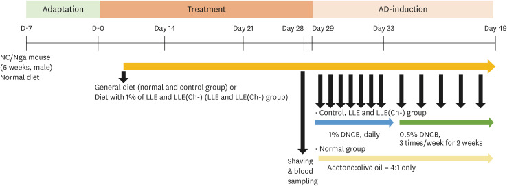
Animal experiments to measure atopic efficacy using DNCB in NC/Nga mice were based on the studies of Nam et al. [18] and Cha et al. [19]. The protocol for the animal experiments was approved by the Animal Experimental Ethics Committee of Chungnam National University (CNU-01123). Sixteen 6-week-old male NC/Nga mice (Orient Bio Co., Ltd., Seongnam, Korea) were used for the experiments after acclimation to the animal room for 1 week. At the beginning of the experiment, the animals were weighed and randomly divided into four groups: normal, control, LLE, and LLE(Ch-) (n = 4 per group). This was done to ensure that there was no difference in average weight between the groups. Experimental animals were raised in a micro ventilation caging system that was automatically maintained at a temperature of 23 ± 2°C and humidity of 55 ± 5%; the light/dark cycle was maintained at 12-h intervals. During the experiment, the mice were fed ad libitum. Normal feed was supplied to the normal and control groups, and feed containing 1% of each extract was supplied to the LLE and LLE(Ch-) groups, for seven weeks. At four weeks of feeding the experimental diet, blood was collected from the tail to measure the serum IgE concentration of mice before the induction of AD. For the application of DNCB, hair was removed with an animal epilator from the lower part of the dorsal neck to the upper part of the tail of the experimental animal. After 24 h of epilation, 200 µL of a 1% DNCB solution dissolved in an acetone-olive oil mixture (4:1) was applied to each mouse on the epilation sites of the control, LLE and LLE(Ch-) groups, once a day for one week. From the second week, 200 µL of 0.5% DNCB was applied to each mouse three times a week for 2 weeks. For the normal group, an acetone-olive oil mixture that did not contain DNCB was used. Throughout the experimental period, animal body weights were measured weekly, and vitality of the mice was monitored. On the last day of the experiment, blood was collected from the inferior vena cava of the animals after anaesthesia with isoflurane. A dorsal skin sample was excised immediately after sacrifice of the mice. An aliquot of the whole blood was used for DNA fragmentation analysis, and the remaining blood was centrifuged at 4°C and 2,000 ×g for 10 min to separate the serum, which was stored at −70°C for further experimentation (Fig. 1).
Measurement of serum IgE and cytokine concentrations
Concentrations of serum IgE, TNF-α, IL-6, and IL-4 were measured using a mouse ELISA kit (BD Biosciences, San Diego, CA, USA). The final absorbance was measured at 450 nm with an ELISA reader (microplate absorbance spectrophotometer; Bio-Rad Laboratories, Inc., Hercules, CA, USA), and the amount of IgE and cytokines produced were calculated based on a standard curve.
Lymphocyte DNA fragmentation analysis
The experiment was performed using the modified method described by Tice et al. [20]. A suspension of 5 µL of whole blood and 75 µL of low melting agarose (LMA) was dispensed onto a slide previously coated with 0.5% normal melting agarose and coagulated for 10 min in the dark at 4°C. When the gel solidified, LMA was re-applied and allowed to solidify. Then, the slide was immersed in an alkaline lysis buffer (2.5 M NaCl, 100 mM Na2 ethylenediaminetetraacetic acid [EDTA], 10 mM Tris) containing 1% Triton X-100 for 60 min at 4°C, followed by an electrophoresis buffer (300 mM NaOH, 10 mM Na2 EDTA, 10 mM Tris pH 13) for 40 min, and electrophoresis was performed. After electrophoresis was completed, the slide was washed three times in 0.4 M Tris buffer (pH 7.5), immersed in 95% ethanol for 5 min, and dried. The nuclei on the slide were stained with 20 µg/mL ethidium bromide, and 50 DNA samples were randomly selected per slide and observed under a microscope (DM 2000; Leica Co., Wetzlar, German). The degree of DNA fragmentation was analysed using a Komet 5.5 image analysing system (Andor, Belfast, Northern Ireland). The degree of DNA damage was expressed as the length of the comet tail (Tail length), the percentage of the tailed DNA (Tail% DNA) and the tail moment. The tail moment (TM) was calculated by the formula as follows:

Given that the strength of the fluorescent signal of migrated DNA is proportional to the degree of DNA damage, a longer and brighter DNA tail implied a higher level of DNA damage. Therefore, DNA damage was quantified by measuring the distance (Tail length) between nuclear genetic material and the resulting “tail.” Further, tail moment, a variable that takes into account both tail DNA amount and distribution, i.e., the length of the tail and the proportion of DNA in the tail, was also determined.
Histological examination
Haematoxylin & eosin (H&E) staining (Sigma-Aldrich Co.) and toluidine blue (Sigma-Aldrich Co.) staining were performed to observe epidermal thickness and mast cell invasion in the dorsal tissue of NC/Nga mice with DNCB-induced atopy. After the experimental animals were sacrificed, the dorsal skin tissue was collected and fixed in 10% neutral formalin solution. The fixed tissue was embedded in a paraffin block and the block was sliced to a thickness of 5 µm to obtain a tissue section, deparaffinized with xylene, and subjected to a hydration step. The slides were then stained with H&E or toluidine blue. After staining, each tissue section was subjected to a dehydration process, sealed, and observed using an optical microscope (×40; Olympus, Tokyo, Japan). Each slide was divided into 5 sections for observation, and the average epidermal thickness was calculated.
HaCaT cell culture
The HaCaT cell line, a human-derived skin keratinocyte, was kindly donated by professor Eun Mi Park (at the Department of Food and Nutrition, Hannam University, Daejeon, Korea). The cell culture medium was Dulbecco’s modified Eagle medium (DMEM; Gibco, Grand Island, NY, USA) containing 10% foetal bovine serum (FBS; Gibco) and 1% penicillin-streptomycin (Pen Strep; Gibco). Cells were cultured at 37°C in a 5% CO2 incubator (BB15; Thermo Fisher Scientific Inc., Waltham, MA, USA). Before the experiment, a water-soluble tetrazolium (WST) assay was performed to evaluate the cytotoxicity of the ethanol extract of L. barbarum leaves. HaCaT cells were seeded into a 96-well plate at a concentration of 1 × 10⁶ cells/well, cultured for 24 h, and then treated with the leaf ethanol extracts at concentrations of 15.6, 31.3, 61.5, 125, 250, 500, and 1,000 µg/mL, and cultured for 24 h. After incubation, 10 µL of EZ-Cytox WST assay reagent (Dogenbio Co., Ltd., Seoul, Korea) was added and incubated for 3 h; the absorbance was measured at 450 nm using a microplate spectrophotometer (BioTek Instrument, Winooski, VT, USA).
Evaluation of chemokine expression
HaCaT cells were dispensed at a concentration of 1 × 10⁶ cells/mL into a 6-well plate and incubated for 24 h; a mixture of TNF-α and IFN-γ was then added at a concentration of 10 ng/mL to stimulate the cells. Simultaneously, the cells were treated with L. barbarum leaf ethanol extract at concentrations of 15.6, 125, and 1,000 µg/mL. After incubation for 24 h, the supernatant was removed, cells were washed twice with phosphate-buffered saline, and RNA was extracted using a tri-reagent (Biofact Co., Daejeon, Korea). Complementary DNA synthesis was performed using an RT-kit (M-MLV, RNase H-, Biofact Co.). The mRNA expression level in the cells was measured using a 2× Real-Time PCR Master Mix kit (Biofact Co.). Initial denaturation was performed at 95°C for 2 min, followed by denaturing at 95°C for 10 s, annealing and extension at 58°C for 30 s, and total amplification was performed for 35 cycles. The 1% agarose gel (Biofact Co.) was stained with Reds Safe (iNtRON Biotechnology Inc., Seongnam, Korea), followed by electrophoresis, and a gel document system (Printgraph GX; Atto, Tokyo, Japan) was used to analyse chemokine expression. Glyceraldehyde 3-phosphate dehydrogenase, a housekeeping gene, was simultaneously measured, and the amount of RNA was determined using ImageJ (NIH open-source software, available from website https://imagej.nih.gov/ij/download.html). The primers used in the experiments are listed in Table 1. The blots represent the results of three independent experiments.
Table 1
Reverse transcriptase-polymerase chain reaction primer sequences

Statistical analysis
The experimental results were statistically processed using SPSS/Windows 24.0 (Statistical package for the Social Science; IBM Corp., Armonk, NY, USA), and all experimental results are expressed as mean ± SD. The Kruskal-Wallis H test was conducted to test the non-normal distribution data and the Wilcoxon’s t-test was performed to examine the significant difference between the averages of each group. Mouse body weight was compared between groups using the non-parametric test methods at 4–7 weeks of the experiment.
RESULTS
Bodyweight change of mice
Until the induction of atopy, the bodyweight of animals in all groups continued to increase, and there was no difference in the mean values between the groups. However, the bodyweight after induction of atopy varied between the groups (Table 2). The animals in the normal group had an average weight of 32.75 ± 3.35 g at seventh weeks, which increased by 9.1% in 3 weeks. However, the body weights of the control, LLE, and LLE(Ch-) group decreased by 17.3%, 21.6%, and 12.4%, respectively, compared to before induction of atopy. The final body weight was significantly lower in all three groups compared to the normal group (P < 0.05), but there was no difference between the 3 experimental groups.
Table 2
Bodyweight change of NC/Nga mice

Serum IgE and cytokines levels
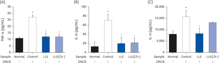
There was no difference in serum IgE concentrations between groups at the beginning of the experiment. However, after DNCB application, the IgE concentration in the control group (18.14 ± 6.85 µg/mL) increased significantly compared to that in the normal group (0.11 ± 0.05 µg/mL), confirming that the atopic response was induced (Fig. 2). The serum IgE concentration in the experimental group decreased by 37.7% and 34.6% in the LLE and LLE(Ch-) groups, respectively, compared to that of the control group. However, there was no difference between the LLE group and LLE(Ch-) group. Serum TNF-α concentration in the control group (27.11 ± 6.85 pg/mL) was approximately 1.5-fold higher than that in the normal group (10.86 ± 0.99 pg/mL) (P < 0.05). Serum TNF-α levels decreased by approximately 55% in both the LLE and LLE(Ch-) groups, compared to that of the control group. IL-6 concentration was significantly higher in the control group (70.68 ± 12.86 pg/mL) than in the normal group (13.33 ± 9.62 pg/mL) (P < 0.05) and decreased by about 72% in the LLE group and approximately 69% in the LLE(Ch-) group compared to the control group (P < 0.05). The concentration of serum IL-4 increased by approximately 2-fold in the control group (15.73 ± 2.75 µg/mL) compared to that in the normal group (8.03 ± 1.53 µg/mL) and decreased by 47% in the LLE group compared to that in the control group (Fig. 3). The concentration of serum IL-4 in the LLE(Ch-) group showed no difference from the control group.
Fig. 2
Effect of the Lycium barbarum leaves ethanol extracts (LLE, LLE[Ch-]) on serum IgE level in atopic dermatitis induced NC/Nga mice by DNCB.
Data are presented as mean ± SD values.
LLE, Lycium barbarum leaves with chlorophyll; LLE(Ch-), Lycium barbarum leaves without chlorophyll; IgE, immunoglobulin E; DNCB, 2,4-dinitrochlorobenzene.
*,†Compared with normal group and control group, respectively at P < 0.05 by Kruskal-Wallis H test and Wilcoxon’s t-test.
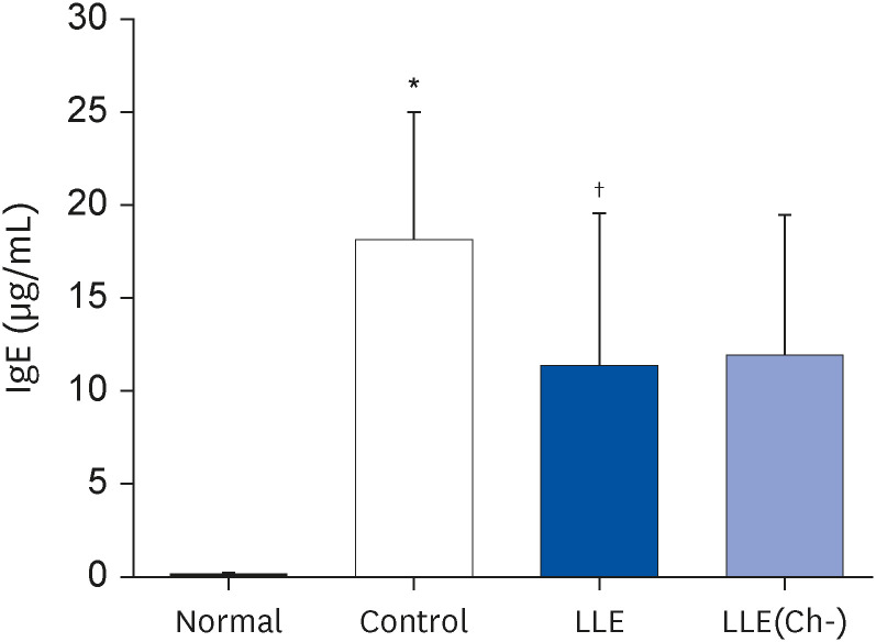
Fig. 3
Effect of the Lycium barbarum leaves ethanol extracts (LLE and LLE[Ch-]) on (A) serum TNF-α, (B) IL-6, and (C) IL-4 concentration in NC/Nga mouse with atopic dermatitis induced by DNCB.
Data are presented as mean ± SD values.
LLE, Lycium barbarum leaves with chlorophyll; LLE(Ch-), Lycium barbarum leaves without chlorophyll; TNF, tumor necrosis factor; IL, interleukin; DNCB, 2,4-dinitrochlorobenzene.
*,†Compared with normal group and control group, respectively at P < 0.05 by Kruskal-Wallis H test and Wilcoxon’s t-test.
DNA fragmentation of lymphocytes
The tail DNA (26.17 ± 3.99%) and tail length (71.97 ± 10.33 µm) of the control group showed 31% and 41% increase, respectively, compared to those in the normal group (Table 3). In the LLE group, only tail DNA (20.52 ± 1.74%) was significantly decreased compared to that in the control group. In contrast, tail DNA (14.99 ± 1.88%), tail length (51.26 ± 2.55 µm), and tail moment (10.38 ± 1.05) of the LLE(Ch-) group were all significantly lower than those in the control group.
Table 3
Levels of DNA damage expressed as tail DNA, tail length, and tail moment in mouse whole blood treated with 100% ethanol extracts of Lycium barbarum leaves

Data are presented as mean ± SD values.
LLE, Lycium barbarum leaf extract with chlorophyll; LLE(Ch-), Lycium barbarum leaf extract without chlorophyll.
*Compared with normal group at P < 0.05 by Kruskal-Wallis H test and Wilcoxon’s t-test.
†Compared with control group at P < 0.05 by Kruskal-Wallis H test and Wilcoxon’s t-test.
‡Compared with LLE group at P < 0.05 by Kruskal-Wallis H test and Wilcoxon’s t-test.
Histological test
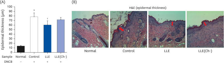
The dorsal skin thickness was 14.3 ± 1.26 μm in the normal group, and it increased to 78.3 ± 8.34 μm in the control group after DNCB application (P < 0.05) (Fig. 4). The dorsal epidermis in the LLE group (60.5 ± 9.00 µm) was significantly thinner than that of the control group at 60.5 ± 9.00 µm, but there was no significant change in the LLE(Ch-) group.
Fig. 4
Effect of Lycium barbarum leaf ethanol extracts (LLE and LLE[Ch-]) on (A) epidermal thickness of NC/Nga mouse. (B) The histopathologic features (×40) of the dorsal epidermis in NC/Nga mouse.
LLE, Lycium barbarum leaves with chlorophyll; LLE(Ch-), Lycium barbarum leaves without chlorophyll; DNCB, 2,4-dinitrochlorobenzene; H&E, haematoxylin & eosin.
*,†Compared with normal group and control group, respectively at P < 0.05 by Kruskal-Wallis H test and Wilcoxon’s t-test.
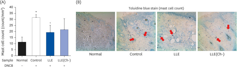
The number of mast cells significantly increased in the control group (31.6 ± 2.3 counts/mm2) compared to that in the normal group (11.4 ± 4.1 counts/mm2; P < 0.05) (Fig. 5). In the LLE group, the number of mast cells (19.4 ± 5.79 counts/mm2) reduced by about 39% compared to that in the control group. In the LLE(Ch-) group, the number of mast cells (21.6 ± 8.97 counts/mm2) tended to decrease by about 32% compared to that in the control group, but the difference was statistically insignificant due to the inter-individual difference.
Fig. 5
Effect of Lycium barbarum leaf ethanol extracts (LLE and LLE[Ch-]) on (A) mast cell count in dermis of NC/Nga mouse. (B) The histopathologic features (×40) of mast cell infiltrations in the dermis of NC/Nga mouse.
LLE, Lycium barbarum leaves with chlorophyll; LLE(Ch-), Lycium barbarum leaves without chlorophyll; DNCB, 2,4-dinitrochlorobenzene.
*,†Compared with normal group and control group, respectively at P < 0.05 by Kruskal-Wallis H test and Wilcoxon’s t-test.
Chemokine production in HaCaT cells
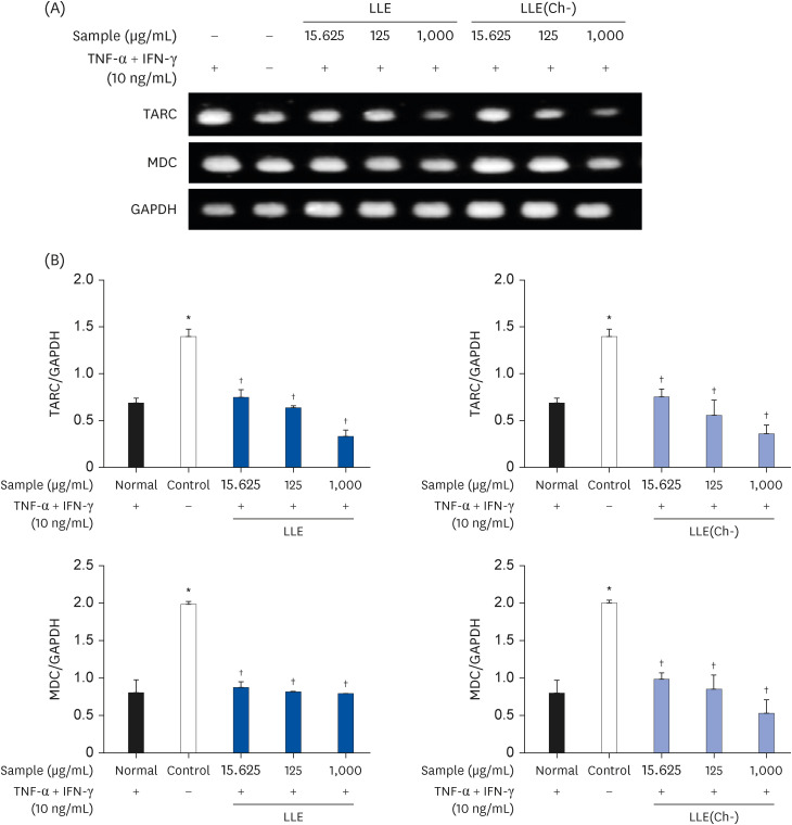
HaCaT cells were treated with LLE and LLE(Ch-) for 24 h; cytotoxicity was not observed up to a concentration of 1,000 μg/mL (Fig. 6). Therefore, in this study, all extracts were tested in a concentration range of 15.6–1,000 μg/mL. The mRNA expression level of MDC in the control group treated with TNF-α and IFN-γ was significantly higher than that in the control group (P < 0.05) (Fig. 7). In contrast, in the group treated with LLE and LLE(Ch-), the expression levels of TARC and MDC significantly decreased at all concentrations compared to those in the control group, and the final level was not different from those in the normal group.
Fig. 6
Effect of Lycium barbarum leaf ethanol extracts (LLE and LLE[Ch-]) on HaCaT cell viability.
Cells were treated with varying concentrations (15.6, 31.3, 62.5, 125, 250, 500, and 1,000 µg/mL) of Lycium barbarum leaf ethanol extracts for 24 h. Cytotoxicity was measured using a WST assay.
LLE, Lycium barbarum leaves with chlorophyll; LLE(Ch-), Lycium barbarum leaves without chlorophyll; WST, water-soluble tetrazolium.
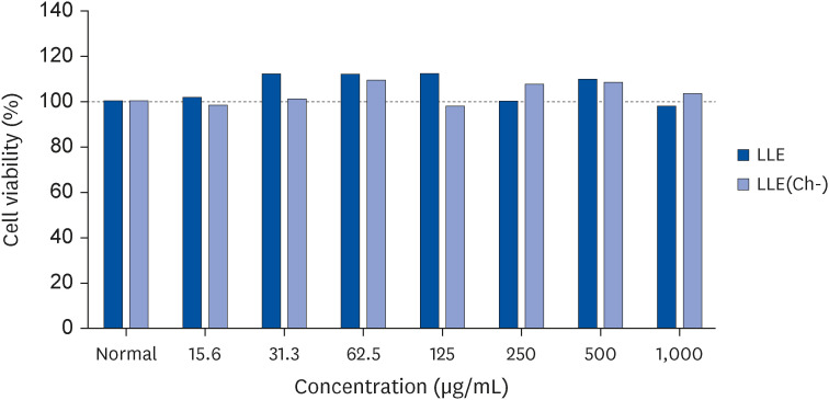
Fig. 7
Effect of the Lycium barbarum leaf ethanol extracts (LLE and LLE[Ch-]) on expression of TARC and MDC in HaCaT cells stimulated by TNF-α and IFN-γ.
(A) and (B) show mRNA expression analysis results. Cells were treated different concentrations (15.625, 125, 1,000 µg/mL), then with or without TNF-α + IFN-γ (10 ng/mL) for 24 h. TARC, MDC, and GAPDH mRNA were analyzed using RT-PCR with specific primers. Data are presented as the mean ± SD (n = 3).
LLE, Lycium barbarum leaves with chlorophyll; LLE(Ch-), Lycium barbarum leaves without chlorophyll; TARC, thymus and activation-regulated chemokine; MDC, macrophage-derived chemokine; TNF, tumor necrosis factor; IFN, interferon; GAPDH, glyceraldehyde 3-phosphate dehydrogenase; RT-PCR, reverse transcriptase-polymerase chain reaction.
*,†Compared with normal group and control group, respectively at P < 0.05 by Kruskal-Wallis H test and Wilcoxon’s t-test.
DISCUSSION
The purpose of this study was to verify the protective effect of L. barbarum leaf ethanol extract and chlorophyll-removed ethanol extract against AD and to examine its potential for development as a functional material. A symptom of AD is an increase in serum IgE due to the activation of Th2 cells in the peripheral blood, which is known to be proportional to the clinical severity of AD [2122]. The results of the present study showed that the serum IgE concentration in the LLE group and the LLE(Ch-) group was significantly lower than that in the control group. Serum IgE has been reported to cause allergic reactions in response to antigens [6]. When IgE-bound mast cells bind to antigens, they undergo degranulation and release mediators such as histamine and various cytokines, thereby promoting an inflammatory response [23]. The results of this study showed that L. barbarum leaf ethanol extracts could alleviate allergic reactions.
AD is caused by an overactive immune response due to an imbalance in the Th1/Th2 cell ratio and bias in the response of Th2 cells. However, as the chronic stage progresses, the activity of Th2 and Th1 cell cytokines such as TNF-α and IFN-γ increases, further promoting the inflammatory response of the skin [2425]. Here we observed that the levels of inflammatory cytokines TNF-α and IL-6 were significantly reduced in NC/Nga mice fed L. barbarum leaf ethanol extracts. In particular, in the LLE group, the concentration of IL-4 in the blood was also significantly reduced, suggesting that Th2 cell-derived IL-4 secretion was suppressed and serum IgE secretion was reduced. However, in the LLE(Ch-) group, only the IL-4 showed a tendency to decrease. Thus, the effect on Th2 cell activity was rather low. TNF-α is an important mediator of the skin inflammatory response produced in keratinocytes, macrophages, and mast cells, and it affects atopic skin by acting on cell growth, differentiation, and necrosis [2426]. In addition, IL-6 promotes Th2 cell differentiation by inducing IL-4 gene expression, while CD4+ T cells are activated, and inhibit IFN-γ and Th1 cell differentiation by inducing the expression of the suppressor of cytokine signalling-1 [27]. IL-4, a cytokine secreted by activated Th2 cells, plays a key role in the activation of mast cells by increasing IgE levels through B-cell growth and differentiation [28].
In the inflammatory response, an excessive increase in free radicals causes the expression of inflammatory cytokines, which aggravates the inflammatory response and induces damage in cell membranes, DNA, and organelles [29]. In particular, when atopic patients are continuously exposed to an oxidative stress environment, it causes fatal tissue damage, and there is a possibility that atopy may be further exacerbated by the breakdown of the skin barrier [30]. Omata et al. [31] described a phenomenon in which the balance of free radicals in the body is disrupted or oxidative stress is increased in infancy in relation to atopic pathophysiology. They reported that maintaining an appropriate level of active oxygen concentration in the body could be helpful in the treatment of atopy. Analysis of the lymphocyte DNA of NC/Nga mice in this study revealed that the DNA protective effect was greater in the LLE(Ch-) group than in the LLE group. The tail length and tail moment of the LLE group tended to decrease. However, the statistical significance of this observation could not be established owing to the wide differences in outcome values between the different animals. Bae et al. [17] measured the degree of DNA damage after administration of L. barbarum leaf ethanol extracts or chlorophyll-removed ethanol extract in BALB/c mice with an lipopolysaccharide-induced inflammatory response and found that there was no significant difference between the 2 groups. In another study examining the antioxidant efficacy of L. barbarum leaf ethanol extract in HepG2 cells, it was shown that the antioxidant activity of chlorophyll-removed ethanol extract of L. barbarum leaves was higher than that of the ethanol extract of L. barbarum leaf before the removal of chlorophyll [16]. In that previous study, the yield, chlorophyll content, and total polyphenol content of the 2 ethanol extracts of L. barbarum leaf (LLE and LLE[Ch-]) were reported. As 99.8% of chlorophyll was removed from LLE(Ch-), the yield decreased from 20.5% to 16.45%, and the total polyphenol content more than doubled (140.5 ± 7.45 mg gallic acid equivalent [GAE]/extract g vs. 312.9 ± 10.2 mg GAE/extract g) [16]. Also, the content of flavonoid, a type of polyphenol, increased from 34.77 ± 1.29 mg catechin equivalents (CE)/g to 42.75 mg CE/g (data not shown). This study is the result of an experiment using the same sample as in the study by Kim et al. [16]. Concerns about a decrease in antioxidant efficacy were predicted by removing chlorophyll from the ethanol extract of L. barbarum leaf; however, the antioxidant activity increased as the polyphenol content increased.
The most common symptoms of AD are hyperkeratosis, dryness, and infiltration of inflammatory cells, caused by excessive cell proliferation resulting from abnormal cell division, which is accompanied by, causing intracellular oedema of the epidermis (spongiosis) and an increase in the thickness of the epidermis [3233]. In the present study, more rashes occurred in the control group than in the normal group, and the epidermis was thickened toward the dermis, but the thickness of the epidermis was significantly reduced in the LLE group. However, there was no statistically significant difference in epidermal thickness in the LLE(Ch-) group compared to that in the control group.
Mast cells play an important role in biological defence in innate and acquired immunity, but when excessively activated, they can trigger allergic inflammation and chronically prolong the inflammatory response. In particular, histamine and serotonin secreted by mast cells have been reported to exacerbate dermatitis as pruritus-causing substances [3435]. The significant decrease of the number of mast cells in the LLE group suggests that the ethanol extract of L. barbarum leaf can alleviate the clinical symptoms of AD through the action of mast cells. However, there was no difference between the LLE group and the LLE(Ch-) group.
We wanted to report the expression of related chemokines to elucidate the mechanism of the results of animal experiments. TARC and MDC are important biomarkers of Th2-dominant inflammatory skin diseases such as AD; this is because they bind to CCR4 expressed by Th2 cells and exacerbate the inflammatory response by migrating and infiltrating Th2 cells to the inflammation site [36]. In the serum of patients with AD, concentrations of TARC and MDC were significantly elevated, and the serum levels of these proteins could be correlated with the severity of AD [37]. The results of this study showed that the expression levels of TARC and MDC decreased at all concentrations in the group treated with LLE and LLE(Ch-) in HaCaT cells induced atopic response to cytokines. Therefore, ethanol extracts of L. barbarum leaves are considered to be effective in alleviating the symptoms of AD by suppressing the expression of the chemokines TARC and MDC related to AD.
In this study, the overall atopy alleviation effect of the ethanol extract of L. barbarum leaves was likely due to the anti-inflammatory effect of polyphenols and flavonoid compounds contained in Licyum’s leaves. A previous study that was conducted at our laboratory showed that the polyphenol content of LLE(Ch-) is very high and its antioxidant activity is higher than that of LLE [16]. However, its protective effect on skin inflammation in AD-induced animals was not evident compared to that of LLE. This could be attributed to the removal of anti-inflammatory substances and other moisturizing ingredients during the process of chlorophyll removal from L. barbarum leaves. Reportedly, L. barbarum contains various antioxidants, such as polyphenol-based rutin and flavonoids, and among these, rutin and betaine are present in large amounts in its leaves [3839]. Further, it has been reported that the polysaccharides in L. barbarum have moisturising properties [404142], which possibly, are involved in the complex mechanism of alleviating AD. Recently, potential anti-inflammatory hydroxycinnamic acid amides, such as kukoamine A [18], kukoamine B [16], and lyciumin [42], were reported as anti-inflammatory metabolites present in Lycium roots. However, the activity of these substances in L. barbarum leaf extract is still unclear.
In this study, the experimental results confirmed that the ethanol extract of L. barbarum leaves relieved atopic symptoms haematologically and histologically and suppressed the chemokine expression of TARC and MDC in cytokine-stimulated human keratinocytes. Therefore, the ethanol extract of L. barbarum leaves could be used as a functional material for alleviating AD with anti-inflammatory and antioxidant effects similar to goji berry. In addition, chlorophyll-removed ethanol extract of L. barbarum leaves also showed similar efficacy; thus, it can also be used as a functional material even after removing the pigment. However, since the anti-inflammatory effect of the chlorophyll-removed ethanol extract of L. barbarum leaves is slightly lower than that of the ethanol extract without the chlorophyll removal, further research is needed to increase the anti-inflammatory effect of this material.
This study had some limitations. In particular, LLE(Ch-) was obtained by removing chlorophyll after LLE preparation, and the contents of all the components and active substances in the two ethanol extracts were not accurately analysed. Therefore, the results obtained are representative of a simple comparison of the physiological activities of the two extracts. Therefore, in future, it would be necessary to investigate the gain and loss of active ingredients owing to the chlorophyll removal process.
 XML Download