Journal List > J Korean Med Sci > v.38(50) > 1516085066

Seo, Hong, and Park: Rare Cause of Exertional Angina
A 76-year-old woman was admitted to our clinic with exertional chest pain for 2 weeks.
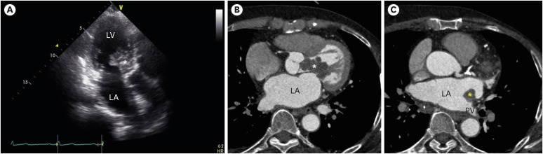
Transthoracic echocardiography revealed a multi-lobulated hypoechoic mass occupying the left ventricular (LV) outflow tract with dynamic systolic protrusion into the aortic root (Fig. 1A; Supplementary Videos 1 and 2). The about 4 cm sized mass was attached to the anterior leaflet of the mitral valve on the LV side. The mass occupied the LV outflow tract, leading to an increased mean pressure gradient of 42 mmHg. Owing to the mass effect, the anterior leaflet of the mitral valve partially closed during diastole, leading to an obstruction of the mitral inflow with a mean pressure gradient of 5 mmHg.
Cardiac computed tomography (CT) showed that the LV mass exhibited enhancement on early post-contrast image (Fig. 1B). An additional small mass, measuring 1.5 cm, was observed on the left atrial wall at the opening of the pulmonary vein (Fig. 1C).
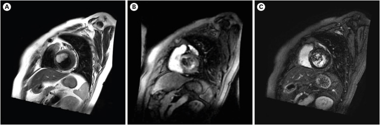
Cardiac magnetic resonance imaging revealed high signal intensity of the masses on T2-weighted imaging (Fig. 2A). During dynamic post-contrast examination, the LV mass showed heterogenous enhancement (Fig. 2B). During the delayed phase, the LV mass showed late gadolinium enhancement (Fig. 2C).
With positron emission tomography CT, other primary malignancies were excluded. The maximum standardized uptake value of the mass was 3.7.
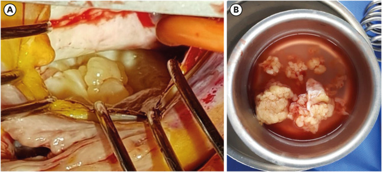
Based on multimodal imaging, the mass was suspected to be primary cardiac sarcoma. The patient underwent surgery for removal of all the cardiac masses (Fig. 3A and B). Histopathologic examination revealed all the cardiac masses as cardiac sarcoma with marked myxoid change. Immunohistochemical staining was positive for CD34, myogenic differentiation 1 and transducer-like enhancer of split 1. Due to extensive myxoid change, the subtype of cardiac sarcoma could not be specified.
Primary cardiac sarcoma is an extremely rare type of intracardiac mass.1 Cardiac masses can be effectively identified and characterized by multimodality imaging.2 The present case suggests the utility of multimodal imaging in the evaluation of cardiac mass.

Ethics Statement

This report was reviewed and approved for publication with exempt of informed consent by the Institutional Review Board (IRB) of Ajou University Hospital (IRB approval No. AJOUIRB-EX-2023-286).

Notes

Disclosures: The authors have no potential conflicts of interest to disclose.

Author Contributions:

  • Conceptualization: Hong YS, Park JS.

  • Data curation: Hong YS.

  • Writing - original draft: Seo KW.

  • Writing - review & editing: Park JS.

References

1. Peters PJ, Reinhardt S. The echocardiographic evaluation of intracardiac masses: a review. J Am Soc Echocardiogr. 2006; 19(2):230–240. PMID: 16455432.
2. Basso C, Rizzo S, Valente M, Thiene G. Prevalence and pathology of primary cardiac tumours. Cardiovasc Med. 2012; 15(1):18–29.

SUPPLEMENTARY MATERIALS

Supplementary Video 1

Transthoracic echocardiography, apical three-chamber view.

Supplementary Video 2

Color Doppler echocardiography.
Fig. 1

Images of masses. (A) Transthoracic echocardiography showed a multi-lobulated shaped mass (white asterisk) attached to the anterior leaflet of the mitral valve on the left ventricular side. (B) Cardiac computed tomography showed that the mass (white asterisk) exhibited enhancement on early post-contrast image. (C) Another small mass (yellow asterisk) was observed on the left atrial wall at the opening of pulmonary vein.

LV = left ventricle, LA = left atrium, PV = pulmonary vein.
jkms-38-e421-g001
Fig. 2

Cardiac magnetic resonance images, mid-ventricular short axis view. (A) T2-weighted image. (B) Dynamic post-contrast examination. (C) The delayed phase.

jkms-38-e421-g002
Fig. 3

Intra-op findings. (A) Multi-lobulated masses were noted. (B) Resected masses.

jkms-38-e421-g003
TOOLS
Similar articles