Abstract
IgG4-related disease is a multi-organ immune-medicated condition that mimics malignancies, infections, and inflammatory disorders. This report presents the case of a 62-year-old man with a history of repeated cellulitis and ulcers on his lower extremities who was diagnosed incidentally with IgG4-related disease during treatment. The patient’s unhealed ulcer was initially treated by debridement and aseptic dressing several times, but the wounds showed no improvement. Lower extremity venous ultrasound revealed unusually enlarged LN around the great saphenous vein (GSV) near the inguinal area and severe varicosity in both truncal veins. Lymphoscintigraphy was performed according to the ultrasound results, which showed secondary lymphedema in both legs. IgG4-released disease was diagnosed in an excisional biopsy of the lymph node near the great saphenous vein during stripping and ligation of the truncal vein. After continuous wound debridement and the administration of steroids and immunosuppressants, the patient's leg ulcer improved after approximately three months.
IgG4-related disease is a multi-organ immune-medicated condition that mimics malignant, infectious, and inflammatory disorders.(1-4) It occurs in nearly every organ system, including the biliary tract, salivary glands, periorbital tissues, kidneys, lungs, lymph nodes, meninges, aorta, breast, prostate, thyroid, pericardium, and skin.(4,5) Regardless of the site of disease, histopathological features show striking similarities across the entire organ.(6) Therefore, IgG4-related disease is like another systemic disease in which multiple organ manifestations lead to the same histopathological features. Most of the reported cases of IgG4-related disease that involved blood vessels have been primary large-vessel vasculitis (giant-cell or Taka-yasu’s arteritis), sarcoidosis, Erdheim-Chester disease, lymphoma, and infectious aortitis.(7) We present a case of IgG4-related disease in which a patient treated for chronic leg ulcer accompany by lymphedema and chronic venous disease.
A 62-year-old man with a history of repeated cellulitis due to a decade-old open wound on both lower extremities presented with unhealed chronic ulcers. He had been diagnosed with stage three chronic renal failure due to lupus nephritis and had received several debridement procedures at a plastic surgery clinic on his leg wounds for years.
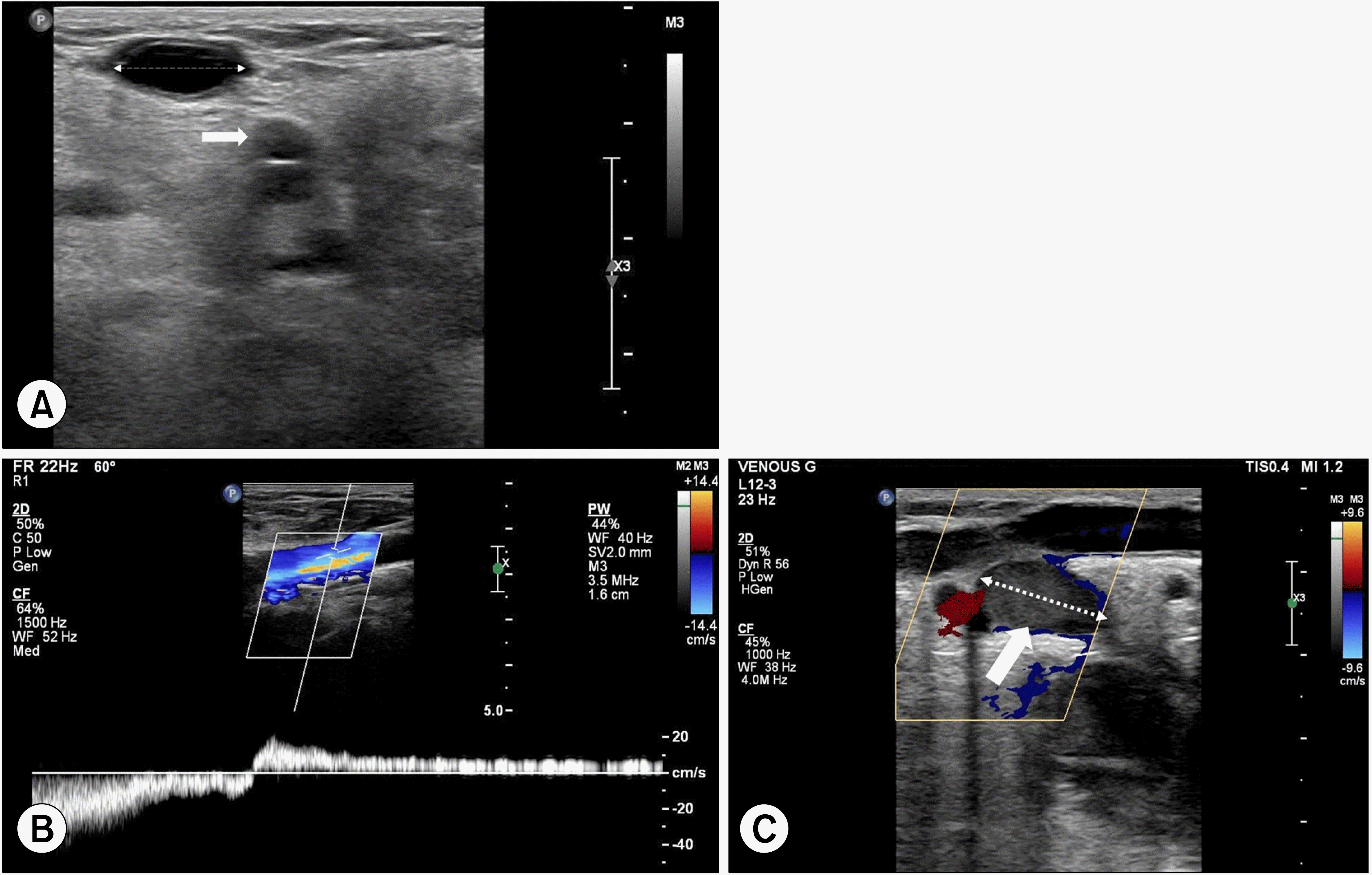
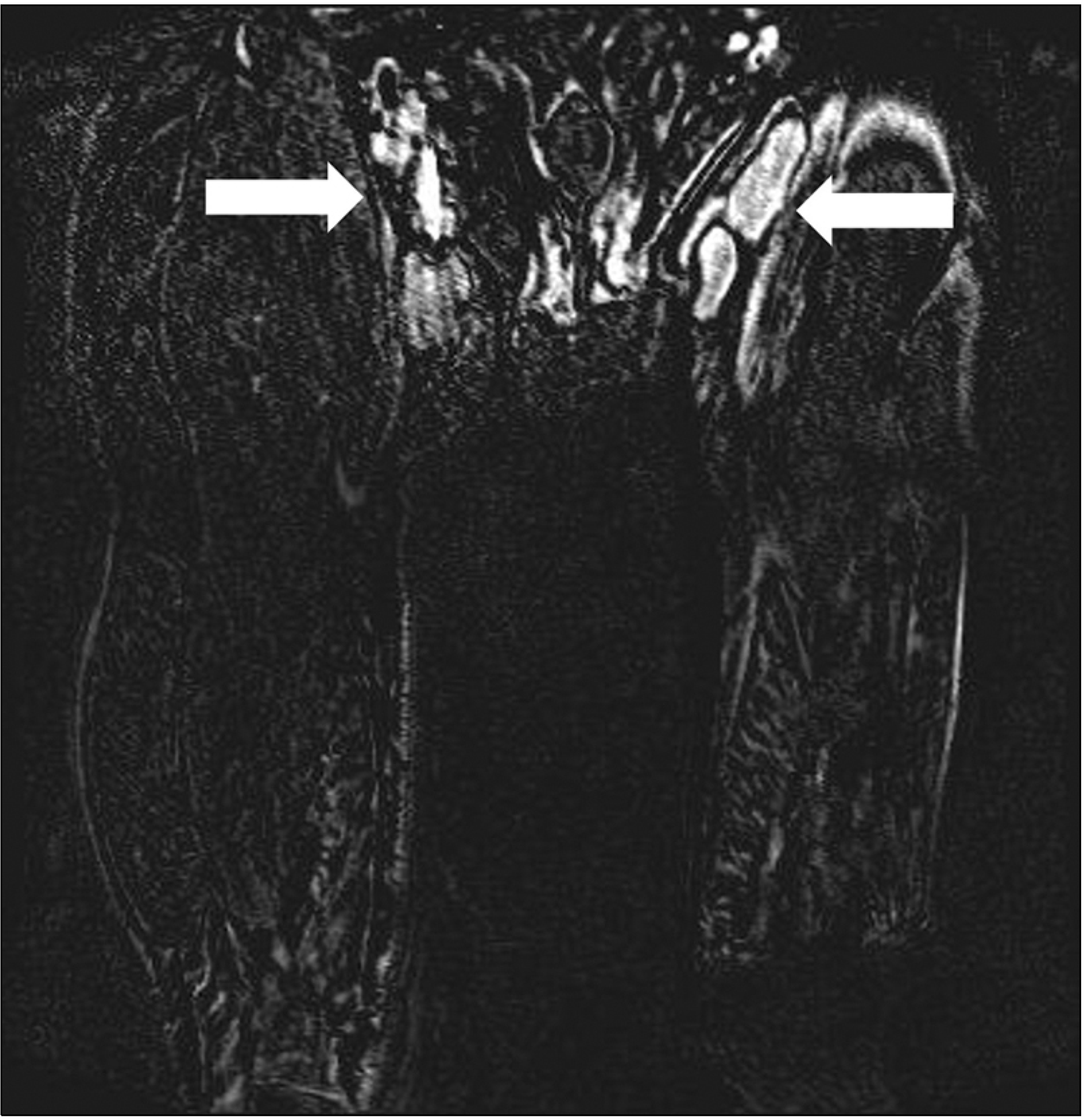
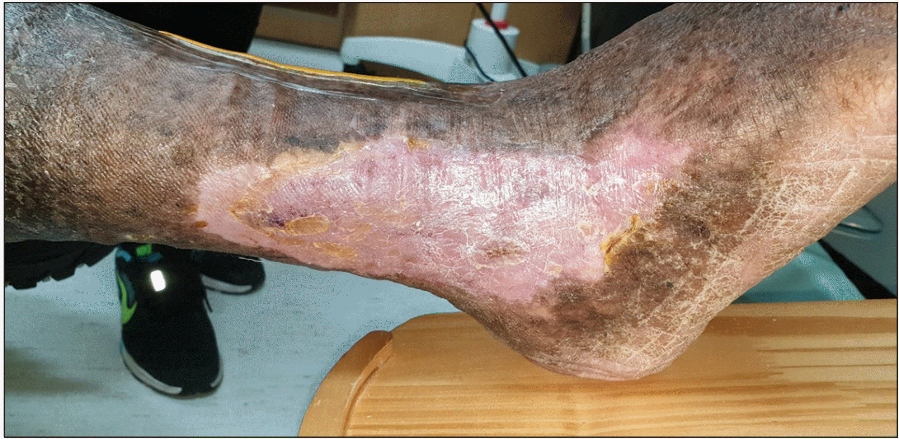
Upon the patient’s arrival at our institution, severe leg ulceration with lipodermatosclerosis and edema were apparent on his both lower legs (Fig. 1). Lower extremity venous duplex ultrasound showed dilatation both great saphenous vein (GSV; diameter > 10 mm) and by the Valsalva maneuver, reflux of both GSV, small saphenous vein (SSV), and perforator veins ware observed for more than 5 seconds. Additionally, multiple enlarged lymph nodes (LNs) were observed around the GSV below the saphenofemoral junction (Fig. 2). In the subsequent magnetic resonance image, it was confirmed that multiple enlarged LNs (3-4.5 cm in diameter) existed around the GSV (Fig. 3). Lymphoscintigraphy showed primary lymphedema with delayed lymphatic drainage in both legs (Fig. 4). Wound debridement was performed several times along with negative pressure vacuum therapy, but there was no discernable improvement in healing and edema. We determined that leg ulcer was caused by chronic venous disease and that stripping of the truncal vein was necessary. In addition, it was considered appropriate to perform an excisional biopsy of the abnormally enlarged lymph node by the ultrasonographic results. The patient underwent sterile draping of both legs under general anesthesia, and then stripping and ligation of both truncal veins was performed with massive wound debridement (Fig. 5). An additional incision was made around the inguinal area to ligation the collateral veins of the GSV, and an excisional biopsy was performed on the largest lymph node.
The pathologic findings indicated IgG4 positive plasma cell infiltrates in the lymph nodes and IgG4 elevation (2,410 mg/L) in serology, which is indicative of IgG4 related disease. The paracortex and medulla of lymph node are expanded and infiltrated by numerous plasma cells. The IgG4 positive plasma cells infiltrates diffusely in lymph nodes (IgG4 > 100 positive cells/HPF, CD20: positive on B cells) (Fig. 6).
Debridement and compression treatment were continuously performed after the surgery, and the patient was medicated with oral prednisolone and mycophenolic acid after consulting with the department of rheumatology. Three months after surgical operation, the edema and unhealed ulcers which had not healed for years showed complete resolution (Fig. 7).
IgG4-related disease is a systemic condition, which is recognized from result of extrapancreatic manifestations of patients with autoimmune pancreatitis in 2003.(8) This disease shares many similarities with sarcoidosis and some forms of systemic vasculitis, as well as other proteinaceous diseases, in which histopathological findings are consistent across a wide range of organ systems.(9) IgG4-related disease has been discribed in variable organ systems: the biliary system, periorbital tissues, salivary glands, kidneys, lungs, lynph nodes, meninges, aorta, breast, prostate, thyroid glands, pericardium, and skin.(7,10-12) The reason for reporting this case is because it is a very rare case of lymphedema caused by lymph node invasion of the lower extremities without invading the lymphatic organs of the head, neck or abdomen.
Much remains unknown about the behavior of IgG4 in vivo, however, and whether its role in IgG4-related disease is primary or secondary.(7) It is well known that fibrosis and inflammatory reactions caused by immune-mediated mechanisms are known to occur due to infiltration of IgG4 antibodies Autoimmune and infectious conditions are potentially immunological triggers for IgG4-related disea-se.(6) Interleukins 4, 5, 10, and 13 and transforming growth factor beta (TGF-β) are overexpressed through an immune response in which type 2 helper T cells predominate and subsequently activate regulatory T cells. These cytokines result in eosinophilia, increased serum IgG4 and IgE concentrations, and characteristic fibrosis progression. Massi-ve infiltration of inflammatory cells leads to organ damage. Infiltration of inflammatory cells leads to swelling and organ dysfunction in the affected area. Epithelial damage may be caused by tissue Inflammation and immune complex deposition.
Many previous researchers claim that the diagnosis of IgG4 related disease is mostly made through tissue examination of involvement.(13) In some patients, the disease is localized to a single organ for many years. Other patients have known or subclinical involvement of other organs in addition to primary organ involvement. In this case, the patient was treated several times for leg swelling and ulcers due to long-standing lymphedema, but a clear diagnosis was not made. Abnormally enlarged lymph nodes were discovered through ultrasound, a basic test performed for leg edema, and accurate diagnosis and treatment could be achieved by electrical diagnostic processing.
Most IgG4-related disease respond well to gluco-corticoids. Response to therapy may differ depending on the affected organ, but the effects are usually very quick.(7,14,15) Azathioprine, mycophenolate mofetil, and methotrexate are used adjunctively to induce additional immunosuppression and to spare the effects of long-term glucocorticoid use.(16,17) In our case, oral glucocorticoids were used immediately after surgery, and mycophenolate mofetil was added as we began to taper the glucocorticoids, which lead to complete resolution of the ulcers and edema. When there is a leg ulcer, determining the primary cause through ultrasound plays a major role for the diagnosis and treatment proceeds.
There may be limitations in determining the exact association between IgG4-related disease and chronic venous ulcers. With this in mind, however, there is value in this case as we were able to successfully treat a patient suffering from venous ulcers and leg edema for many years with both surgical intervention and glucocorticoid and mycophenolate acid therapy.
IgG4 related disease is a condition in which IgG4-related immune cells and factors invade various organs of the body and cause various symptoms. It is very rare for it to invade the lymph nodes of the legs, causing ulcers and edema. Ultrasound, which is commonly performed when there are leg ulcers and edema, is a useful means of revealing the cause of leg edema or ulcers and the possibility of accompanying diseases.
ACKNOWLEDGEMENTS
This paper was supported by Fund of Biomedical Research Institute, Jeonbuk National University and Hospital.
REFERENCES
1. Mahajan VS, Mattoo H, Deshpande V, Pillai SS, Stone JH. 2014; IgG4-related disease. Annu Rev Pathol. 9:315–47. DOI: 10.1146/annurev-pathol-012513-104708. PMID: 24111912.

2. Umehara H, Okazaki K, Masaki Y, Kawano M, Yamamoto M, Saeki T, et al. 2012; A novel clinical entity, IgG4-related disease (IgG4RD): general concept and details. Mod Rheumatol. 22:1–14. DOI: 10.3109/s10165-011-0508-6. PMID: 21881964.

3. Stone JH, Khosroshahi A, Deshpande V, Chan JK, Heathcote JG, Aalberse R, et al. 2012; Recommendations for the nomenclature of IgG4-related disease and its individual organ system manifestations. Arthritis Rheum. 64:3061–7. DOI: 10.1002/art.34593. PMID: 22736240. PMCID: PMC5963880.

4. Kamisawa T, Zen Y, Pillai S, Stone JH. 2015; IgG4-related disease. Lancet. 385:1460–71. DOI: 10.1016/S0140-6736(14)60720-0. PMID: 25481618.

5. Akiyama M, Kaneko Y, Takeuchi T. 2019; Characteristics and prognosis of IgG4-related periaortitis/periarteritis: a systematic literature review. Autoimmun Rev. 18:102354. DOI: 10.1016/j.autrev.2019.102354. PMID: 31323364.

6. Zen Y, Nakanuma Y. 2011; Pathogenesis of IgG4-related disease. Curr Opin Rheumatol. 23:114–8. DOI: 10.1097/BOR.0b013e3283412f4a. PMID: 21045701.

7. Stone JH, Zen Y, Deshpande V. 2012; IgG4-related disease. N Engl J Med. 366:539–51. DOI: 10.1056/NEJMra1104650. PMID: 22316447.

8. Kamisawa T, Funata N, Hayashi Y, Eishi Y, Koike M, Tsuruta K, et al. 2003; A new clinicopathological entity of IgG4-related autoimmune disease. J Gastroenterol. 38:982–4. DOI: 10.1007/s00535-003-1175-y. PMID: 14614606.

9. Deshpande V, Gupta R, Sainani N, Sahani DV, Virk R, Ferrone C, et al. 2011; Subclassification of autoimmune pancreatitis: a histologic classification with clinical significance. Am J Surg Pathol. 35:26–35. DOI: 10.1097/PAS.0b013e3182027717. PMID: 21164284.
10. Stone JH, Khosroshahi A, Hilgenberg A, Spooner A, Issel-bacher EM, Stone JR. 2009; IgG4-related systemic disease and lymphoplasmacytic aortitis. Arthritis Rheum. 60:3139–45. DOI: 10.1002/art.24798. PMID: 19790067.

11. Dahlgren M, Khosroshahi A, Nielsen GP, Deshpande V, Stone JH. 2010; Riedel's thyroiditis and multifocal fibrosclerosis are part of the IgG4-related systemic disease spectrum. Arthritis Care Res (Hoboken). 62:1312–8. DOI: 10.1002/acr.20215. PMID: 20506114.

12. Saeki T, Saito A, Hiura T, Yamazaki H, Emura I, Ueno M, et al. 2006; Lymphoplasmacytic infiltration of multiple organs with immunoreactivity for IgG4: IgG4-related systemic disease. Intern Med. 45:163–7. DOI: 10.2169/internalmedicine.45.1431. PMID: 16508232.

13. Smith B, Carroll MB. 2012; Persistent lymphadenopathy due to IgG4- re-l-a-ted disease. Case Reports Immunol. 2012:158208. DOI: 10.1155/2012/158208. PMID: 25383229. PMCID: PMC4207595.
14. Kamisawa T, Shimosegawa T, Okazaki K, Nishino T, Wata-nabe H, Kanno A, et al. 2009; Standard steroid treatment for autoimmune pancreatitis. Gut. 58:1504–7. DOI: 10.1136/gut.2008.172908. PMID: 19398440.

15. Kamisawa T, Okazaki K, Kawa S, Shimosegawa T, Tanaka M. Research Committee for Intractable Pancreatic Disease and Japan Pancreas Society. 2010; Japanese consensus guidelines for management of autoimmune pancreatitis: III. Treatment and prognosis of AIP. J Gastroenterol. 45:471–7. DOI: 10.1007/s00535-010-0221-9. PMID: 20213336.

16. Hart PA, Kamisawa T, Brugge WR, Chung JB, Culver EL, Czakó L, et al. 2013; Long-term outcomes of autoimmune pancreatitis: a multicentre, international analysis. Gut. 62:1771–6. DOI: 10.1136/gutjnl-2012-303617. PMID: 23232048. PMCID: PMC3862979.

17. Kamisawa T, Egawa N, Inokuma S, Tsuruta K, Okamoto A, Kamata N, et al. 2003; Pancreatic endocrine and exocrine function and salivary gland function in autoimmune pancreatitis before and after steroid therapy. Pancreas. 27:235–8. DOI: 10.1097/00006676-200310000-00007. PMID: 14508128.

Fig. 1
Diagnostic duplex scan sh-ow-ed a dilatated great saphenous vein (dott arrow; diameter 11 mm) and perforator vein (white arrow) (A). Spectral Doppler showed reflux (>5 seconds) of the both trucal veins (B). Additionally, abnormally enlar-ged lymph nodes (large wite arrow; diameter 3.8 cm) were observed around the proximal great saphenous vein and saphenfemoral junction (C).

Fig. 2
The lower extremity magnetic resonance image without enhancement (due to chronic renal failure stage 3) showed numerous lymph nodes enlarged (white arrow) to 3-4.5 cm near the saphenofemoral junction.

Fig. 3
Severe unhealed chronic ulceration, swelling and soft tissue deformity present throughout the both ankles at the first visit to the out patient clinic.

Fig. 4
(A) Removed great and small saphenous veins of both legs. (B) Chronic unhealed ulcers immediately after doing massive debridement for leg wound.

Fig. 5
Lymphoscintigraphy present lyphatic flow to the right inguinal area was observed better than to the left inguinal area, and in the 1-hour (A) and 5-hour (B) delayed images, lymph nodes activity was well observed in both inguinal areas, but was observed more prominently in the right inguinal area. In the 1-hour delayed image, dermal backflows were observed in both legs and were more prominent in the right lower leg than in the left leg.

Fig. 6
Histologic findings of IgG4- re-lated disease of lymph node. The paracortex and medulla of lymph node are expanded (A) and infiltrated by numerous plasma cells (B). (C) and (D) IgG4-positive plasma cells infil-trates diffusely in lymph node. Ori-ginal magnification: (A) and (C); ×100, (B) and (D); ×400.


 PDF
PDF Citation
 Print
Print


 XML Download