

 PDF
PDF Citation
 Print
Print


INTRODUCTION
Endodontic retreatment is a clinical procedure to repair the failure of a previous endodontic treatment by providing a new chemical preparation, new instrumentation, and refilling of the root canals. To provide a new filling that is as homogeneous as possible, access is made for removal of the old filling material and for subsequent decontamination of the root canal system with irrigating solutions [12]. For unsuccessful endodontic cases, endodontic retreatment should be considered as a first option [34].
Ideally, the endodontic retreatment should completely remove any preexisting filling material since it may contain microorganisms and may also act as a physical barrier, blocking the action of irrigation solutions and proper sealing of the new filling. However, several studies have shown that complete removal of the filling material is not possible, especially in complex anatomies [56789101112131415].
Various techniques have been used to remove filling materials, including hand files and rotary instruments to hasten the procedure [16]. Different rotary file systems manufactured with nickel-titanium alloys have been analyzed for their filling-removal abilities according to effectiveness, safety, and agility [51016]. However, there has been no consensus in the scientific literature regarding which endodontic retreatment method is most effective. Although some studies have shown that rotary systems achieve better results, the manual techniques should not be discarded, as other studies have shown that the cleanest root canal walls occurred when treated with hand files [17181920]. Other studies have also shown no difference between endodontic retreatments done with hand or rotary files [1921].
Currently, reciprocating (RCP) motion file systems are commonly used for root canal instrumentation because they require less time, provide quality and simplicity of use, and generally enable the use of a single instrument [22]. However, few studies have evaluated the effectiveness of these instruments during endodontic retreatment procedures [1123242526]. There is currently no consensus about which instrument kinematics are most efficient for endodontic retreatment, and no study has assessed the effect of using the same instrument with different kinematics (RCP motion and continuous counterclockwise rotation [CCR]) in an endodontic retreatment procedure.
To reduce the biases that could occur due to the use of instruments with different characteristics, this study evaluated one instrument, the WaveOne Primary file, in both RCP and CCR. We analyzed the work time required to remove the root canal filling material, and micro-computed tomography (CT) was used to assess its effectiveness in removing the filling material. The null hypothesis was that there would be no difference in the efficacy of the endodontic retreatment procedure between the movements used with the WaveOne Primary file.
MATERIALS AND METHODS
The sample size was calculated using G*Power version 3.1 software for Mac (Heinrich Heine, Universität Düsseldorf, Düsseldorf, Germany) with the F-test family for 1-way analysis, setting the following parameters (based on the study by Crozeta et al. [23]): an effect size of 1.5, an alpha type error at 5%, and a power beta at 95%. A total sample of 20 teeth (n = 10) was calculated.
For this study, 20 central mandibular incisors (extracted due to unrestorable caries or periodontal disease) previously stored in 0.1% thymol solution at 5°C were used. The teeth met our inclusion criteria if they had a complete root formation and a single root canal (type I according to Vertucci’s classification [27]), determined by radiographs performed in a mesiodistal direction. The use of teeth for research purposes was approved by the Faculdade de Odontologia de Bauru-USP (CAAE 31745214.1.0000.5428).
The complete access cavities were formed using 1012 and 3080 diamond burs (KG Sorensen, Cotia, SP, Brazil). The working length was established by introducing a size 10 K-file (Dentsply Sirona, Ballaigues, Switzerland) until it could be seen through the apical foramen, and then retracted 1 mm.
The root canals were instrumented using R25 Reciproc files (VDW, Munich, Germany) with a VDW Silver Reciproc electric motor (VDW) according to manufacturer-defined programming. To prepare the cervical, middle, and apical thirds in sequence, the file was introduced into the root canal with 3 in-and-out pecking motions until reaching the working length. After each instrument insertion, the canals were irrigated with 2 mL of 2.5% sodium hypochlorite through an irrigation tip (NaviTip, Ultradent, South Jordan, UT, USA). After the final canal instrumentation, 2 mL of 17% ethylenediaminetetraacetic acid was instilled for 4 minutes, followed by a final irrigation with saline solution. The root canals were dried with R25 paper points (VDW).
The modified Tagger hybrid root canal filling procedure was used [28]. An R25 gutta-percha point (VDW) with adequate tug-back, coated with AH Plus sealer (Dentsply Sirona) was inserted into the root canal at the working length. Lateral compaction was performed using a spreader and 3 auxiliary gutta-percha points (Dentsply Sirona). Thermomechanical compaction was performed using a size 45 McSpadden condenser (Dentsply Sirona) inserted into the root canal apically up to 5 mm from the working length, then worked in a CCR at 8,000 rpm and kept in this position for 2 seconds. Next, the condenser was removed slowly with gentle pressure on 1 side of the root canal wall. A hand plugger was immediately used to perform the vertical condensation of the plasticized gutta-percha. The excess material was seared off and condensed with a hand plugger 1 mm below the canal orifice. The crowns were sealed with a temporary restorative material. The teeth were stored at 37ºC and 100% humidity for 15 days. Periapical radiographs of each tooth were taken by digital radiography (Kodak RVG 500, Eastman Kodak Co., Rochester, NY, USA) in the mesiodistal direction to confirm the apical extent and homogeneity of the root canal filling.
The samples were scanned using a micro-CT system (SkyScan 1174, Bruker, Kontich, Belgium) with 50 kV X-ray tube voltage, 800 μA anode current, and a voxel size of 16.8 μm with 0.7° rotation step parameters. The digital data were further elaborated using reconstruction software (NReconv1.6.4.8, Bruker). CTan software (CTan v1.11.10.0, Bruker) was used to measure the initial volume (mm3) of the root canal fillings, and the volume at 9 mm (1–10 mm from the apical dentin end) was recorded. The teeth were selected based on their initial volume and were randomly divided into 2 groups (n = 10) with similar characteristics to ensure homogeneity between the groups.
The endodontic retreatments were performed using WaveOne Primary files (Sirona Maillefer) and an electric motor X Smart Plus (Dentsply Sirona). Two groups were established according to the kinematics used in the instrument: group 1 was treated with an RCP-activated file with a respective file program in the electric motor, and group 2 was treated with a CCR file with a speed of 300 rpm and a torque of 1.4 N.
For both groups, 1 mL of xylene solvent was put into the pulp chamber for 1 minute before starting the first instrument penetration. The root canals were emptied with 3 steps of instrument insertion (cervical, middle, and apical thirds) until reaching the working length. Next, a brushing motion of the instrument was used to promote removal of the filling material. The duration of the endodontic retreatment was recorded in seconds with a digital timer, and all procedure errors were registered.
After completion of the endodontic retreatment procedures, the teeth were rescanned by micro-CT, using the same parameters that were used to scan, reconstruct, and measure the volume of the initial filling material. The remaining filling material was expressed as a percentage of the initial root filling volume. The paired t-test was used to analyze the data. Prism 5.0 software (GraphPad Software Inc., La Jolla, CA, USA) was used to analyze all data, and the significance level was set at 5%.
RESULTS
Endodontic retreatment using a WaveOne Primary instrument resulted in a shorter procedure time, with no significant difference between the 2 kinematic methods used. The mean procedure time was 322 seconds for the RCP group and 327 seconds for the CCR group (p < 0.05). Six instrument fractures were recorded: 1 with RCP movement and 5 with continuous rotation. New specimens replaced the samples when a procedural error occurred.
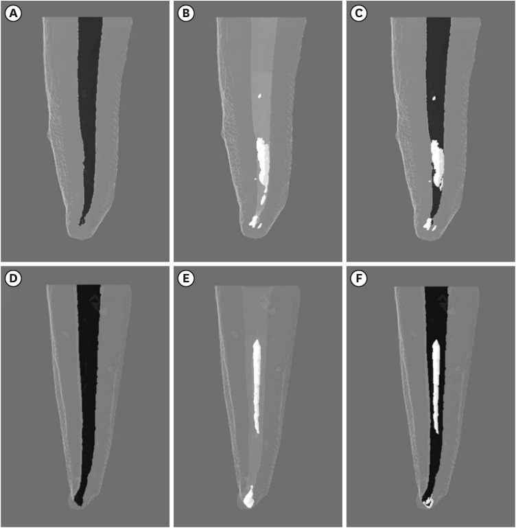
The endodontic retreatment protocols evaluated in this study did not completely remove the filling material from inside the root canals, and no statistically significant differences were observed between the 2 groups. The mean percentage of residual obturation material was 9.94% in the RCP group and 15.94% in the CCR group. Figure 1 shows 3-dimensional pre- and post-endodontic retreatment images of the representative specimens, illustrating the amount and spatial location of the residual filling material (Figure 1).
Figure 1
Three-dimensional pre- and post-endodontic retreatment images of representative specimens instrumented with reciprocating (A-C) and rotary (D-F) systems, illustrating the obturation (A, D) amount, spatial location of the remaining filling material (B, F), and superposition of the images (C, F).
DISCUSSION
Several protocols have been suggested for endodontic retreatment, with manual stainless steel files, mechanical nickel-titanium instruments, or a combination of both [78101213161920]. However, no method has yet fully met the goal of the procedure. Several studies have shown that total removal of the filling material is not possible [1567891213], which was corroborated by the results of our study (i.e., no specimen had 100% of the filling material removed). The anatomical complexity of root canal systems makes them difficult to clear, even with the use of auxiliary solvents and ultrasonic irrigation [79101115].
The null hypothesis was proven in this study, because there were no statistically significant differences between the 2 kinematic methods employed, based on the proportion of obturator material removed and the working time. The working time required for infilling root canals is a very important variable, since a faster procedure enables more time for a thorough cleaning and decontamination of the root canal system.
A previous study compared the effectiveness of a RCP file (Reciproc, VDW) and a rotary retreatment file (Mtwo retreatment, VDW) for the removal of filling material during root canal retreatment and reached the same conclusion; namely, there was no difference between the 2 kinematic methods in their ability to remove filling material [12]. Other studies have indicated that a mechanical preparation with nickel-titanium instruments consumed less time in endodontic retreatment, regardless of method [1116].
The WaveOne Primary file was used with agility in both RCP motion and CCR, and required a similar amount of time for both movements. However, both times were longer than reported by a previous study that used the same instrument in a RCP motion [11]. These differing results can be attributed to the obturation technique used. The present study used a thermoplastic technique (modified Tagger hybrid), which promotes greater compaction of the filling materials and results in greater penetration and bond strength in the intra-radicular dentin when compared with a single cone technique [29].
As in other studies, analysis of the remaining filling material was performed using micro-CT [69101113]. This was a non-destructive technique that allowed for high-definition analysis through 3-dimensional models. Our results showed that the WaveOne Primary file, used in an RCP motion (as recommended by the manufacturer for root canal preparation), provided more than 90% removal of the obturator material, corroborating the results of a previous study [11]. However, when the instrument was used in a counterclockwise rotation, there was no significant difference in the percentage of remaining filling material.
It is important to note that there were accidents during the endodontic retreatment procedures. There were more fractures when the instruments were used in CCR, probably due to torsional failure. The instrument was designed to be used in an RCP motion.
CONCLUSIONS
WaveOne Primary files performed comparably in both RCP movement and CCR in terms of working time and remaining obturation material. However, neither technique was able to completely remove the filling material during the endodontic retreatment procedures. The RCP motion technique did prove to be safer, with fewer fractured instruments.
 XML Download