

 PDF
PDF Citation
 Print
Print


INTRODUCTION
Severely damaged crowns are often observed in endodontically treated teeth, requiring preparation of the root canal to receive a post in order to promote retention of the crown restoration while maintaining the integrity of the canal walls [1]. Numerous recommendations have been made about the characteristics of post space preparation, including specific techniques for the removal of filling material and root canal enlargement [12]. Increased diameters [3] and longer post lengths [2] have been shown not to increase post retention or fracture resistance. In fact, the post space diameter should not exceed one-third of the root diameter [3], and the remaining dentin thickness should not be less than 1 mm [4].
Different techniques have been proposed to remove preexisting root canal filling material in order to create a post space, including the use of chemical solvent, heated instruments, and Largo-Peeso reamers, Gates-Glidden burs, or other types of rotary instruments [5]. Mechanical removal of gutta-percha is efficient and probably the most commonly used technique, but it can result in excessive wear of the root canal walls [126]. In addition, numerous momentary stress concentrations in dentin may be created during root canal enlargement because of the contact between the instrument and dentin walls [7]. The use of larger [18] or stiffer [7] instruments results in more contact with the wall and, therefore, more friction and stress concentration, which is transmitted through the root and may damage the dentin, leading to incomplete cracks or craze lines that may develop into vertical root fractures (VRFs), a clinical complication that may affect long-term tooth survival [9].
Despite the high rates of VRFs in teeth with intraradicular posts [10], little attention has been paid to the effect of post space preparation on the development of dentin defects. In fact, only 1 study [6] has focused on the effect of post space preparation on the formation of root defects. In that study, post space preparation with drills of the Rebilda post system (VOCO, Cuxhaven, Germany) had a significant impact on crack propagation [6]. As far as we know, no studies have evaluated the effect of other post space preparation drills on the occurrence of root dentin defects. Therefore, the aim of this study was to investigate the incidence of root dentin defects after the use of Gates-Glidden, Largo-Peeso, Exacto, and WhitePost drills for post space preparation. The null hypothesis tested was that the development of root dentin defects would not be affected by 1) the post space preparation drill or 2) the root canal level.
MATERIALS AND METHODS
Sample selection and preparation
A total of 120 freshly extracted bovine mandibular incisors with fully formed roots, which were morphologically similar in size and shape, and single straight root canals with comparable widths, about 9 mm from the apex (measured on radiographs taken from the buccolingual and mesiodistal views) and with a cervical diameter of approximately 1 mm, were selected for this study and stored in distilled water until use. Teeth with calcified or flared canals and anatomic irregularities were discarded. A total of 96 teeth were decoronated with a double-faced diamond disc (KG Sorensen, Barueri, SP, Brazil) operated perpendicular to the longitudinal axis of the teeth, leaving standardized root sections of 14 mm in length. All roots were examined for the presence of preexisting external defects at ×20 magnification (Expert DN; Müller Optronic, Erfurt, Germany), and those presenting any cracks, fractures, or craze lines were excluded and replaced by similar teeth. To simulate the periodontal ligament [11], the roots were covered with a silicone impression material (Aquasil; Dentsply Maillefer, Ballaigues, Switzerland). Specimens were then placed in a polyvinyl chloride pipe (14 × 20 mm) and embedded in acrylic resin blocks.
Root canal instrumentation and filling
Twelve roots served as controls with no intervention. The 84 root canals remaining were selected for the intracanal interventions. They were explored with sizes 10 and 15 K-files (Dentsply Maillefer), which were inserted passively until their tips became visible at the apical foramen. A total of 24 roots that showed patency larger than an ISO size 15 file was discarded. The remaining 60 roots were randomly divided into 5 groups, according to the endodontic procedures (instrumentation, filling, and post space preparation) (n = 12 per group).
For root canal instrumentation, the cervical and middle thirds of each specimen were prepared sequentially with Gates-Glidden (Dentsply Maillefer) sizes 1 (0.50 mm) and 2 (0.70 mm) until resistance was found on the canal walls. The apical thirds were instrumented to a working length of 13 mm using a crown-down technique with ProTaper Next rotary instruments (Dentsply Maillefer), with a sequence of X1 (size 0.17/0.04), X2 (size 0.25/0.06), X3 (size 0.30/0.07), and X4 (size 0.40/0.06). Each instrument was used for the instrumentation of 3 root canals and operated at a rotational speed of 300 rpm and 2.9 Ncm torque with an endodontic electric motor (X-Smart Plus; Dentsply Maillefer).
At each instrument change, the root canals were irrigated with 3 mL of 1.0% sodium hypochlorite (NaOCl; Pharm, Phloraceae, Cuiabá, MT, Brazil) by using a syringe and a 31-gauge needle (NaviTip; Ultradent, South Jordan, UT, USA). Final irrigation was performed with 3 mL of 17% ethylenediaminetetraacetic acid (EDTA; Biodinâmica, Ibiporã, PR, Brazil) for 3 minutes, with the final 2 minutes under active agitation with an endodontic file, followed by 3 mL of 1.0% NaOCl. The roots were then dried with sterilized paper points (Dentsply Maillefer) and filled with gutta-percha points (Dentsply Maillefer) and AH Plus (Dentsply Maillefer), which was mixed according to the manufacturer's instructions using the lateral condensation technique. Excess gutta-percha and sealer were removed with heated condensers (Paiva; SS White, Rio de Janeiro, RJ, Brazil), and the root canal openings were sealed with temporary cement (White Cimpat; Septodont, São Paulo, SP, Brazil). The specimens were stored at 37°C and 100% humidity for 1 week.
Post space preparation
After the storage period, of the 60 roots that were endodontically treated, 48 were divided into 4 groups (G3–G6; n = 12 each), according to the post space preparation protocol.
The temporary restorative material was removed, and heated condensers (Paiva; SS White) were used to remove the initial root canal filling. The post spaces were prepared sequentially until a standardized depth of 9 mm was achieved, leaving 4 mm of gutta-percha in the apical third, according to the manufacturer's instructions. Post space preparation drills of each group were selected, aiming to standardize the apical diameter at about 1.1 mm. The drills were used in a low-speed handpiece (Kavo Ind. Com. Ltda., Joinville, SC, Brazil) at 15,000 to 20,000 rpm. The root canals were irrigated with 1.0% NaOCl after each drill change and dried with sterilized paper points (Dentsply Maillefer).
A single endodontist with 10 years of experience performed all clinical procedures. All roots were stored in distilled water during the experimental procedures in order to avoid dehydration.
The investigated groups, the operative steps, and the post space preparation protocols are presented in Table 1. The PSP protocols for the G5 and G6 groups were performed as recommended by the respective manufacturers.
Table 1
Operative steps of the control and experimental groups (n = 12) and the post space preparation protocols

Sectioning and root dentin analysis
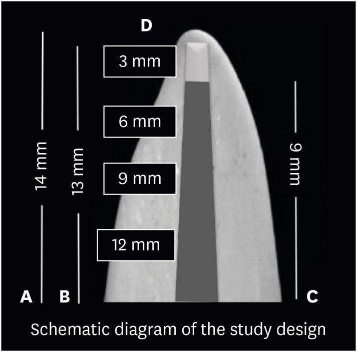
The roots were removed from the resin blocks and horizontally sectioned at 3, 6, 9, and 12 mm from the apex with a double-faced diamond disc (4-inch diameter × 0.012-inch thickness × 1/2 inch; Arbor, Extec, Enfield, CT, USA) and a low-speed saw under water cooling (Isomet 1000; Buehler, Lake Bluff, IL, USA). The slices were analyzed under a stereomicroscope (Expert DN; Müller Optronic, Erfurt, Germany) at ×25 magnification, and digital images of each section were captured. The occurrence of defects was recorded as “no defect,” “fracture,” and “other defects” [12].
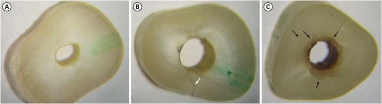
“No defect” was defined as the absence of any type of line in the outer surface of the root and on the inner wall of the root canal. “Fracture” was defined as a line that extended from the root canal space to the outer surface of the root. “Other defects” were defined as all other observed lines that did not extend from the root canal to the external surface of the root, including craze lines (lines extending from the outer surface into the dentin that did not reach the canal lumen), and partial cracks (lines that extended from the canal wall into the dentin without reaching the outer surface of the root) (Figure 1). A total of 48 images were examined in each group. A blinded, previously calibrated examiner analyzed all images. The same examiner read the images twice, with an interval of 1 week between readings. The study design is presented in Figure 2.
Statistical analysis
The results were expressed as the number and percentage of roots with defects in each group. The χ2 test was used to compare the presence of defective roots between the control and experimental groups using Minitab, version 17.0 (Minitab Inc., State College, PA, USA). The level of significance was set at p < 0.05. Intraexaminer agreement in the reading of images was evaluated by the kappa coefficient in 10% of the sample.
RESULTS
The kappa value was 0.83. Of the 288 slices that were analyzed, 114 (39.6%) had root dentin defects (other defects and fractures). No defects were observed in G1 (root canals without any intervention). When considering the formation of other defects, G5 had significantly more cracks and craze lines than G1, G2, and G3 (p < 0.05), but not significantly more than G4 and G6 (p > 0.05) (Table 2). When considering fracture formation, G5 had significantly more defects than G1, G2, G3, and G4 (p < 0.05), but not significantly more than G6 (p > 0.05) (Table 3).
Table 2
Number and percentage of other defects in the different cross-section slices.

*G1, control; G2, root canal instrumentation and filling; G3, post preparation with Gates-Glidden drills; G4, post preparation with Largo Peeso reamers;G5, post preparation with Exacto drill; G6, post preparation with WhitePost drill. †Capital letters compare groups in vertical columns and lowercase letters compare groups in horizontal rows.
Table 3
Number and percentage of root fractures in the different cross-sectional slices.

*G1, control; G2, root canal instrumentation and filling; G3, post preparation with Gates-Glidden drills; G4, post preparation with Largo Peeso reamers; G5, post preparation with Exacto drill; G6, post preparation with WhitePost drill. †Capital letters compare groups in vertical columns and lowercase letters compare groups in horizontal rows.
DISCUSSION
This study aimed to investigate whether the occurrence of root dentin defects is influenced by the type of drill used for post space preparation. To the authors' knowledge, no other study has focused on the formation of root dentin defects after PSP with Largo-Peeso reamers and Gates-Glidden, Exacto, and WhitePost drills. The results showed that both the post space preparation protocol and root canal level influenced the development of root dentin defects. Therefore, the null hypotheses were rejected.
Although various methods have been proposed to investigate the effect of intracanal procedures on the formation of dentin defects, such as thermography, optical coherence tomography, and scanning electron microscopy [13], much of the knowledge of this topic is derived from investigations using micro-computed tomography (micro-CT) [1415161718] and/or root-sectioning methods [1112192021]. Micro-CT is an accurate, non-destructive 3-dimensional method [17] that allows quantitative and qualitative volumetric assessments of specimens before and after any intracanal procedure [1622]. With this method, each tooth may serve as its own control [17]. In addition, hundreds of dentin slices can be analyzed per specimen, thereby enabling the full extension of the defects to be tracked [1314]. However, it is expensive, time-consuming, and often unavailable to the general research community [24]. Furthermore, Pedullà et al. [21] suggested that the increase in temperature from the use of high-resolution micro-CT scans may provoke dehydration and subsequently an increase in existing defects, which may affect the results of the study. Lim et al. [23], however, observed that the resolution and contrast of micro-CT are not always enough to identify all root defects because of the restricted signal-to-noise ratio. Root-sectioning methods allow direct inspection by optical microscopy of defects in slices proceeding from several levels of the canal [1215]. Swift results are provided without the need for post-processing and much smaller datasets are required, making it possible to screen many samples in a short time [24]. This simplifies tracking the appearance and evolution of cracks involving sample motion, which is not possible with micro-CT [24]. However, sectioning methods have some limitations, such as their destructive nature [2122] and 2-dimensional, single-moment observations of only a few slices per tooth, which may result in the chance of missing some previous defects along the root [15]. Furthermore, it is not possible to assess the precise moment at which the defect is developed [14]. Unfortunately, an ideal experimental model to evaluate the presence of dentin defects has not yet been established [1822]. Therefore, more attention should be drawn to the development of new methods to understand the phenomenon of dentin defect formation better.
An understanding of the cause-and-effect relationship between endodontic procedures and dentin defect formation has been obtained from studies using teeth with several root and root canal morphologies, such as lower incisors [16], premolars [121824] and molars [111525], and upper premolars [26] and incisors [27]. This fact makes it difficult to compare the results [2127], since there is wide variation in tooth anatomy, such as canal length, width, and shape; dentin hardness; calcification; size and location of the apical constriction and angle; radius length; and the location of curvatures [28]. Another aspect that deserves attention is that previous publications examining root defects have not provided appropriate data on the hydration conditions of the teeth (storage conditions and time), tooth condition before extraction, trauma, age and sex of the donors, and variables related to the lifetime of the tooth in vivo (masticatory function, excursive interferences, or any parafunctions) [131729].
In this study, we used bovine teeth since human teeth are difficult to obtain for academic dental investigation [29]. Bovine teeth are easier to handle due to their dimensions, enable better uniformity in terms of age and root canal space, and decrease the possibility of transmitting diseases [30]. It is worth noting that the use of bovine teeth makes it possible to unify different features and to use a lower number of animals, reducing confounding factors related to dentin [30]. Previous investigations have also demonstrated that human and bovine teeth have similar biological, chemical, and physical properties [31], which could make bovine teeth possible substitutes for human teeth in root dentin defect evaluations. In the current study, bovine incisors with straight canals were chosen to permit the use of tapered post space preparation drills, as the effect of these drills on the development of root dentin defects has been inadequately investigated [6]. When choosing bovine teeth, older teeth were desirable due to the possibility of a greater morphologic resemblance to human teeth [31].
For all teeth included in this study, the outer surface of the roots was inspected preoperatively with an operative microscope to assess the presence of cracks and/or fractures. One could argue that some defects might have been internal and not visible externally. However, no defects were noticed in the teeth with no intervention (control group), which is consistent with several studies that used a similar method [192732]. Therefore, it is reasonable to interpret all root dentin defects observed in the present study as having resulted from the intracanal procedures, rather than from the sectioning method [12]. It is important to consider that no auxiliary tools were used to improve the visualization of defects. Recent studies have demonstrated that illumination conditions may influence the capacity to detect root dentin defects [182122]. Coelho et al. [22] observed that the use of light-emitting diode (LED) transillumination enhances the visualization of dentin defects, especially in control groups (uninstrumented roots). This is one of the limitations of the present study, and further sectioning studies should be performed with the aid of LED transillumination to reassess the potential of dentin defect formation resulting from different intracanal procedures.
It has been demonstrated that specimen storage conditions may affect the biomechanical properties of root dentin [14152224]. Therefore, in the present study, all specimens were kept in distilled water throughout the entire experiment [152427]. In addition, to reproduce periodontal ligament and bone and consequently stress absorption [12], the roots used in the experiment were covered with a silicone impression material and embedded in acrylic resin blocks [112133]. Although it is very difficult to imitate the complexity of the anatomic and biologic aspects of the periodontal ligament, and no artificial material can accurately reproduce its viscoelastic properties [12], periodontal ligament reproduction is important since it may influence the results of studies investigating the effects of intracanal forces on the formation of root dentin defects [33]. Additionally, in the present study, the specimens were equally allocated among the groups according to their root canal widths at the 9-mm level [2133]. A disparity in dentin thickness would cause important variations in strength, and hence in response to stress distribution, during intracanal procedures [26]. Moreover, uniformity was obtained in the groups by including only teeth with a canal width near the apex consistent with a size 15 K-file and leaving roots of about 14 mm in length [21].
Previous studies have shown a clear correlation between root canal instrumentation and the development of root dentin defects [7111215171920212227], attributing the harmful effects to the instrument's cross-sectional geometry, alloy, heat treatment, cutting efficiency, size, tip configuration, variable pitch and taper, flute form, number of instruments used, and kinematics [714172125]. In this study, root canal instrumentation was performed using the ProTaper Next system, which works in continuous rotation and consists of 5 files [25]. ProTaper Next instruments are made of the M-Wire alloy, which is produced by a pioneering thermal treatment protocol and has the advantage of instrument flexibility and enhanced resistance to cyclic fatigue [25]. Its off-centered rectangular projection creates a swaggering movement, which reduces the screw effect and unsafe taper lock by reducing contact between the instrument and root canal wall [14]. These instrument characteristics may result in less stress generation inside the root canal, and hence less damage to root dentin [34].
The irrigants used during root canal preparation can also induce root dentin defects [32]. There is evidence that high concentrations of NaOCl can alter the elastic modulus and flexural strength of root canal dentin [2635]. Ari et al. [35] observed reduced root dentin microhardness after contact with NaOCl at concentrations of 2.5% and 5.25%, but not at low concentrations. In this study, 1.0% NaOCl solution was employed for irrigation to reduce the possible alterations to the mechanical characteristics of dentin during intracanal procedures [20].
Despite the extensive investigations on the impact of root canal filling procedures on defect formation [121519], contradictory findings have been reported on the influence of the lateral compaction technique [12151926]. Shemesh et al. [12] assessed the occurrence of dentin defects after root filling with lateral compaction or passive compaction techniques and reported a higher incidence of dentin defects in teeth filled by lateral compaction. Adorno et al. [26] evaluated the effects of instrumentation and filling (lateral compaction associated or not with vertical compaction) on crack initiation and propagation in apical dentin and reported a significant effect of root canal filling only on crack propagation, with no significant difference between the 2 techniques in crack propagation. Shemesh et al. [19] compared the incidence of dentin defects after root canal instrumentation, lateral compaction, and continuous wave compaction of gutta-percha, and no difference was observed in the occurrence of defects between the 2 filling procedures. De-Deus et al. [15] reported that the cold lateral compaction technique was not linked to the development of new root defects. In this study, the lateral condensation technique was the method of choice for root canal obturation, since it is broadly used to fill the root canal system, does not necessitate an exceptional or expensive apparatus, and is considered safe in controlling the apical extension of the filling [1219]. The results of the present study showed a lower incidence of defects (12.5%) than those of previous studies, which demonstrated an occurrence of defects after root canal instrumentation and filling ranging from 15.99% to 30.00% [1519], which may be explained by the difference among studies in the methods used [13].
Different drills have been developed and suggested for post space preparation [636]. In the present study, Gates-Glidden #1, #2, #3, and #4 drills and Largo-Peeso #1, #2, and #3 reamers were compared with Exacto #3 and WhitePost #2 drills. Only drills that act in the straight portion of the canal were selected. Gates-Glidden drills and Largo-Peeso reamers were chosen because they are widely used in preflaring and post space preparation [836], while Exacto and WhitePost drills were chosen because of their use during the placement of a fiber post inside the root canal, and because no studies have investigated the effects of these burs on dentin defect formation. According to their manufacturers, Exacto and WhitePost drills consist of stainless-steel drills, with a conical shape and inactive tips. The main differences between these drills are related to their cervical and tip diameters. The Exacto drill has a tip diameter of 1.10 mm and a cervical diameter of 1.60 mm, whereas the WhitePost drill has diameters of 1.05 mm and 1.80, respectively. The results revealed that Exacto drill and Largo-Peeso reamers promoted significantly more “other” types of defects than Gates-Glidden drills, while no significant differences were noted among the Largo-Peeso reamers, Exacto drill, and WhitePost drill (Table 2). Furthermore, teeth instrumented with the Exacto and WhitePost drills were the only ones in which root fractures were observed (Table 3). These results may be credited to the highly-tapered design of Exacto and WhitePost drills, to the stiffness of the larger instruments [67], and to unnecessary dentin removal [36]. It has been demonstrated that fiber post space preparation drills are more aggressive than other instruments [6]. It is also noteworthy that the recommendations for Exacto post space preparation involve the use of heated devices to remove part of the root canal filling. The use of these instruments, even with light pressure, can promote higher coronal stress concentration, which could lead to a higher risk of dentin defect development [2637]. It will be useful to evaluate whether comparable root dentin defects will be present after post space preparation with drills having different features. Moreover, professionals must be conscious of the possibilities of harm during post space preparation with Exacto drill.
Although dentin defects were observed at all 4 levels of the root canal, a higher number of root fractures and other defects occurred coronally than apically. This is consistent with the results of previous studies [242737] and may be attributed to the increase in tensile stress and in the regions of stress concentration associated with post space preparation. It is important to highlight that PSP may cause root dentin defects, which can remain confined inside to the root canal walls. Unfortunately, the root-sectioning method used in the present study does not provide information about the root canal lumen [13]. Even though this ex vivo study did not reflect clinical situations, the results may explain some failures observed in endodontically treated teeth with intraradicular posts. Nevertheless, further studies using rapid, reliable, and accurate experimental methods are essential to obtain a better knowledge of the influence of different PSP protocols on the formation of root dentin defects.
CONCLUSIONS
Post space preparation with Largo-Peeso reamers and Gates-Glidden, Exacto, and WhitePost drills induced damage to the root dentin. However, Gates-Glidden drills tended to cause fewer root dentin defects than other drills. A higher number of root dentin defects occurred coronally than apically.
 XML Download