

 PDF
PDF Citation
 Print
Print


INTRODUCTION
Anatomical complexities, such as flattened root canals, make the shaping and cleaning of root canal system even more difficult [1234]. Nickel-titanium (NiTi) instruments promote a centric preparation, allowing circular morphology with less contact on the buccal and lingual walls of the root canal [5]. An uninstrumented surface favors the maintenance of biofilm and debris accumulation, which can affect the prognosis of endodontic treatment [67].
New NiTi instruments with different heat treatments, designs, kinematic, and diameters have been developed to allow proper root canal preparations, removal of debris and microorganisms, and prevention of excessive dentin removal [58910111213141516]. NiTi instruments with control memory (CM) heat treatment have higher flexibility and cyclic fatigue resistance, improving their shaping ability, and reducing the preparation time [17181920].
ProDesign Logic System (PDL; Easy Equipamentos Odontológicos, Belo Horizonte, MG, Brazil) is manufactured with a NiTi CM alloy. This rotary system is composed of glide path files (taper 0.01) and shaping files (tapers 0.03, 0.04, 0.05, and 0.06). These instruments have a modified S-shaped cross-section, variable helical angles with cutting edges, and an inactive tip [2122]. PDL has shown a similar ability for root canal preparation to Reciproc, Protaper Next and Waveone Gold systems [18].
HyFlex EDM (HEDM; Coltene/Whaledent, Altstätten, Switzerland) is a NiTi CM instrument subjected to the electrical discharge machining (EDM) process, which increases its resistance to cyclic fatigue and flexibility [2324]. HEDM has different tapers and cross-sections (triangular in the coronal portion, trapezoidal in the middle portion and quadrangular in the apical portion), providing better cutting efficiency and removal of debris [22]. HEDM improved the centralization of the canal when compared with Protaper Next [19].
Preparation systems with different kinematics do not prepare all the root canal walls [569]. Therefore, the use of auxiliary procedures, such as ultrasonic activation, may improve root canal cleaning [25]. The physical effect of ultrasonic tips contributes to disinfection and debris removal from the root canals [312]. The mechanical action of ultrasonic tips favors the cleaning of flattened areas during root canal preparation or retreatment [2627].
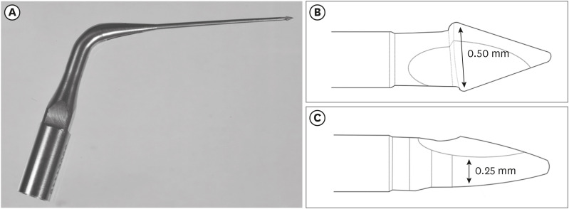
Flatsonic ultrasonic tip (Helse Ultrasonic, Santa Rosa de Viterbo, São Paulo, SP, Brazil) is manufactured with stainless steel and has an inverted arrow-shaped tip with a flattening that corresponds to the diameter of an ISO 25 instrument. Flatsonic tip improves the cleaning of oval root canals and flattened distal canals of mandibular molars when used as a complementary preparation [2628]. In addition, an increase in the removal of debris from oval root canals was observed when 2 instruments with distinct diameters were used [29]. However, no studies have evaluated the preparation of flattened root canals using a combination of the Flatsonic tip and a small-diameter instrument.
Thus, the aim of this study was to evaluate the percentages of volume increase, debris, and uninstrumented surface area in flattened root canals after preparation with PDL or HEDM and the effectiveness of a complementary protocol combining the Flatsonic tip (F) and a small-diameter instrument (PDL 25/0.03). The first null hypothesis was that the rotary systems would have a similar capacity for flattened root canal preparation. The second null hypothesis was that the FPDL protocol would have no influence on the percentage of debris and uninstrumented surface area in flattened root canals.
MATERIALS AND METHODS
Sample size calculation
G*Power 3.1.7 for Windows program (Heinrich Heine University, Düsseldorf, Germany) was used for the sample calculation. An alpha-type error of 0.05 and power (1–beta) of 0.80 were established for all the variables. For the t-test in 2 independent groups, previous studies that used micro-computed tomography (micro-CT) to evaluate the preparation of root canals with similar morphology were used to determine the specific effect for each variable: percentage of volume increase of the root canals, 0.9779; debris, 1.2638; and uninstrumented surface area, 1.3909 [230]. For the t-test of dependent means, a previous study was used to determine the effect size for debris (2.7110) and uninstrumented surface area (2.0064) [28]. The resulting sample size was 12 specimens per group. A sample of 16 root canals per group was planned, taking into consideration the risk of loss of teeth during implementation of the methodology.
Sample selection
After approval from the Ethics Committee for Research (Protocol CAAE No. 98685518.4.0000.5416), 32 human maxillary second premolars with single root canals that had a completely formed apex, absence of endodontic treatment, and extensive restorations were selected and stored in a glass bottle containing 0.1% thymol solution at 5°C. The specimens were radiographed (Kodak RVG 6100 Digital Radiography System, Marne-la-Vallée, France) in the buccolingual and mesiodistal directions and scanned with a micro-CT device (SkyScan 1176, Bruker-microCT, Kontich, Belgium) using the following parameters: 80 kV, 300 μA, rotation 180°, rotation step 0.5, frame averaging 3, Cu + Al filter, exposure time of 125 ms, and voxel size of 35 µm. The inclusion criteria were applied, and the homogeneous distribution of the samples was confirmed. The root canals were considered flattened when the buccolingual diameter was 4 or more times larger than the mesiodistal diameter at 9 mm from the radiographic apex [3132]. The teeth were divided into 2 experimental groups (n = 16), with stratified random sampling considering the preoperative volume of the root canals.
Conventional access to the root canals was created using high-speed diamond burs (No. 2, KG Sorensen, São Paulo, SP, Brazil). The canals were explored using a size 10 K-file (Dentsply Sirona, Ballaigues, Switzerland) until its tip became visible in the apical foramen. The working length (WL) was established 1 mm short of the apical foramen, as confirmed by digital radiography. The teeth were mounted in an appliance with acrylic resin, and enveloped in condensation silicone (Oranwash, Zhermack SpA, Badia Polesine, Italy) to simulate the periodontal ligament.
Preparation and complementary procedures of the root canal
Preparation and complementary procedures were performed by an endodontic specialist using a surgical microscope with ×13 magnification (DF Vasconcellos, Valença, RJ, Brazil). Each group (n = 16) was instrumented using the rotary systems, as follows:
1. ProDesign Logic
PDL 30/0.01 was operated using a VDW.SILVER motor (VDW GmbH, Munich, Germany) with rotary motion at a speed of 350 rpm and a torque of 1 N·cm, and PDL 30/0.05 was used at 950 rpm and a torque of 4 N·cm, according to the specifications of the manufacturer.
2. HyFlex EDM
HEDM 10/0.05 was operated using a VDW.SILVER motor with rotary motion at a speed of 300 rpm and a torque of 1.8 N·cm, and HEDM 25/0.08 was used at 500 rpm and a torque of 2.5 N·cm, according to the specifications of the manufacturer.
All instruments were introduced into the root canal using an in-and-out motion up to the WL. For both groups, the irrigation protocol was performed using 2.5 mL of 2.5% sodium hypochlorite (NaOCl) (Ciclo Farma, Serrana, SP, Brazil) after the use of each instrument, using a 5-mL syringe (Ultradent Products, South Jordan, UT, USA) with a Navitip 30-G needle (Ultradent Products) at 2 mm short of the WL, using an in-and-out motion, with continuous flow and aspiration. Final irrigation was performed with 5 mL of 2.5% NaOCl, 2.5 mL of 17% EDTA (Biodinâmica, Ibiporã, PR, Brazil) and 5 mL of physiological solution.
3. Preparation with Flatsonic and ProDesign Logic 25/0.03
In the complementary preparation, all root canals were prepared by Flatsonic (Figure 1) coupled to the ultrasound appliance Ultrawave XS (Ultradent Products) at a frequency of 50 Hz and power of 25%. This tip was activated inside the root canal, at 4 mm to the WL, in the direction of the flattened area (buccal and lingual direction) performing 3 cycles of 30 seconds. Each cycle was performed as follows: 15 seconds in the direction of the buccal wall, with 10 seconds performed without air/water cooling and 5 seconds with air/water cooling in accordance with the manufacturer's specifications. The same procedure was performed for the lingual wall. In each ultrasound cycle, 2.5 mL of 2.5% NaOCl was used (1.25 mL before and 1.25 mL after each cycle). After use of the Flatsonic, the PDL 25/0.03 instrument was operated using a VDW.SILVER motor, with rotary motion, at 950 rpm and a torque of 2 N·cm, in accordance with the manufacturer's specifications. The instrument was introduced into the root canal up to the WL, after which brushing motions were made on the buccal and lingual walls (flattened areas in the apical third) with an amplitude of 3 mm, totaling 3 motions on each wall. The irrigation and final irrigation protocols were performed in accordance with the criteria previously described for preparations with NiTi systems.
Micro-computed tomography analysis
The specimens were scanned before and after preparation and after complementary preparation using a SkyScan 1176 micro-CT device. The parameters used were 80 kV, 300 μA, rotation step 0.5, rotation 180°, frame averaging 4, Cu + Al filter, an exposure time of 2000 ms, and a voxel size of 8.74 µm. The images of each sample were reconstructed using NRecon software (v.1.6.4.7, Bruker-microCT). For superimposition of the samples throughout all the experimental stages, geometric realignment was performed by means of the 3D registration function of the Data Viewer software (v.1.5.1, Bruker-microCT). The images were analyzed using the CTAn software (v.1.14.4, Bruker-microCT) and 3-dimensional images were created using CTVox software (v.3.2, Bruker-microCT).
The initial volume, final volume, and final surface after preparation were obtained. Based on these values, the percentage of volume increase of the root canals (% volume increase of the root canals), percentage of debris (% debris) and percentage of uninstrumented surface area (% uninstrumented surface area) were calculated using the following formulas [29]:


Statistical analysis
The normality of the data was assessed with the Shapiro-Wilk test. The data for the percentage of volume increase of the root canals did not present a normal distribution, while the data for the volume of the canal, debris, and uninstrumented surface area showed normal distributions. To compare the percentage of volume increase of the root canals, the Kruskal-Wallis and Dunn tests were used for between-group comparisons, and the paired Wilcoxon test was used for comparisons in the same group before and after complementary preparation with Flatsonic and PDL 25/0.03. In analyses of the data for the volume of the canal, debris, and uninstrumented surface area, the non-paired t-test was used for comparisons between groups, analysis of variance and the Tukey test were used for comparisons between thirds in the same group, and the paired t-test was used for comparisons between the same groups before and after complementary preparation with the Flatsonic and PDL 25/0.03 (α = 0.05).
RESULTS
There was no significant difference in the percentage of volume increase of the root canals, debris, and uninstrumented surface area between PDL and HEDM in the entire canal (p > 0.05). However, the PDL group had a higher percentage of debris than the HEDM group in the middle and apical thirds (p < 0.05) (Table 1).
Table 1
Initial volume of the root canal (mm3), volume increase of the root canal (%), debris (%), and uninstrumented surface area (%) of flattened root canals of maxillary second premolars prepared with ProDesign Logic (PDL) or HyFlex EDM (HEDM)

Different superscript lowercase letters indicate statistically significant differences between the groups. Different superscript uppercase letters indicate statistically significant differences among the thirds of the same preparation for each analysis: mean and ± standard deviation for parametric data (analysis of variance and Tukey tests, 5% significance), and median, minimum and maximum values for non-parametric data (Kruskal-Wallis and Dunn, 5% significance)
*Analysis of variance; †Kruskal-Wallis.
After application of FPDL as a complementary protocol, there was a significant decrease of debris and uninstrumented surface area after preparation with HEDM and PDL in the entire canal and in all thirds (p < 0.05). The FPDL protocol had the lowest percentage of debris in the middle and apical thirds and uninstrumented surface area in the apical third when combined with HEDM (p < 0.05) (Tables 2 and 3; Figures 2 and 3).
Table 2
Means and standard deviations of debris (%) of flattened root canals of maxillary second premolars prepared with ProDesign Logic (PDL) or HyFlex EDM (HEDM) before and after complementary preparation with Flatsonic and PDL 25.03 (FPDL)

Table 3
Means and standard deviations of uninstrumented surface (%) of flattened root canals of maxillary second premolars prepared with ProDesign Logic (PDL) or HyFlex EDM (HEDM) before and after complementary preparation with Flatsonic and PDL 25/0.03 (FPDL)

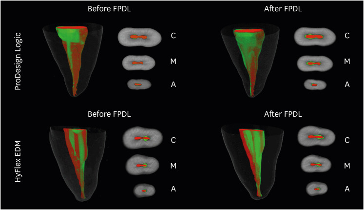
Figure 2
Three-dimensional reconstructions and cross-sectional views of the cervical (C), middle (M), and apical (A) thirds of representative images of flattened root canals of the maxillary second premolars prepared by ProDesign Logic or HyFlex EDM before and after complementary preparation with Flatsonic and PDL 25.03 (FPDL), showing uninstrumented surfaces (red) and instrumented surfaces (green).
DISCUSSION
Poor cleaning of the root canals is associated with endodontic treatment failure [33]. NiTi instruments and a supplementary ultrasonic tip have been observed to be useful for the preparation of oval and long oval root canals [2634]. However, flattened root canals represent a challenge for endodontic treatment because their buccolingual diameter is 4 or more times larger than the mesiodistal diameter [31]. This is the first study to compare the FPDL complementary protocol after using 2 different NiTi rotary instruments to prepare flattened root canals.
The first null hypothesis was partially rejected. The percentage of uninstrumented surface area was similar between PDL and HEDM, with mean values of 57.02% and 49.41%, respectively. In agreement with these findings, various studies have shown that it is difficult to clean the walls of flattened root canals using NiTi instruments [128]. Siqueira et al. [35] showed that oval and flattened root canals presented 10% to 80% of uninstrumented surface area after preparation. Similar preparations using different rotary instruments with different cross-sections have been observed since their oval morphology does not allow the instruments to touch all the root canal walls [5930]. The uninstrumented surface of root canals favors the maintenance of debris and bacterial biofilm, contributing to persistent infections [635].
The PDL and HEDM instruments produced a similar percentage of debris in the entire root canal. However, the HEDM group had less accumulation of debris in the middle and apical thirds than the PDL group, even after the use of Flatsonic and PDL 25/0.03. PDL and HEDM are heat-treated instruments with some differences, such as the surface treatment, cross-sectional geometry, and taper [192223]. Although the use of instruments with the same diameter is recommended for comparisons between different preparations [3637], the present study showed no difference between the systems in the increase in volume after preparation, even when using 2 rotary systems with different diameters and tapers. However, the greater taper (0.08) in the apical 4 mm and the trapezoidal and quadrangular cross-section of the HEDM 25/0.08 may have contributed to the lower percentage of debris and cleaning in the middle and apical thirds of the flattened root canals [22].
The proposed protocol using the Flatsonic ultrasonic tip and PDL 25/0.03 significantly decreased the percentage of debris and uninstrumented surface after preparation with PDL and HEDM. Therefore, the second null hypothesis was rejected. Previous studies demonstrated that the combination of mechanized systems with Flatsonic improved the cleaning of oval and flattened root canals [2628]. The 0.25 mm diameter and design of the Flatsonic tip favored its physical and mechanical effect in the cervical and middle thirds. The mechanical action of Flatsonic allowed greater contact with the flattened areas and consequently, a greater effect of the irrigating solution. The acoustic microcurrent and cavitation promoted by ultrasonic tips decrease accumulated debris [12].
Enlargement of the canal during the initial preparation favored the effect of the Flatsonic tip. The PDL and HEDM preparations promoted access of the Flatsonic tip to the buccal and lingual flattened areas. The improved cleaning in the apical third was favored by using the PDL 25/0.03 instrument with a brushing motion. The lower taper and brushing motion favored contact with areas that were difficult to access in the apical third. Instruments with smaller diameter enhance cleaning in oval root canals [29]. In addition, instruments with smaller tapers are associated with less unnecessary dentin removal, especially in the cervical third [38]. The protocol using HEDM and FPDL promoted a lower percentage of uninstrumented surface area (25.99%) than with PDL and FPDL (42.99%). The HEDM preparation probably contributed to a greater effect of the Flatsonic tip and 25/0.03 instrument. The present study showed an increase of approximately 14% in the apical third of the root canals after preparation with both systems. This apical enlargement promotes a lower percentage of non-instrumented surface and decreased debris [3940]. Therefore, the small apical enlargement, promoted by both rotary NiTi systems in this study, may have overestimated the effect of the complementary protocol.
Appropriate preparation of the root canal is critical for achieving a favorable prognosis of endodontic treatment [33]. However, new mechanized NiTi instruments are not able to reach all the root canal walls, precluding effective cleaning. In the present study, the proposed protocol with the use of the Flatsonic tip and PDL 25/0.03 showed favorable results in the preparation of flattened root canals. In addition, the combination of HEDM and FPDL seems to be a better protocol than PDL and FPDL to reduce debris and the uninstrumented surface area in flattened root canals.
CONCLUSIONS
Preparation of flattened root canals using NiTi instruments resulted in a large percentage of debris and uninstrumented surface areas, without significant differences between the systems. The combination of a Flatsonic ultrasonic tip and a small-diameter instrument enhanced the cleaning of flattened areas of the root canal. The protocol using the HEDM, Flatsonic tip, and 25/0.03 instrument presented better cleaning in flattened root canals.
 XML Download