

 PDF
PDF Citation
 Print
Print


INTRODUCTION
Endodontic regeneration (ER) is the most recent treatment option for immature teeth with pulpal necrosis. ER requires effective disinfection of the root canal, which can be achieved through irrigation solutions and intracanal medicaments [123456]. Various intracanal medicaments have been advocated for use in ER, including calcium hydroxide (Ca[OH]2), triple antibiotic paste (TAP) (equal amounts of metronidazole, ciprofloxacin, and minocycline), and double antibiotic paste (DAP) (equal parts of metronidazole and ciprofloxacin) [78910].
Several studies have demonstrated clear evidence of significant tooth discoloration after TAP application [111213]. Furthermore, the discoloration was mainly attributed to the presence of minocycline within TAP rather than the metronidazole or ciprofloxacin [111314]. Therefore, some studies suggested sealing the dentine within the pulp chamber with dental adhesive before TAP application [131516]. Other studies recommended using minocycline-free antibiotic medicaments such as DAP, or replacing the minocycline present in TAP with non-tetracycline antibiotics [1214]. Indeed, recent studies demonstrated that the application of DAP or Ca(OH)2 did not cause any significant tooth discoloration [1417]. However, other studies have shown that Ca(OH)2 and high concentrations of DAP can also lead to significant tooth discoloration [181920]. Minocycline was proposed to chemically bind into dentine and enamel via Ca2+ chelation [21]. In contrast, Ca2+ ions dissociated from Ca(OH)2 were suggested to physically incorporate within dentinal tubules and react with hydroxyapatite-forming crystals to form white or yellow color changes in teeth [19].
Multiple studies have recommended using low antibiotic concentrations to minimize the discoloration effect and optimize the biological environment within the root canal system [2223242526]. Loading low antibiotic concentrations into a methylcellulose hydrogel system was proposed to offer an injectable version of these intracanal medicaments with controlled concentrations in an attempt to create a biocompatible delivery system [252728]. However, no previous study has explored the ability of these hydrogel systems loaded with low concentrations of DAP or TAP to induce tooth discoloration. The aim of this study was to evaluate the effects of medium and low concentrations of TAP and DAP (1 and 10 mg/mL) loaded into an aqueous methylcellulose system on crown discoloration and explore the ability of a 1-step self-etch adhesive bonding agent to prevent crown discoloration. The null hypothesis was that low and medium concentrations of hydrogel-based DAP and TAP medicaments would cause clinically perceivable tooth discoloration at all measured time points regardless of the use of an adhesive bonding agent.
MATERIALS AND METHODS
Sample preparation
Intact human third molars, free of cracks, fractures, caries, abrasions, and visible discoloration (n = 160), were selected following local Institutional Review Board (IRB) guidelines (IRB # 1408889870). The teeth were stored at 4°C in 0.1% thymol solution and were used no more than 6 months after extraction [2930]. Soft tissue was removed by hand scaling and polishing performed by a lab-polishing machine (Handler, Westfield, NJ, USA) with pumice (Henry Schein, Melville, NY, USA). Teeth were horizontally sectioned 1 mm apical to the buccal cementoenamel junction using a water-cooled low-speed diamond saw (Buehler, Lake Bluff, IL, USA) to create crowns with a 1 mm extension to the roots. Access cavity preparation was not performed and pulpal tissues were removed with a spoon excavator. Additionally, the internal axial walls of the pulp chambers were mechanically debrided through retrograde access by using an ultrasonic endo tip #3 (ProUltra, Dentsply, Johnson City, TN, USA) attached to an endodontic ultrasonic (ASI, Englewood, CO, USA). Then, 5 mL of 1.5% sodium hypochlorite (NaOCl) followed by 5 mL of 17% ethylenediaminetetraacetic acid (EDTA) (Vista, Racine, WI, USA) was used to rinse each pulp chamber using 27-gauge needles. Finally, each pulp chamber was irrigated with 5 mL of sterile water for 1 minute. Molar human teeth rather than anterior teeth were used to enhance the selection process of intact human teeth with no discoloration and improve the ability to remove all pulpal tissue from the retrograde access without the need for the traditional access opening, which can interfere with color measurements.
Samples were randomly divided into 8 groups based on the type and concentration of intracanal medicament (n = 20 per group): a no-treatment control group, Ca(OH)2, a typical clinical concentration of TAP (1,000 mg/mL), a medium concentration of TAP (10 mg/mL), a low concentration of TAP (1 mg/mL), a typical clinical concentration of DAP (1,000 mg/mL), a medium concentration of DAP (10 mg/mL), and a low concentration of DAP (1 mg/mL). Furthermore, each group was subdivided into 2 subgroups: with and without dental adhesive (n = 10 per subgroup).
Intracanal medicament preparation
Intracanal medicaments were prepared according to previous studies [2728]. To prepare the commonly used clinical concentration of TAP, 1,000 mg of United States Pharmacopeia grade antibiotic powders compounded with equal portions of metronidazole, ciprofloxacin, and minocycline (Champs Pharmacy, San Antonio, TX, USA) was mixed with 1 mL of sterile water. To prepare the commonly used clinical concentration of DAP, 1,000 mg of United States Pharmacopeia grade antibiotic powder compounded with equal portions of metronidazole and ciprofloxacin (Champs Pharmacy) was mixed with 1 mL of sterile water. To prepare the medium or low concentrations of antibiotic medicaments, 1,000 or 100 mg of DAP or TAP powder was dissolved independently in 100 mL of sterile water. Then, 8 g of methylcellulose powder (Methocel 60 HG, Sigma-Aldrich, St. Louis, MO, USA) was gradually added to the 100 mL solution of TAP or DAP and mixed for 60 minutes using a magnetic stir bar to obtain a final homogenous paste with a 10 mg/mL or 1 mg/mL concentration of TAP or DAP. Commercial Ca(OH)2 intracanal dressing was also used (UltraCal XS, Ultradent, South Jordan, UT, USA).
Application of dental adhesive and intracanal medicaments
The internal walls of the pulp chambers for half of the samples in each group were coated with a 1-step self-etch adhesive bonding agent (G-aenial, GC, Alsip, IL, USA) before application of the intracanal medicament. The bonding agent was applied according to the manufacturer's instructions. In summary, the bonding agent was applied for 10 seconds, then air-dried for 5 seconds. Next, a light-emitting diode light-curing unit (Ivoclar Vivadent Inc., Amherst, NY, USA) was used at a distance of 1 mm for 10 seconds through the retrograde access opening. Intracanal medicaments were then delivered into the pulp chamber using 1 mL disposable syringes (BD, Franklin Lakes, NJ, USA) with intracanal capillary tips (UltraCal XS, Ultradent). All internal walls of each pulp chamber were completely covered with 0.05 mL of the corresponding intracanal medicament. After that, the retrograde access was sealed with a 2 mm thickness of A3 shade regular composite (Kerr, Orange, CA, USA) using a plastic hand instrument. The composite was applied on the external surface of the root with extreme caution to avoid pushing the composite into the pulp chamber. Each group was then incubated in 100% humidity at 37°C for 4 weeks.
After 4 weeks, the pulp chambers of all samples were re-accessed using a round diamond bur to remove the retrograde composite. The pulp chamber was then rinsed with 5 mL of 17% EDTA followed by 5 mL of sterile water through the retrograde access to remove the intracanal medicaments. The retrograde access was then resealed with A3 shade regular composite as described earlier. All samples were then subjected to artificial aging (5,000 thermal cycles), which corresponds to approximately 6 months of in vivo functioning [31]. For each cycle, the samples were alternated between 2 water baths of 5°C and 55°C with a dwelling time of 30 seconds at each temperature extreme (Thermocycler, SD Mechatronik, Feldkirchen-Westerham, Germany) [32].
Color measurements
Color measurements were performed at the following intervals for each sample: prior to intracanal medicament application (baseline), 1 day after application, 1 week after application, and 4 weeks after application, as well as after thermo-cycling, which was performed directly after the 4-week reading point. Crown color measurements were recorded with a Spectrophotometer Vita Easyshade (Vident, Brea, CA, USA) under standard lab illuminations. To standardize the area of color measurement and the path of application of the spectrophotometer during repeated measurements, a close-fit custom-made cap with a buccal circular hole 6.2 mm in diameter was prepared using clear, soft thermoplastic sheets 2 mm thick (UltraCal XS, Ultradent). The hole was made by a Soft Tissue Puncher with a 6.2 mm diameter (Omnia Spa, Parma, Italy). The diameter of the hole matched the diameter of the spectrophotometer tip that was used for color measurement.
Each sample was mounted into its own custom-made cap before each color measurement. The device was calibrated at each time interval and the color measurements were reported by using the Commission Internationale de l'Eclairage L*a*b* system. The value of L* is lightness (from 0 [black] to 100 [white]), and the values of a*and b* are the red-green axis (from +80 [red] to −80 [green]) and the yellow-blue axis (from +80 [yellow] to −80 [blue]) in the chromaticity parameter, respectively. The mean value of 3 measurements of the total color change (ΔE) was calculated at each time interval by the spectrophotometer. ΔE describes the color difference between the time point before intracanal medicament placement and each subsequent time point measurement. The ΔE of each sample was calculated by the following equation:
The proposed acceptance for color matching that was adopted in this study was 3.7 ΔE units (perceptibility threshold), beyond which the differences were considered clinically perceivable [33].
Statistical analysis
The 1-sample t-test was used to determine whether the color change for each treatment-adhesive bonding agent combination for each time point was significantly different from 3.7, the perceptibility threshold. Repeated-measures analysis of variance (ANOVA) was used to evaluate the effects of treatment, adhesive, and time on color change; 2-way and 3-way interactions were included in the model. A 5% significance level was used.
RESULTS
Color change within adhesive groups
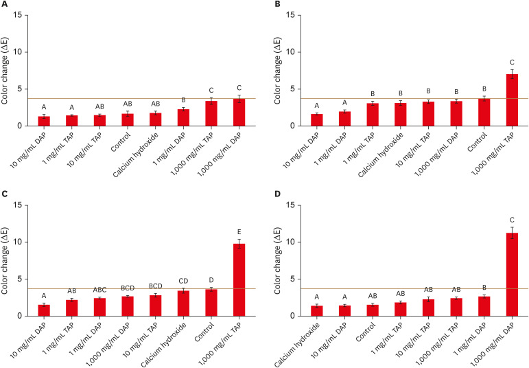
Figure 1A illustrates that the 10 mg/mL DAP (p = 0.0218) or TAP (p < 0.0001), 1 mg/mL TAP (p = 0.0441), no treatment (p < 0.0001) and Ca(OH)2 (p = 0.0002) groups had significantly lower color changes than the other groups after 1 day. After 1 week, the 10 mg/mL DAP (p = 0.0014) and 1 mg/mL DAP (p = 0.0136) groups had significantly lower color changes than other groups, while the 1,000 mg/mL TAP group had significantly higher color change than all groups (p < 0.0001) (Figure 1B). The 4-week data illustrate that the 10 mg/mL DAP (p = 0.0393), 1 mg/mL DAP (p = 0.0347) and TAP (p = 0.0265) groups had significantly lower color changes than the other groups, while the 1,000 mg/mL TAP group had a significantly higher color change than all groups (p < 0.0001) (Figure 1C). Following thermal cycling, the Ca(OH)2 (p = 0.0236), 1,000 mg/mL DAP (p < 0.0001), 10 mg/mL DAP (p = 0.0284) or TAP (p < 0.0001), 1 mg/mL TAP (p < 0.0001) and no treatment groups (p < 0.0001) had significantly lower color changes than other groups, while the 1,000 mg/mL TAP group had a significantly higher color change than all groups (p < 0.0001) (Figure 1D).
Figure 1
Mean ± standard error of color changes in different treatment groups with adhesive after intracanal medicament application for (A) 1 day, (B) 1 week, (C) 4 weeks, and (D) after thermal cycling. Treatments with the same superscript letter were not significantly different at p < 0.05. The red horizontal line represents the perceptible threshold (ΔE = 3.7).
Color change within non-adhesive groups
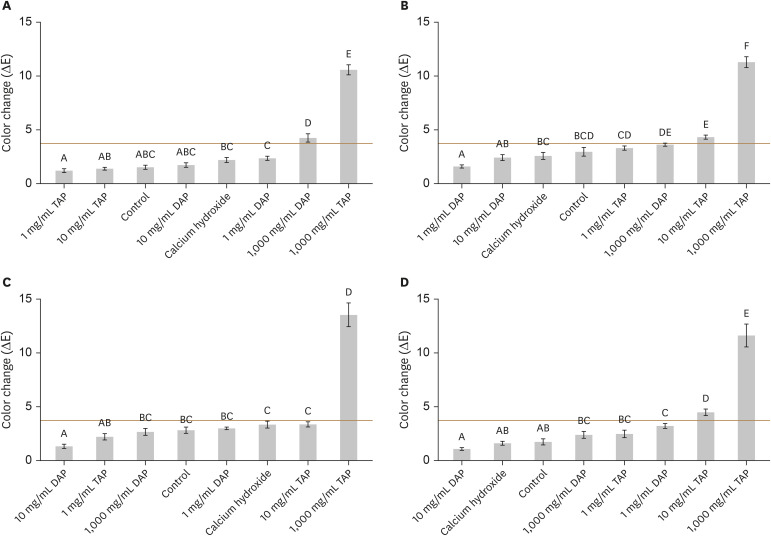
As shown in Figure 2A, the 1 mg/mL TAP (p = 0.0181), 10 mg/mL TAP (p = 0.0272) or DAP (p < 0.0001) and no treatment groups (p < 0.0001) had significantly lower color changes than the other groups after 1 day. After 1 week, the 1 mg/mL DAP (p = 0.0267) and 10 mg/mL (p = 0.0482) DAP groups had significantly lower color changes than the other groups (Figure 2B). The 4-week data demonstrate that the 10 mg/mL DAP (p = 0.0185) and 1 mg/mL TAP (p = 0.0405) groups had significantly lower color changes than the other groups (Figure 2C). Following thermal cycling, the 10 mg/mL DAP (p = 0.0194), Ca(OH)2 (p = 0.004), and no treatment (p = 0.0089) groups had significantly lower color changes than the other groups (Figure 2D). At all time points, the 1,000 mg/mL TAP group had significantly higher color changes than all groups (p < 0.0001) (Figure 2).
Figure 2
Mean ± standard error of color changes in different treatment groups without adhesive after intracanal medicament application for (A) 1 day, (B) 1 week, (C) 4 weeks, and (D) after thermal cycling. Treatments with the same superscript letter were not significantly different at p < 0.05. The red horizontal line represents the perceptible threshold (ΔE = 3.7).
The effect of adhesive on color change
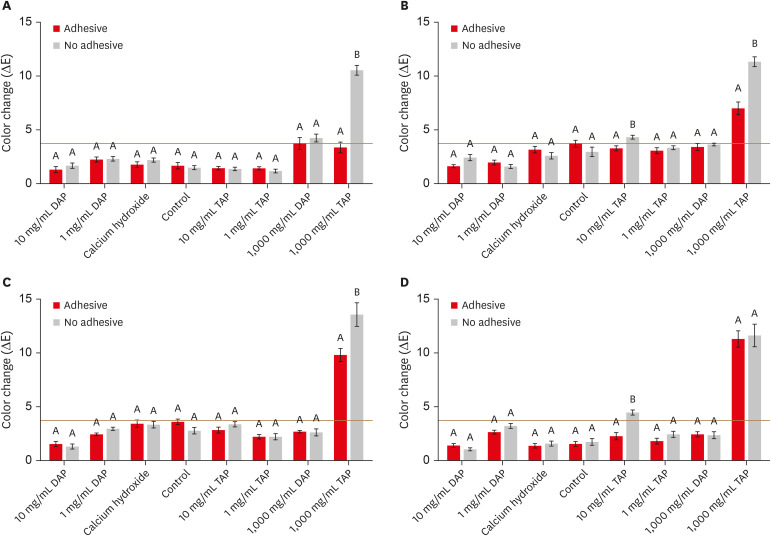
Figure 3A-3C demonstrate that the 1,000 mg/mL TAP group with adhesive had significantly lower color changes than the 1,000 mg/mL TAP group with no adhesive after 1 day, 1 week, and 4 weeks (all p < 0.0001). Furthermore, the 10 mg/mL TAP group with adhesive had significantly lower color changes than 10 mg/mL TAP with no adhesive after 1 week (p = 0.0206) and after thermal cycling (p = 0.0001) (Figure 3B and 3D). However, all other treatment groups had no significant differences in color change according to whether the adhesive was used or not (p > 0.05) (Figure 3).
Figure 3
Mean ± standard error of color changes in the adhesive and no adhesive treatment groups after intracanal medicament application for (A) 1 day, (B) 1 week, (C) 4 weeks, and (D) after thermal cycling. The adhesive and no adhesive treatment groups with the same superscript letter were not significantly different at p < 0.05. The red horizontal line represents the perceptible threshold (ΔE = 3.7).
Perceivable color change
The following treatment groups demonstrated significant increases (p < 0.05) and clinically perceivable color changes (ΔE > 3.7): 10 mg/mL TAP with no adhesive after 1 week (Figure 2B) and after thermal cycling (Figure 2D), 1,000 mg/mL TAP with adhesive after 1 and 4 weeks as well as after thermal cycling (Figure 1B-1D), and 1,000 mg/mL TAP with no adhesive after all time points (Figure 2). Finally, the statistical analyses demonstrated no significant interactions between the tested variables (treatment, adhesive, and time) in 2-way and 3-way ANOVA.
DISCUSSION
Many factors can cause crown discoloration during or after ER. These include the types and concentrations of the intracanal medicaments, the type of intracanal cemental barrier, and the presence or absence of blood inside the root canal system [1112182223]. This study evaluated the effects of low and moderate concentrations of TAP and DAP (1 mg/mL and 10 mg/mL) loaded into an aqueous methylcellulose system on crown discoloration with or without using an adhesive bonding agent. The use of a methylcellulose-based antibiotic system can facilitate the delivery and distribution of intracanal medicaments. Furthermore, it may modify the physiochemical interactions between the active antibiotic ingredients and the internal wall of dentine [28].
Our current study showed that both high and low concentrations of DAP and TAP demonstrated a significant increase in color change at the majority of time points. However, only 1,000 mg/mL TAP demonstrated consistent and significant discoloration that was clinically perceivable (ΔE > 3.7) regardless of adhesive use, which generally agrees with previous studies [132234]. Therefore, the null hypothesis was rejected because the moderate and low concentrations of hydrogel-based DAP and TAP medicaments did not cause clinically perceivable tooth discoloration at all measured time points regardless of the use of an adhesive bonding agent. Instead, the low and moderate concentrations of TAP generally showed concentration dependent discoloration that was not clinically perceivable, except for 10 mg/mL TAP without adhesive following 1 week and thermal cycling. Previous studies found clinically perceivable discoloration following the use of 0.1 or 1 mg/mL TAP [222435]. The lower discoloration observed in our study for 1 mg/mL TAP in comparison to previous studies could be attributed to the methylcellulose hydrogel system, which may minimize the diffusion and/or bonding of minocycline to internal dentine walls.
The vast majority of DAP groups at 1 and 10 mg/mL did not demonstrate discoloration that was clinically perceivable, in general agreement with recent studies [2336]. However, the higher concentration of DAP caused a significant and/or clinically perceivable discoloration at some time points for both the adhesive and non-adhesive groups. Previous studies have also found that higher concentrations of DAP can lead to discoloration that is clinically perceivable [1837]. However, the current study as well as previous studies agreed that DAP can cause significantly less discoloration than TAP [1436]. In contrast, Ca(OH)2 caused no clinically perceivable discoloration regardless of the use of adhesive. Previous studies have also suggested minimal or no tooth discoloration when Ca(OH)2 was used [1735]. It is also worth mentioning that the use of a bonding agent before Ca(OH)2 or any tested concentration of DAP did not significantly decrease tooth discoloration at any of the tested time points.
The reasons for using the 1-step self-etch adhesive bonding agent (G-aenial) are the simplicity of the application technique, the high bond strength, and the good sealing abilities to dentine [3839]. These abilities arise from the presence of 4-methacryloxyethyl trimellitic acid (4-META), which has a hydrophilic (carboxylic) group that is suitable for dentine bonding [39]. In addition, there are possible chemical interactions between hydroxyapatite and 4-META [40]. The additional chemical components of the G-aenial bonding agent include urethane dimethacrylates, triethylene glycol dimethacrylates, phosphoric acid ester monomer, silicon dioxide, photo-initiator, acetone, and distilled water [383940]. One of the interesting findings of this study was that the use of bonding agent significantly decreased tooth discoloration in the 1,000 mg/mL TAP group up to the 4-week time point. However, the use of adhesive did not significantly improve discoloration after thermal cycling when 1,000 mg/mL TAP was used, although the use of adhesive significantly improved discoloration after thermal cycling when 10 mg/mL TAP was used. This can be explained by the ability of thermal cycling to accelerate the chemical degradation of the bonded interface, which led to diffusion of TAP into dentine. When lower concentrations of TAP are suspended within a methylcellulose hydrogel system, the ability of active ingredients to diffuse after thermal cycling could be minimized. A previous study examined the effect of DAP on tooth discoloration after a 6-month aging period [23]. Nevertheless, none of the previous studies used a thermal cycling aging system, which is more clinically relevant. The repetitive fluctuation of temperatures during thermal cycling can accelerate the hydrolysis and breakdown of an adhesive agent. Thermal cycling can also facilitate the contraction/expansion of the adhesive agent, which eventually leads to cracks within the adhesive agent and percolation of the residual intracanal medicaments. It is worth noting that a recent study explored the ability of 2 single-step restorative materials to minimize discoloration after minocycline-based intracanal medicament [16]. The study proposed that both materials were able to minimize tooth discoloration after 4 weeks, which generally agrees with our study. Unfortunately, the study did not examine the ability of the restorative materials to minimize discoloration after long-term aging or thermal cycling.
One of the limitations of this study is that the buccal surface thickness of the crowns could not be standardized. The thickness of an object might have an effect on the diffusion and reflection of light, which may affect the color readings. In order to minimize the effect of different thicknesses in the present study, a close-fit custom-made cap was used for each sample before each color reading. In our study, we chose not to access the canal through the traditional access cavity to avoid any interference with the color measurements. Additionally, we wanted to standardize the application process of the adhesive bonding agent. If we chose not to apply the adhesive on the roof of the pulp chamber, there would have been a higher chance of application variability between samples. It is also important to note that the clinically perceivable color change point (ΔE = 3.7) is not an absolute but rather an average number [33]. Therefore, there might be a possibility of clinically detectable discoloration at lower or higher values than ΔE = 3.7.
CONCLUSIONS
Within the limitations of this study, it can be concluded that both Ca(OH)2, as well as 1 or 10 mg/mL of DAP loaded into a methylcellulose system, did not cause clinically perceivable tooth discoloration. The use of a bonding agent before Ca(OH)2 or any tested concentration of DAP did not significantly decrease tooth discoloration at any of the tested time points. However, the use of adhesive agent was able to significantly decrease tooth discoloration in the 1,000 mg/mL TAP group up to 4 weeks.
 XML Download