

 PDF
PDF Citation
 Print
Print


I. Introduction
Surgical removal of a third molar (M3) is one of the most common procedures in daily dental practice. This procedure accounts for 95% of teeth removed in patients between 19 and 21 years of age in the United States1. Surgeons advocate the surgery for various reasons such as prophylaxis, orthodontics, and treatment of pathology associated with impacted teeth2. Some researchers have advised early treatment to avoid postoperative complications.
The advisability and timing of surgical removal of asymptomatic M3s is uncertain among dental practitioners3. Early removal almost always involves developing tooth germs, so the term ‘germectomy’ was introduced. The role of the oral surgeon is to select the appropriate time to remove the M3 germ, affording fewer treatment sequelae and good outcomes3.
II. Definitions of Germectomy
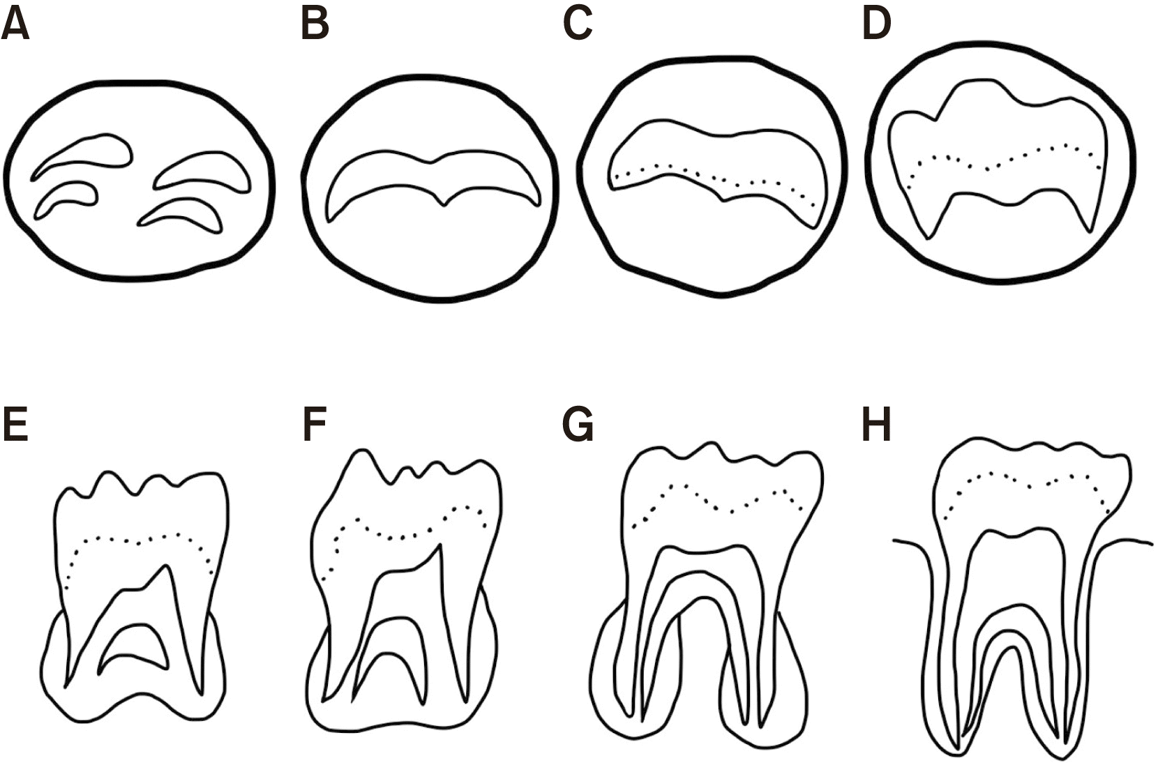
Third molar crypt formation begins at 3 to 4 years of age. Calcification starts at 7 to 10 years of age and is complete by 12 to 16 years of age. Eruption of the M3 begins between 17 and 21 years of age4. Studies have defined germectomy in various terms5-13.(Table 1) One study described germectomy as the removal of M3s in patients between 9 and 16 years of age5. In contrast, the degree of root formation was defined in several other studies6-14. Some authors defined germectomy as surgical removal of M3s in patients with formation of only the tooth crown14. Several others have described the surgery as removal of the developing bud prior to anchoring of the roots in the jaw11. A white paper by the American Association of Oral and Maxillofacial Surgeons regarding M3 data described germectomy as the removal of a tooth that has one-third (or less) root development with a radiographically discernible periodontal ligament7. This definition was adopted by Sivolella et al.8 in 2011. Most authors defined the tooth in the procedure as having one-third or less of its root6,10. Recently, a systematic review advocated Demirjian’s classification of types B, C, and D for the definition of germectomy13,15,16.(Fig. 1)
In summary, two distinct criteria are used to determine germectomy—the chronological age of patients and the anatomical appearance and stage of root formation. These definitions can be used interchangeably as differences in treatment are not clinically significant.
III. Advantages of Germectomy
Opinions differ regarding the indications for germectomy. Some surgeons favor early surgical removal for various reasons. First, early surgical removal would obviate any related risks such as dental caries, pulpitis, pericoronitis, or cystic lesions6,9,17,18. Second, early removal could reduce postoperative complications compared to delayed surgical removal. Less frequent alveolar osteitis and nerve injury are observed in patients with removed M3 for both the inferior alveolar nerve and lingual nerve6,19 as the alveolar bone of teenagers is more flexible. Several authors have stated the simplicity of the procedure and reported a decreased operation time compared to conventional surgical removal at a later age9. Moreover, younger patients tend to have fewer systemic conditions than older patients19. Accordingly, the morbidity associated with early germectomy seems to be less serious than with delaying removal to an older age. Third, orthodontic advantages have been discussed including prevention of crowding from M3 eruption and creation of space without the need for premolar extraction6,17.
Anterior crowding represents a longstanding controversy regarding the presence of M3s. In a recent systematic review and in other literature, the issue is debated13,20,21. Mandibular second molar (M2) impaction is another reported indication. One study determined impacted M3s to be associated with impacted M2s, while another study found that M3 germectomy did not benefit the M2 uprighting process. Meanwhile, the authors of the systematic review supported the idea of M3 germectomy in cases of impacted M2 since delaying M3 removal would only put the M2 at greater risk of dental caries or periodontitis and/or complicate M2 eruption13. Furthermore, germectomy of M3s was justified to gain space in cases requiring molar distalization. For these reasons, some orthodontists consider early surgical removal of M3 to reduce unstable dental arch alignment.
A prior systematic review evaluated the indications for M3 removal in syndromic or incompliant patients. They suggested early removal in cases of morphologic abnormalities, such as Ekman-Westborg-Julin syndrome associated with macrodontic multituberculate impacted M3, self-mutilating behavior, and/or recurrent epilepsy that can be triggered by pain from impacted M322.
IV. Disadvantages of Germectomy
In contrast to mandibular M3, germectomy of maxillary M3 is more difficult and time-consuming because of the higher tooth bud position17. Several authors have reported surgical removal of maxillary M3 in young patients with displacement of the tooth bud into the infratemporal fossa23-26. One author stated that M3 removal presents operative challenges, and germectomy should be limited to mandibular tooth buds27. Most maxillary distalization caused superior and posterior movements of unerupted M3. The results suggest that distalization in adolescents with unerupted M3s may be achieved without early germectomy28. A systematic review revealed that the effect of the maxillary M2 and M3 eruption stage on maxillary molar distalization was nominal29, creating more risk than benefit for germectomy of maxillary M3.
Some authors did not support the idea of germectomy, while others argued that early extraction was beneficial, even though it did not have to be as early as germectomy. There were no significant differences in complications among patients 12-18 years of age30. Formation of two-thirds of the tooth roots formed is a preferred time for germectomy to avoid morbidities associated with eruption of M3 and an easier procedure because some portion of the tooth would have emerged out of the bony crypt. Several studies found that postoperative complications increase only in patients older than 23-24 years5,19.
Another reason why surgeons do not strongly support germectomy is that the orthodontic advantages are not definitive. The relationship between anterior crowding and M3s has not been established. A randomized, controlled trial in 1998 found that removal of M3 cannot be justified to reduce or prevent late incisor crowding31. Cassetta and Altieri32 found that germectomy of M3 did not simplify uprighting of impacted M2, calling into question the orthodontic purposes for early removal of M3. Furthermore, early germectomy of developing tooth buds could deprive M3s of the potential as future tooth reservoirs for autotransplantation, especially in syndromic patients prone to multiple tooth loss17,22.
With so many contradicting opinions, some authors have proposed a middle ground of appropriate age to determine the fate of M3s. Chiapasco et al.5 proposed waiting until 17-24 years of age, while Haddad10 offered a narrower range of 18-20 years of age. Taken together, the optimal age range for M3 evaluation without increasing the accompanying risks of M3 eruption and postoperative complications was between 18-20 years of age5,10.
V. Considerations of Germectomy Time
The best chronological age for germectomy has yet to be determined. Seven to ten years of age has proven to be a difficult time for surgery due to poor patient compliance and reluctant parental consent. Moreover, the prognosis of M2 is difficult to evaluate in this age range because of the lack of certain criteria for space discrepancy evaluations33.
The surgical removal of M3s when M2s have yet to erupt can be difficult. Some clinicians do not recommend this surgery for patients between 11-12 years of age because of their uncooperativeness during surgery. Moreover, flap designs and suturing can be complicated33.
Patients between 13 and 14 years of age appear to be the best candidates for germectomy, as the M2 is fully erupted, patient compliance is satisfactory, and the dental volume is sufficient to allow minimally invasive surgery33.
VI. Surgical Techniques
Details of the germectomy procedure regarding anesthetic technique, preoperative preparation, flap designs, ostectomy, and odontectomy have been described in several reports.
1. Preoperative imaging
Bisconte et al.33 suggested using cone-beam computed tomography (CBCT) to evaluate tooth germ root formation and dental volume, while other reports recommended against it. Panoramic films seem adequate for assessing the tooth and its relationship to the inferior alveolar canal before surgery. CBCT could be beneficial in complex cases or when panoramic films do not provide good visualization of all vital structures.
2. Preoperative preparation
The use of antibiotic prophylaxis was studied in 2009. Two grams of amoxicillin taken one hour before germectomy was observed to yield better surgical results than no antibiotic prophylaxis. Postoperative pain and analgesic use were reduced in the antibiotic prophylaxis group. In addition, delayed wound infections, which occurred commonly at 2-3 weeks postoperatively, also decreased. Fever and swelling did not differ between the two groups12.
3. Anesthetic technique
There was a concern regarding the psychological effects of germectomy on young patients, even though later studies observed acceptable psychological status of both the parents and patients17. For this reason, surgical removal of M3 under sedation or general anesthesia (GA) must be considered for some patients. A pilot study assessed patient preference for anesthetic techniques used in germectomy. Most patients preferred local anesthesia (LA) to GA and LA with sedation. The study also found that bilateral removal of M3s under LA was well tolerated by patients17. Finne and Klämfeldt6 utilized diazepam oral sedation together with LA to perform germectomy and observed no sedative agent complications. Later studies almost always performed germectomy under LA, except for one study using GA because of the young age of the patients.
4. Flap designs
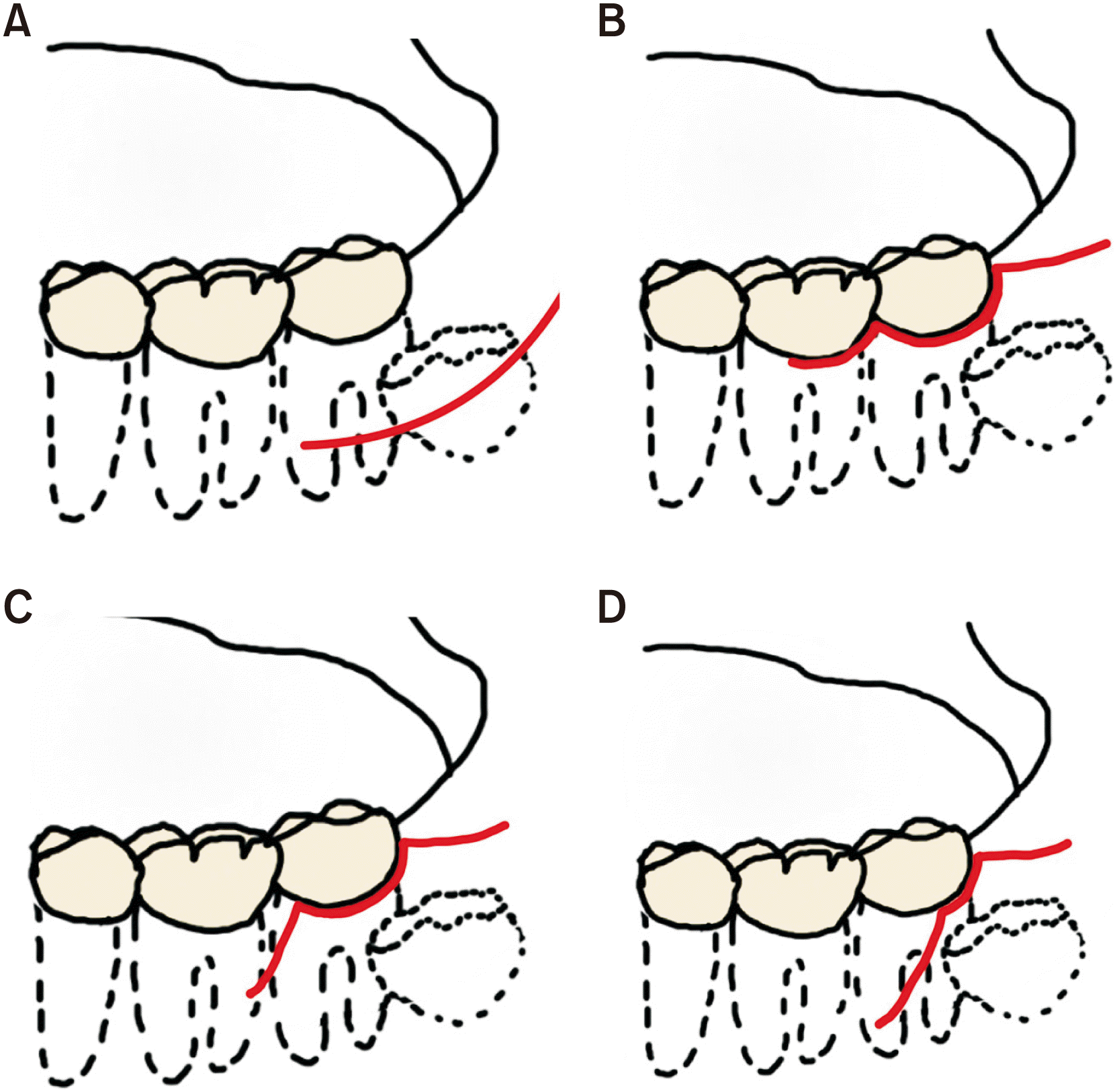
Various flap designs have been mentioned in the literature. The advantages and disadvantages of each design were discussed.(Fig. 2)
A vestibular incision or lateral trephination technique was described. An approximately 25 mm incision was recommended at the mucobuccal fold from the anterior border of the ascending ramus to the mesial root of the first mandibular molar (M1). Lateral trephination with vestibular incisions resulted in a lower infection rate, fewer periodontal pockets distobuccal to the M2, and less frequent unattached gingiva. Bleeding, swelling, and ecchymosis were not different6; possibly resulting from the flap design not involving the marginal gingiva of the M2 and subsequent minimal periodontal damage. Infection was also decreased, possibly because bacterial migration from the gingival sulcus of the M2 into the surgical wound was avoided.
An envelope flap was also frequently mentioned. A sulcular incision extended along the crestal bone at the M3 area. Monaco et al.12 conducted a randomized controlled trial comparing a triangular flap with an envelope flap in germectomy. Surgery time, intraoperative pain, bleeding, swelling, fever, and infection were not significantly different between the groups. Probing depths were larger with the envelope flap at 1 week after surgery12. This may be the result of involvement of a larger amount of marginal gingiva. However, periodontal healing was complete in both groups after 3-6 months.
A triangular incision helps increase visualization, and the position of the involved vertical incision can vary. Healing of the M2 periodontium was studied utilizing a triangular flap with different positions of the vertical incision. Vertical incisions distobuccal and mesiobuccal to the M2 were compared. No differences were observed, and complete recovery of the periodontium was evidenced within 90 days in all cases in both types of vertical incisions18.
To summarize, different flap designs yielded various effects on germectomy. Lateral trepanation could reduce infection, pocket depth, and unattached gingiva, while lingual nerve protection did not significantly limit lingual nerve damage11. The triangular flap did not produce a significant difference compared to the envelope flap, and the position of the vertical incision did not affect the healing of the M2 periodontium.
5. Ostectomy
Typically, a cortical window is created distobuccal to the M234. A series of pits could be created using a round bur to delimit the bone covering the crown when evaluating the resistance between the bone and enamel or follicular space. The covering bone can then be removed using a bone chisel or bur to expose the buccal aspect of the crown to its greatest contour. Care should be taken to maintain the bone posterior to the M2 and to avoid exposure of its distal root.
A split-mouth randomized-controlled trial was conducted to compare ostectomy results by piezoelectric and rotatory methods. The piezoelectric method significantly increased surgery time in the osteotomy step. However, no difference was observed regarding postoperative complications. Patients experienced irritating buzzing in the ipsilateral ear while undergoing piezoelectric surgery, and half of the patients indicated greater comfort with piezoelectric surgery. Meanwhile, patients who preferred the rotatory method cited the shorter surgery duration8.
6. Odontectomy
Tooth sectioning was performed to minimize the excessive removal of bone. Tooth buds could be separated into 2-4 segments by making a second perpendicular section if required12,14,34. If no signs of root formation are observed, the tooth germ should be turned upside down before splitting, and the crown should be secured by a fine elevator to prevent rotation. Tooth sectioning should be incompletely performed to avoid damaging the lingual wall.
Special consideration regarding germectomy has been discussed in the literature. Younger patients tend to exhibit limited mouth opening and have a small tolerance for surgery lasting longer than 30 minutes. Tooth germs are further from the bony cortex if the patients are younger than 11-12 years, hindering access to the tooth germs. Last, a relatively long time passed before communication with the patients and parents to mitigate any psychological reactions17.
7. Wound closure
Most studies about wound closure in M3 surgical removal demonstrated less pain and swelling in secondary closure wounds35,36. All the studies reviewed included both partially erupted and unerupted M3s. Germectomy differed from routine M3 surgical removal in the position of the impacted tooth, cooperation of younger patients, and concerns of the parents. Secondary closure may not be the answer for all germectomy cases. Additional studies should be performed on wound closures in germectomy. For example, insertion of collagen sponges was reported37, but a comparison of its clinical techniques has yet to be completed.
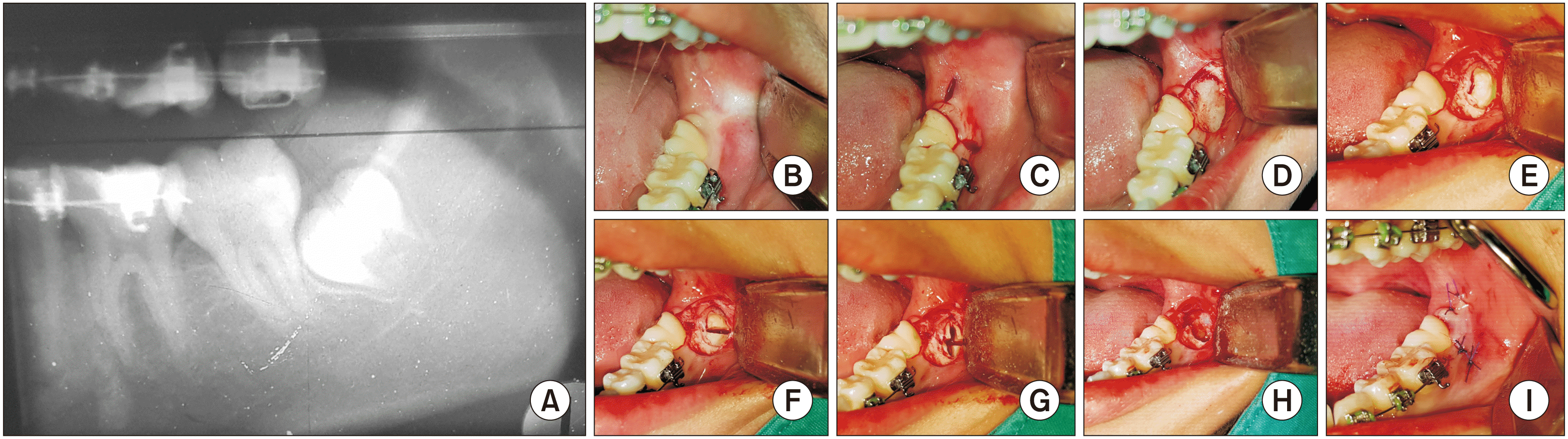
An example of a germectomy case is presented in Fig. 3.
VII. Complications
Oral surgeries are often accompanied by postoperative complications, and germectomy is no exception. General non-specific postoperative complications such as pain, swelling, and trismus were recorded in the literature6,17. Most complications were associated with decreased available space at the ramus posterior to M3 or with Nolla’s third molar calcification less than stage 7 or 1/3 completed root form38.
Nerve paresthesia was one of the main focuses in the literature on germectomy. During the early stages of tooth development, roots are shorter and often further from the inferior alveolar canal. Consequently, nerve paresthesia was believed to be less of an issue in germectomy compared to delayed surgical removal and was not observed in all cases of germectomy5. The lingual nerve was also studied, and no evidence of paresthesia was found after germectomy8,11,30.
Dry socket or alveolar osteitis is another complication frequently reported after removal of M3. In adult patients, the incidence of dry socket is around 4.6%39. Although Bjørnland et al.17 observed dry socket in 1.8% of cases, while Chiapasco et al.5 reported no dry socket in the germectomy group compared to 0.5%-2.1% in other groups.
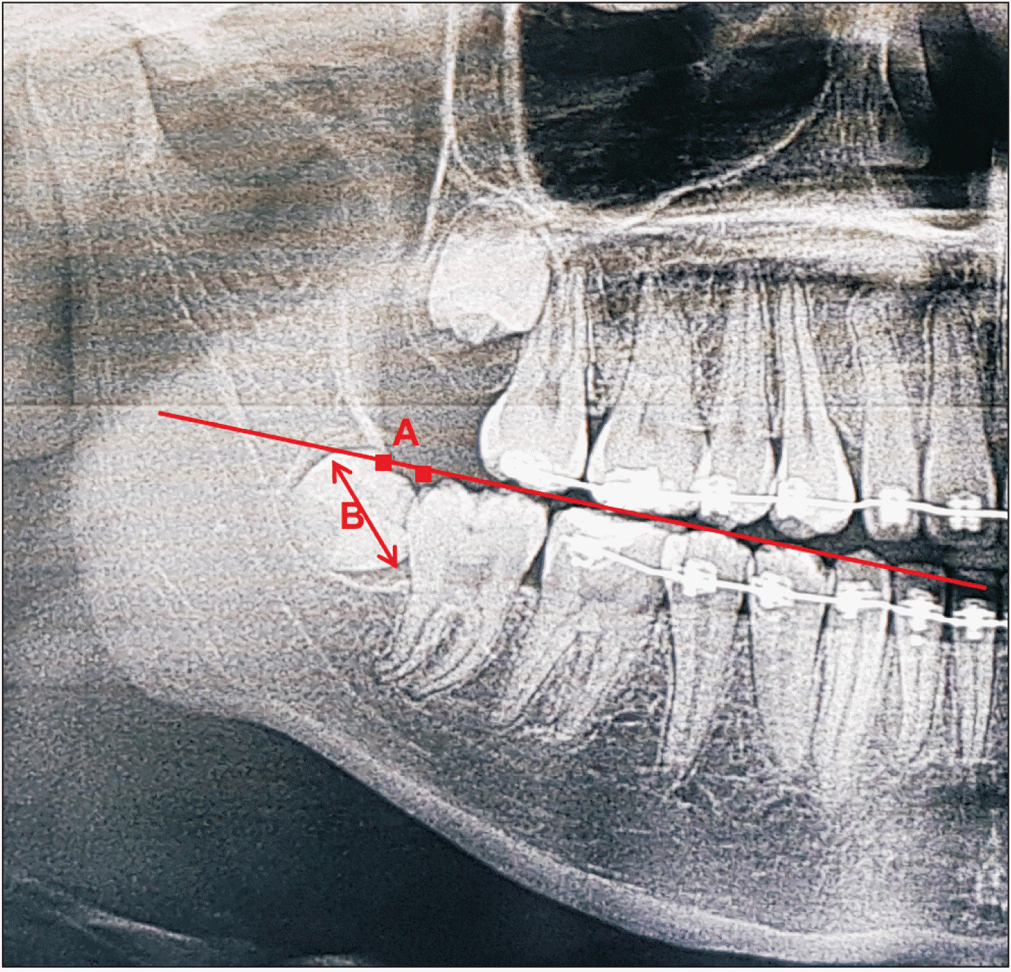
Delayed-onset infection (DOI) was defined as swelling and/or purulent drainage from the alveolus 15 days to 2 months postoperatively34. Another study defined DOI as an inflammatory swelling of the operated area any time after the removal of sutures at 1 week postoperatively40. Antibiotic prophylaxis of 2 g of amoxicillin 1 hour prior to germectomy can reduce DOI significantly14. A lower Ganss ratio was associated with increasing DOI. The Ganss ratio is the comparison between the distal surface of the M2 to the anterior border of the ramus and mesiodistal width of the M334,41.(Fig. 4) Other risks for infection include full soft tissue coverage, complete bony impaction, Pell and Gregory’s class III, deeper position, younger age, and lower Nolla stage40.
VIII. Conclusion
Germectomy is a procedure to remove M3 germs that have either formed only a crown or the crown and no more than one-third of the root length. Surgeons have various opinions on whether germectomy should be performed. It is important to understand the benefits and drawbacks of each treatment plan before developing one for each M3. Communication with patients and their parents about the plan and its outcome must be prioritized. Many surgeons have proposed distinct surgical techniques. It is essential to fully evaluate each aspect of the treatment plan and surgical techniques to be used. Finally, early M3 removal can reduce and eliminate some of the most unpleasant complications of late M3 removal. However, other complications could occur more frequently and patients must be well informed.
 XML Download