

 PDF
PDF Citation
 Print
Print


INTRODUCTION
High-resolution bowel ultrasound (US) has emerged as an important imaging technique in the diagnosis and follow-up of patients with Crohn’s disease (CD) [1-3]. It is a noninvasive, non-ionizing imaging modality which is well tolerated and accepted by patients, and whose accuracy for detecting intramural and extramural extension of the disease is excellent [4,5].
The normal bowel wall has a layered appearance, often referred to as mural echo-stratification. Stratification appears as 5 concentric alternating hyperechoic and hypoechoic layers, where the hyperechoic central layer corresponds to the submucosa and the hypoechoic external layer to the muscularis propria [6,7]. These layers become more prominent in patients with CD, finding which has traditionally been attributed to transmural inflammation and edema [7].
Intramural fat deposits can be found in longstanding diseases. When intramural fat deposition is abundant, it can be macroscopically visible as a yellow submucosal rim on gross specimen. Computed tomography (CT) can demonstrate this submucosal fat accumulation in the bowel wall, called the fat halo sign (FHS), which has been reported to denote a chronic phase of inflammatory disease [8]. In a previously published series, the FHS was seen in the small bowel in 17% of 100 patients with CD [9]. It is important not to misinterpret this layer of submucosal fat as indicative of acute inflammation and submucosal edema [10].
Fat within tissue is echogenic on the US. It is thus not surprising to find thick echogenic submucosa on US images of segments that contain fat [11]. On US, the difference between submucosal thickening due to fat or edema cannot be made.
Wall thickness is an important parameter in evaluating inflammatory activity (all US or magnetic resonance imaging [MRI] scores include wall thickness). On the other hand, it has been shown that the degree of thickening of the intestinal wall correlates with the severity of CD assessed by endoscopy and/or pathology [12,13]. In many US scores severity increases along with wall thickness. US scores, similar to MRI activity scores, are used in many hospitals to assess activity in a given patient and monitor response to treatment. Therefore, should thickening of the wall be mainly due to the presence of fat in the submucosal layer, the activity of the disease could actually be less severe than what is suggested by wall thickness, which might mislead clinical decision-making regarding the treatment plan.
For over more than 15 years performing US in patients with CD, we have seen many cases with submucosal fat on CT that had thick submucosa on US. To the authors’ knowledge, the relationship between submucosal thickness measured on US and the presence of fat in the wall in patients with CD has never been studied.
The objective is to investigate whether there are US features that allow suspecting the presence of submucosal fat deposition in the intestinal wall of patients with CD.
METHODS
1. Study Design
This study is a retrospective cross-sectional analysis of a series of consecutive abdominal CTs performed to patients with CD over a period of 10 years (2011–2020). Between January 1, 2011, and December 31, 2020, we searched the Pathology Picture Archiving and Communication System (Pathology PACS; PathSpeed, GE Medical Systems Integrated Imaging Solutions) database to identify consecutive CD patients who underwent CT. The inclusion criteria were as follows: (1) patients with established CD; (2) with abdominal CT performed during the study period; (3) patients who had undergone US examination 3 months before or after CT. The exclusion criteria were as follows: (1) postoperative CT looking for complications after surgery; (2) exclusive CT colonic involvement.
The indications for CT were suspected obstruction, abscess formation or perforation. In our hospital, routine monitoring of CD patients is performed with US or MRI. CT images were retrospectively analyzed on an image archiving and communication system (PACS) by 2 abdominal radiologists (J.V. and G.M.). Disagreements were classified by consensus. The Institutional Review Board of Doctor Peset University Hospital approved this retrospective study (IRB No. 20.22) and waived the requirement to obtain informed consent.
2. Patients
During the study period, there were 94 patients with a diagnosis of CD and a CT scan. Of these 94 patients, 22 were excluded because CT was performed looking for postsurgical complications and 4 because of exclusive colonic CT involvement. For patients with multiple studies (n = 13), the last CT study was used to analyze. The remaining 68 patients who met the selection criteria comprised the study cohort that underwent further analysis. The group comprised 44 men and 24 women with a mean age of 57 years (range, 18–89 years).
3. Data Collection
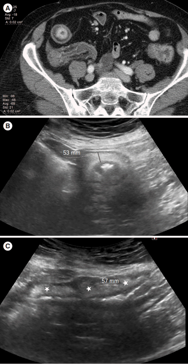
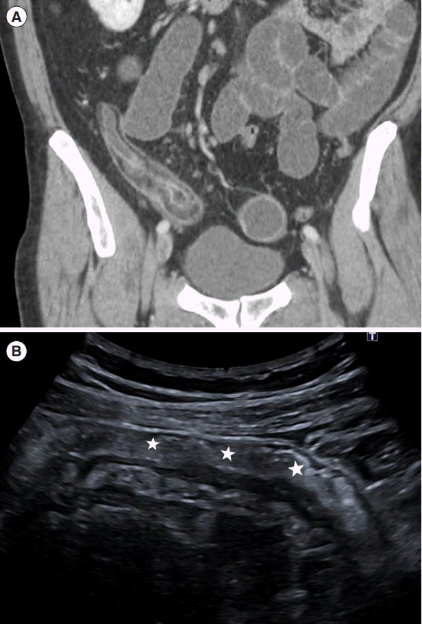
CT studies were retrospectively reviewed by 2 experienced radiologists for the presence of submucosal fat in the bowel wall, defined as a low density < 10 Hounsfield units (HU) intramural line, seen in the entire circumference of the bowel wall in the axial projection (Fig. 1). The density of the fat was measured on magnified views of the bowel segment with a round cursor maximally adapted to the low density. A measurement of less than 10 HU was regarded as positive for the presence of the FHS. The severity of the mural enhancement was reviewed in consensus by the radiologists as mild or severe as can be seen in Fig. 1A, based on subjective assessment.
Off-site, 2 readers, unaware of the diagnosis of FHS on CT, but knowing the segment evaluated in CT, reviewed the US examinations on PACS workstations. The location of the segment (terminal or nonterminal ileum) was verified on the US examination reports. Wall and submucosal thickness were measured on longitudinal sections of the same segment of CT; the average of 3 measures was used for analysis.
Measurements were made on images where a continuous and similar thickness of the submucosa was seen (Fig. 2). We also reviewed the reports of the US examinations via the hospital radiology information looking for signs of inflammatory activity such as vascularization of the wall measured by color Doppler [3], ulcers of the wall or transmural complications (fistulae or abscess) in the evaluated segment.
CT studies were performed on 64-MDCT scanner (Light-Speed VCT; GE HealthCare, Milwaukee, WI, USA) or 16-MDCT scanner (SOMATOM Emotion Siemens, Erlangen, Germany), acquiring slices 1.5 mm thick and with an index of 8 mm, following the administration of 120 mL of intravenous contrast. The sonographic examinations were performed by using several scanners during the period included (Aplio 80, 300 or 500; Toshiba, Tokyo, Japan), employing a 3.5 MHz convex, a 6 to 9 MHz, high-frequency convex probe or a linear 5 to 10 MHz.
4. Statistical Analysis
Basic descriptive statistics were used, which included the mean and standard deviation for continuous variables and the absolute frequency and percentages for qualitative variables. For univariate analysis, the Student t-test was applied for comparing continuous variables and the Fisher exact test was applied for contrasting categorical variables. A two-tailed P value of less than 0.05 was considered to indicate a significant difference.
A receiver operating characteristic curve was constructed to determine the best cutoff US submucosal wall thickness value for predicting submucosal fat in the bowel wall determined on CT. The best cutoff value was determined, while balancing the best sensitivity with the lowest false-positive rate. Statistics were calculated with the SPSS package version 20 (IBM Corp., Armonk, NY, USA).
RESULTS
Demographic and clinical characteristics of the 68 patients with CD included in the study are collected in Table 1. The FHS was present in 22 of the 68 patients (31%) on CT. The locations of the FHS were 18 terminal ileum (81.8%) and 4 nonterminal ileum (18.2%). The FHS was present in 6 out of 20 patients (30%) in the group where disease duration was less than 1 year and in 16 of 48 patients (33.3%) in the group with a longer disease duration (P<0.789).
There were significant differences between submucosal thickness of patients with FHS and patients without FHS (4.19 mm vs. 2.41 mm, P<0.001). Submucosal thickness showed a significant correlation with the presence of fat in the bowel wall on CT (Fisher exact test, P<0.001).
From the receiver operating characteristic curve, a threshold value of 3.1 mm of submucosal thickness had the best sensitivity and specificity to suspect FHS (95.5% and 89.1%, respectively), predictive positive value of 80.8% and negative predictive value of 97.6% (area under the curve, 0.962 ± 0.019; 95% confidence interval, 0.93–1.00), with an odds ratio of 172 (95% confidence interval, 18.80–1,570). All 15 patients (65.2% of patients with FHS) with a submucosal thickness > 3.9 mm had FHS on CT.
Of the 22 patients in whom FHS was detected all but one (n = 21) showed mucosal enhancement on CT as sign of inflammatory activity, 14 intense and 6 mild enhancement. Sixteen of the 22 patients (73%) with submucosal fat showed signs of inflammatory activity on US, color Doppler grade 2 in 13 patients (59%), 1 sinus tract and 2 with mural ulcers. Two of these patients had an enteroenteral fistula in an adjacent segment with mural loss of the echo structure. Seven of the 22 patients (32%) showed dilatation of the loops proximal to the segment with fat in the submucosa as a sign of stenosis.
During follow-up, 7 patients underwent several US examinations (4.42 years, range 3–6 years) with no changes in thickness (4.22 mm vs. 4.16 mm, P<0.111) or color Doppler grade, 3 patients underwent surgery (2 fibrostenosing subtypes and 1 inflammatory). The other 12 patients did not have image controls because the CT in which the submucosal fat was detected had been performed in the last 2 years (2019–2020).
DISCUSSION
A diagnosis of fat within a lesion or in the submucosa of a bowel segment in CD cannot be made with US; however, based on our results, marked thickening of the submucosa on US may suggest fat deposit on the wall and therefore recommend its confirmation with other techniques. In this study, submucosal thickness greater than 3.1 mm showed a sensitivity of 95.5% and a specificity of 89.1% for suspecting FHS and moreover, all patients with a submucosal thickness greater than 3.9 mm had FHS on CT.
In patients with gastrointestinal symptoms, an initial US to detect intestinal pathology and a colonoscopy to diagnose CD are usually performed. After the diagnosis of CD, a dedicated magnetic resonance enterography is performed to complete staging. If intestinal lesions are accessible to US, this technique has been proposed in many hospitals as a surrogate for colonoscopy or MRI in evaluating response to therapy in CD [14]. In these hospitals, MRI is used only for complex cases or when the lesions are not adequately imaged by US (e.g., high body mass index or pelvic location).
Knowledge that a submucosal fat deposit has occurred is important because most US scores, and MRI scores, include wall thickness to evaluate disease activity or severity in CD. Wall thickness is considered as the strongest predictor of disease activity followed by hyperemia on color Doppler US, as shown by several prior studies [4,15,16]. Multiple research have shown excellent correlation of wall thickness on US with activity at colonoscopy [17]. The score by Sævik et al. [18], the only US index validated to date, introduces a simple US activity score for CD that includes intestinal wall thickness and color Doppler grades. Two other scores have been published, both also use wall thickness and color Doppler grade to determine inflammatory activity and severity [19,20]. An increase in thickness is associated in all US scores with an increase in the severity of the disease.
When US is used to monitor the response to therapy, it is important to be aware of the existence of submucosal fat accumulations in CD, because in these cases the increase in bowel wall thickness can also reflect chronic deposition of fat and it could not really indicate the true degree of inflammatory activity or severity. US score could misclassify these segments as inflamed lesions, proof patients into a risk of receiving incorrect treatment.
Therefore, if submucosal thickness increases during follow-up, it is very likely that it is due to the deposition of fat and in these cases, a low-dose CT scan or better an MRI should be performed to confirm mural fat deposition. Fat saturated and non-fat saturated T2 sequences are required to discern between edema or intramural fat deposition, edema demonstrates persistent high signal intensity with both sequences, whereas fat saturation will reduce wall signal intensity that is due to fat infiltration [10].
On the other hand, US detection of bowel wall thickness > 7 mm has been shown to be an independent risk of surgery [21]. It is very likely that this may not be fulfilled in cases where a large part of the wall thickness is due to fat deposition.
In the published series, during treatment monitoring, the thickness of the intestinal wall has shown a very significant decrease 3 months after treatment onset [22,23]. In our series, only a small percentage of patients had US follow-up, with no change in thickness, therefore, it can only be hypothesized that less improvement in bowel wall thickness can be expected within segments with FHS, since fat deposition as a finding of chronic inflammation remains unchanged. To our knowledge, no previous work on sectional imaging techniques has assessed the relationship between the presence of fat in the wall and the decrease of wall thickness during treatment monitoring.
Therefore, in cases of marked submucosal thickening, we should combine this parameter with vascularization, more strictly related to neoangiogenesis and disease activity, allowing us to distinguish the increase in bowel wall thickening due to fat deposition from changes due to acute inflammatory activity. The differentiation between active and inactive disease is then based on the detection of increased vascularity within the intestinal wall. This is done initially with color Doppler, where inactivity will show reduced or no blood flow [24]. In our series, color Doppler grade 2 as a sign of inflammatory activity was detected in 59% of segments with fat deposition of the submucosal layer, where mucosal enhancement on CT was seen in 96% of cases. In segments with FHS there were no cases with severe involvement, ulcers, or fistulas.
It has been published that in some cases color Doppler US is not reliable due to the patient’s body habits or technical factors. In these cases, if there is no color Doppler signal within a thickened intestinal loop with very thick submucosa, the question arises between an inactive disease or a technical failure [24]. In this circumstance, the use of contrast-enhanced US can show transmural enhancement in cases of active disease without a color Doppler signal due to technical failure [24]. This fact was confirmed in Ripollés et al. [25], where the use of contrast detected inflammatory activity in the thickened bowel wall of 46 patients without hyperemia on color Doppler
In our series, the percentage of patients with FHS on CT was greater than previously reported (31% vs. 17%). The advent of improved technology, including a capacity for thinner slices, may account for this fact. Anyway, during the review of the CT studies we realized that the presence of focal fat in the wall was observed in many more cases.
Regarding the time of appearance of FHS, in our study, there was no statistically significant difference in the prevalence of FHS among patients with disease less than 1 year and those with disease duration beyond 1 year. Submucosal fat deposition has been reported to denote a chronic phase of inflammatory disease and its prevalence must be significantly dependent on duration [9]. However, there is an increasing body of literature to suggest that CD diagnosis is often established following considerable diagnostic delay, which is a long period from first symptom onset to diagnosis [26]. Moreover, it has also been reported that FHS can appear as early as 12 days or 6 months in cases who had received corticosteroid therapy [27]. This possible association between corticosteroid therapy and FHS has been previously noted [8].
There are some limitations to our study that should be considered when interpreting our results. The main drawback is that our investigation is a retrospective study, including selection and interpretation bias. It is important to note that since we only included patients who underwent emergency CT, there is a selection bias, therefore, the sample of this study may not reflect the real proportion of patients with CD and fatty wall deposition. Another limitation is that the records of corticosteroid therapy were not obtained; it is possible that a significant number of patients would have received corticosteroid therapy previously for long periods, which explained the higher incidence of fat deposition in this series. Also, we have not studied the diagnostic delay defined as the period of time (in months) from the first symptoms to the diagnosis of CD. On the other hand, we have only been able to assess changes in wall and submucosal thickness in a few patients during treatment follow-up. Lastly, the cutoff points obtained in our study have not been subjected to external validation and, furthermore, they are based on a small sample size, so they should be confirmed in studies with a larger number of patients.
In conclusion, US cannot diagnose fat in the submucosa, but our results suggest that the presence of FHS on the wall can be suspected in cases with marked thickening of the submucosal layer and consequently recommend its confirmation with other techniques. If US submucosal thickness is > 3.1 mm, the activity of the disease must be measured by other parameters such as the color Doppler degree. The detection of submucosal fat on the wall could help avoid incorrect decision-making regarding treatment plan.
 XML Download