Abstract
Phlebosclerotic colitis is a rare form of intestinal ischemia. It is caused by calcified peripheral mesenteric veins and a thickened colonic wall. These characteristic findings can be identified on CT and colonoscopy. A 37-year-old female with a history of long-term herbal medicine use presented with acute lower abdominal pain and vomiting of sudden onset. Colonoscopic findings showed dark-blue discolored edematous mucosa and multiple ulcers from the ascending colon to the sigmoid colon. Abdominal CT findings showed diffuse thickening of the colonic wall and calcifications of the peripheral mesenteric veins from the ascending colon to the sigmoid colon. Based on these findings, the patient was diagnosed with phlebosclerotic colitis. We report this rare case of phlebosclerotic colitis in a healthy young female patient with a history of long-term herbal medicine use and include a review of the relevant literature.
Ischemic colitis, characterized by reduced blood flow to the colon, is a disease that commonly affects the elderly. The main causes of ischemic colitis include atherosclerosis, diabetes, hemorrhagic shock, malignant tumors, pancreatitis, and intestinal obstruction.1-3 Phlebosclerotic colitis is a rare type of ischemic colitis caused by colic venous congestion with calcification of the mesenteric vein.1,3,4 The exact etiopathogenesis remains to be clarified, but it is presumed that a flow defect of the colonic vein due to calcification of the mesenteric vein causes ischemic colitis.1,4 Mesenteric venous calcification is a secondary phenomenon caused by a long-term increase in intraperitoneal pressure as a result of chronic diseases, such as diabetes mellitus, heart failure, renal dysfunction, and hepatocirrhosis.4-6 Other causes include chemical stimulation, drugs, and increased pressure in the large intestinal lumen, but the etiology is still unclear.7-9
The incidence of phlebosclerotic colitis has been reported to be high in East Asian countries, specifically China and Taiwan10,11 and some studies have suggested an association with long-term use of herbal medicines.11,12
In South Korea, cases of phlebosclerotic colitis in the young population without an underlying chronic disease are rare, with only a few reports on the association between phlebosclerotic colitis and long-term herbal medicine use.
We report a case of phlebosclerotic colitis in a healthy young female patient with a history of long-term use of herbal medicine and include a review of the relevant literature.
A 37-year-old female patient who presented with acute abdominal pain and vomiting of sudden onset was admitted to the hospital on the same day. The patient’s history revealed that she had intermittent diarrhea for four years with no underlying disease or history of surgery. She did not smoke and had been drinking two bottles of beer per day for around two days a month, for the past 17 years. The patient had been taking a herbal medicine supplement for weight loss for the past decade. The supplement was composed of Ephedra intermedia, gypsum, Rehmanniae radix, Scutellaria baicalensis, Rheum palmatum, Longan arillus, and Gardenia jasminoides.
At the time of admission, the patient’s vital signs were as follows: blood pressure,133/86 mmHg; pulse rate, 96 beats/min; respiratory rate, 20 beats/min; and body temperature, 36.8℃.
On physical examination, the patient showed acute symptoms with no abdominal swelling; the abdomen was flat and soft. While the patient complained of mild pain across the entire abdomen on pressing, there was no rebound tenderness, palpable mass, or organomegaly, and her bowel sounds were normal. A peripheral blood test indicated a white blood cell count of 17,170/mm3 (87.5% neutrophils), hemoglobin of 12.8 g/dL, hematocrit of 40%, and platelet count of 352,000/mm3. Serum biochemical profile included urea nitrogen 6.0 mg/dL, creatinine 0.75 mg/dL, total protein 7.9 g/dL, albumin 4.9 g/dL, AST 20 IU/L, ALT 11 IU/L, total bilirubin 0.5 mg/dL, amylase 110 IU/L, fasting blood glucose 120 mg/dL, LDH 289 IU/L, and CRP 0.74 mg/dL. The PT was 10.6 s (120.5%). The autoantibody test was positive (1:40), and the antineutrophilic cytoplasmic antibody was negative.
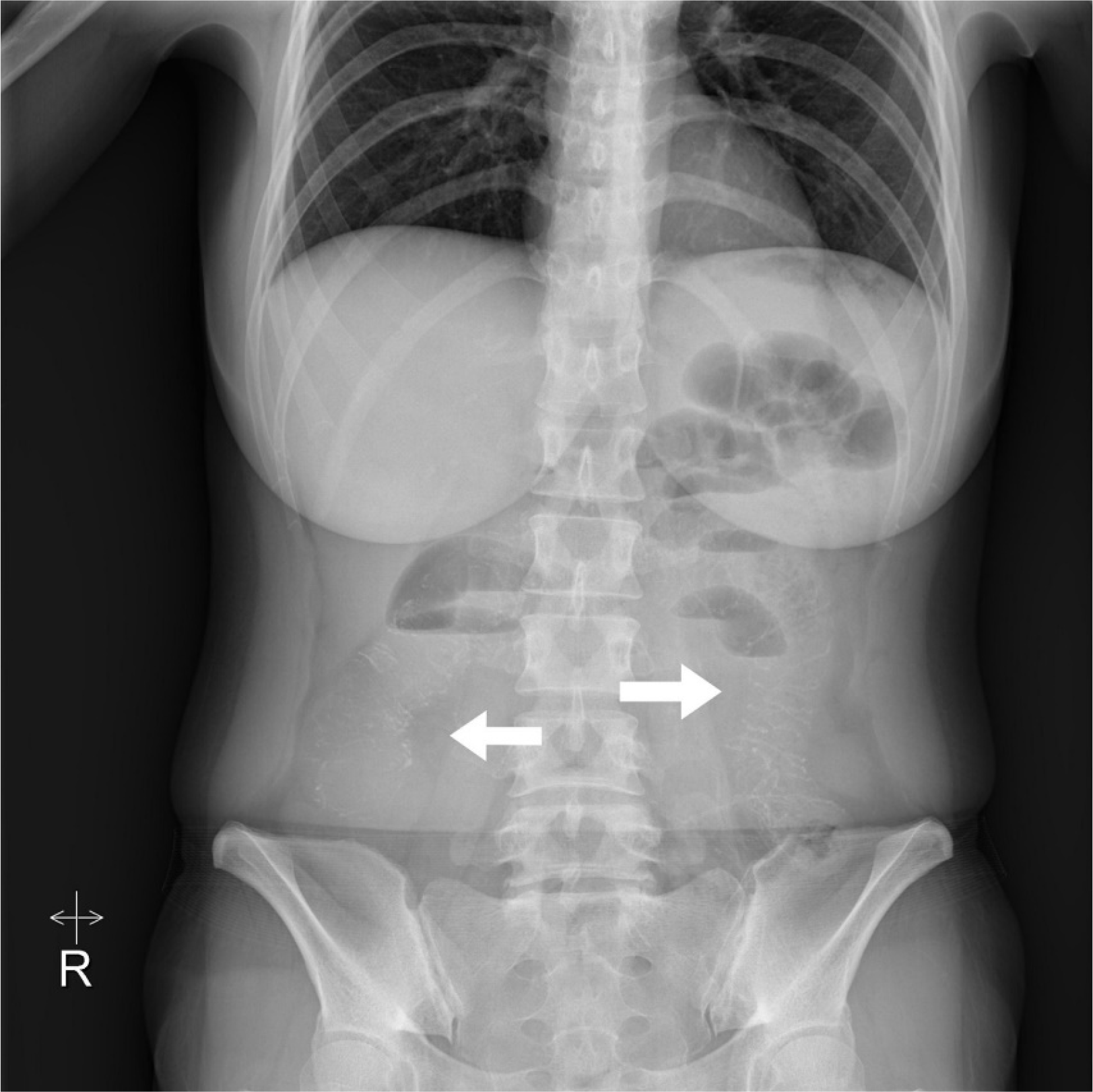
Plain abdominal radiographic findings revealed dilatation of the small intestine and multiple linear calcifications along the intestinal wall in the right lower quadrant, left lower quadrant, and epigastric area (Fig. 1). Abdominal CT findings included diffuse thickening of the colonic wall from the ascending colon to the sigmoid colon and calcifications of the peripheral vessels with consequent dilatation of the jejunal and ileal loops. However, no intestinal volvulus or ischemia was observed (Fig. 2). Colonoscopy indicated dark-blue discolored edematous mucosa and multiple ulcers from the ascending colon to the sigmoid colon (Fig. 3). Congestion and inflammatory cells were observed on the endoscopic biopsy specimen, but no submucosal fibrosis or vascular wall thickening was detected.
Based on the colonoscopic and unique radiological findings, the patient was diagnosed with phlebosclerotic colitis, and conservative treatment, including fasting and fluid infusion, was given. The symptoms improved during hospitalization, and the subsequent oral diet led to favorable results, and the patient was discharged. The patient is being monitored at the outpatient clinic, and no notable symptoms related to colitis have since been detected. A follow-up colonoscopy performed six months after hospital discharge showed lesion improvement, with regression of multiple erosions, ulcers, and mucosal friability and the presence of mild dark-blue discolored mucosa (Fig. 4).
We obtained individual written informed consents for the procedure and for publication.
Phlebosclerotic colitis is a rare type of ischemic colitis of non-thrombolytic venous origin, characterized by the calcification and fibrosis of the peripheral mesenteric vein.6 It is known to occur predominantly in elderly patients and/or patients with underlying diseases.3,6 When it occurs in younger patients, there could be misdiagnosis or delayed diagnosis, especially in the absence of any underlying disease, despite the presence of gastrointestinal symptoms, as in the present case.
The seven cases of phlebosclerotic colitis reported in South Korea to date included three men with chronic diseases such as diabetes, hypertension, renal failure, and liver cirrhosis. Among women, there were no underlying diseases in three patients, while a history of herbal medicine use was observed in one middle-aged patient.13 Additionally, there was a report of phlebosclerotic colitis in a young woman, but there was no obvious risk factor. In the present case, since phlebosclerotic colitis occurred in a young woman without any underlying disease with a long-term history of taking herbal medicine, we hypothesized that the herbal medicine was a potential cause of the disease in this patient.
The symptoms of phlebosclerotic colitis generally include sudden abdominal pain, diarrhea, hematochezia, and vomiting. However, in some cases, the condition is detected by chance during a colonoscopy.3,6,14 In severe cases, hematochezia, intestinal obstruction, or perforation symptoms could appear with chronic anemia.11,15 Case reports suggest that the condition could mainly affect the ascending colon or the entire large intestine, while the clinical features vary according to the area and length of the affected intestinal segment.16 Invasion of the descending colon, in particular, is viewed as a sign of poor prognosis.12
In phlebosclerotic colitis, plain abdominal radiographic findings include multiple thread-like calcifications.1,12 Abdominal CT findings include intestinal dilatation, intestinal wall thickening, and calcification of the mesenteric vein.1,6,12 On a lower gastrointestinal series, the loss of semilunar folds in the intestine, a thumbprint-like shape, irregularity of the intestinal lumen, stiffness, and stenosis are observed. These are common, non-specific features of ischemic colitis of venous origin.1,14 Colonoscopically, dark-blue or dark-purple discolored edematous mucosa that mostly affects the ascending colon, luminal stenosis, extensive inflammation, ulcers, and loss of the semilunar folds in the colon can be detected.17 While the histopathological findings vary, phlebosclerotic colitis is characterized by fibrosis and thickening of the intestinal submucosal layer. The characteristic features include venous wall thickening around the submucosal layer, collagen deposition, fibrosis, and calcification, with intestinal wall thickening and submucosal fibrosis.4,11,12 The present case was diagnosed as phlebosclerotic colitis based on the clinical symptoms, clinical history, and unique colonoscopic and radiological findings.
The treatment of phlebosclerotic colitis can be either conservative or surgical, according to its severity, with most patients showing symptomatic improvement after either treatment.3,18 Intermittent abdominal pain may recur after conservative treatment, but surgical treatment is rarely required. In addition, calcification and fibrosis of the peripheral mesenteric vein can be maintained without significant improvement, and inflammation and ulcers, as presented in the current case, show improvement on follow-up endoscopy. However, the mild dark-blue discolored mucosa often remains. Surgical treatment may be necessary for patients who exhibit paralytic ileus, inflammation across the entire intestinal wall, and peritonitis caused by necrosis and perforation.3,19 Depending on the depth of the invasion, the surgical treatment may involve hemicolectomy, subtotal colectomy, or total colectomy, and the postoperative prognosis has been relatively favorable, according to previous reports.20
Phlebosclerotic colitis is known to occur predominantly in elderly patients with chronic diseases,3,6 while it is uncommon in healthy young individuals without underlying diseases. Hence, phlebosclerotic colitis should be part of the differential diagnosis if dark-blue discolored edematous mucosa are detected during colonoscopy. Furthermore, while the etiopathogenesis of phlebosclerotic colitis remains unclear, unique radiologic findings could contribute to a more accurate diagnosis. Therefore, the calcification of the mesenteric vein should be examined. With early diagnosis, the patient’s prognosis is likely to be favorable.
Several studies published in East Asian countries have suggested an association between phlebosclerotic colitis and prolonged use of herbal medicine. However, the exact mechanism of herbal medicine induced ischemic colonic damage is yet to be determined.10,12 This implies the need for continued research on the etiopathogenesis of phlebosclerotic colitis as well as further studies on its association with the long-term use of herbal medicine. To conclude, we report a rare case of phlebosclerotic colitis in a healthy young female with a history of long-term herbal medicine use in South Korea. Reports of phlebosclerotic colitis in healthy young patients who have taken herbal medicine for a long time are emerging. This suggests that the consumption of herbal medicine may be an important cause leading to this rare condition, in addition to other factors such as an underlying chronic disease or old age, which should be considered in clinical practice.
REFERENCES
1. Yao T, Iwashita A, Hoashi T, et al. 2000; Phlebosclerotic colitis: value of radiography in diagnosis--report of three cases. Radiology. 214:188–192. DOI: 10.1148/radiology.214.1.r00ja01188. PMID: 10644121.
2. Theodoropoulou A, Koutroubakis IE. 2008; Ischemic colitis: clinical practice in diagnosis and treatment. World J Gastroenterol. 14:7302–7308. DOI: 10.3748/wjg.14.7302. PMID: 19109863. PMCID: PMC2778113.
3. Brandt LJ, Boley SJ. 2000; AGA technical review on intestinal ischemia. American Gastrointestinal Association. Gastroenterology. 118:954–968. DOI: 10.1016/S0016-5085(00)70183-1. PMID: 10784596.
4. Choi JM, Lee KN, Kim HS, et al. 2014; Idiopathic phlebosclerotic colitis: a rare entity of chronic ischemic colitis. Korean J Gastroenterol. 63:183–186. DOI: 10.4166/kjg.2014.63.3.183. PMID: 24651592.
5. Kang HY, Noh R, Kim SM, Shin HD, Yun SY, Song IH. 2009; Phlebosclerotic colitis in a cirrhotic patient with portal hypertension: the first case in Korea. J Korean Med Sci. 24:1195–1199. DOI: 10.3346/jkms.2009.24.6.1195. PMID: 19949682. PMCID: PMC2775874.
6. Song JH, Kim JI, Jung JH, et al. 2012; [A case of phlebosclerotic colitis in a hemodialysis patient]. Korean J Gastroenterol. 59:40–43. Korean. DOI: 10.4166/kjg.2012.59.1.40. PMID: 22289953.
7. Chang KM. 2007; New histologic findings in idiopathic mesenteric phlebosclerosis: clues to its pathogenesis and etiology--probably ingested toxic agent-related. J Chin Med Assoc. 70:227–235. DOI: 10.1016/S1726-4901(09)70364-8. PMID: 17591581.
8. Yeh HJ, Lin PY, Kao WY, Kun CH, Chang CC. 2018; Idiopathic mesenteric phlebosclerosis associated with long-term use of Chinese herbal medicine. Turk J Gastroenterol. 29:140–142. DOI: 10.5152/tjg.2018.17072. PMID: 29391325. PMCID: PMC6322607.
9. Hiramatsu K, Sakata H, Horita Y, et al. 2012; Mesenteric phlebosclerosis associated with long-term oral intake of geniposide, an ingredient of herbal medicine. Aliment Pharmacol Ther. 36:575–586. DOI: 10.1111/j.1365-2036.2012.05221.x. PMID: 22817400.
10. Guo F, Zhou YF, Zhang F, Yuan F, Yuan YZ, Yao WY. 2014; Idiopathic mesenteric phlebosclerosis associated with long-term use of medical liquor: two case reports and literature review. World J Gastroenterol. 20:5561–5566. DOI: 10.3748/wjg.v20.i18.5561. PMID: 24833888. PMCID: PMC4017073.
11. Iwashita A, Yao T, Schlemper RJ, et al. 2003; Mesenteric phlebosclerosis: a new disease entity causing ischemic colitis. Dis Colon Rectum. 46:209–220. DOI: 10.1007/s10350-004-6526-0.
12. Minh ND, Hung ND, Huyen PT, et al. 2022; Phlebosclerotic colitis with long-term herbal medicine use. Radiol Case Rep. 17:1696–1701. DOI: 10.1016/j.radcr.2022.02.069. PMID: 35342497. PMCID: PMC8942791.
13. Park JK, Sung YH, Cho SY, Oh CY, An SH. 2015; Phlebosclerotic colitis in a healthy young woman. Clin Endosc. 48:447–451. DOI: 10.5946/ce.2015.48.5.447. PMID: 26473132. PMCID: PMC4604287.
14. Oshitani N, Matsumura Y, Kono M, et al. 2002; Asymptomatic chronic intestinal ischemia caused by idiopathic phlebosclerosis of mesenteric vein. Dig Dis Sci. 47:2711–2714. DOI: 10.1023/A:1021090113274. PMID: 12498290.
15. Kato T, Miyazaki K, Nakamura T, Tan KY, Chiba T, Konishi F. 2010; Perforated phlebosclerotic colitis--description of a case and review of this condition. Colorectal Dis. 12:149–151. DOI: 10.1111/j.1463-1318.2008.01726.x. PMID: 19175648.
16. Chen MT, Yu SL, Yang TH. 2010; A case of phlebosclerotic colitis with involvement of the entire colon. Chang Gung Med J. 33:581–585.
17. Hu P, Deng L. 2013; Phlebosclerotic colitis: three cases and literature review. Abdom Imaging. 38:1220–1224. DOI: 10.1007/s00261-013-0001-0. PMID: 23589075.
18. Yu CJ, Wang HH, Chou JW, et al. 2009; Phlebosclerotic colitis with nonsurgical treatment. Int J Colorectal Dis. 24:1241–1242. DOI: 10.1007/s00384-009-0707-1. PMID: 19390857.
19. Yoshikawa K, Okahisa T, Kaji M, et al. 2009; Idiopathic phlebosclerosis: an atypical presentation of ischemic colitis treated by laparoscopic colectomy. Surgery. 145:682–684. DOI: 10.1016/j.surg.2008.04.015. PMID: 19486773.
20. Fang YL, Hsu HC, Chou YH, Wu CC, Chou YY. 2014; Phlebosclerotic colitis: A case report and review of the literature. Exp Ther Med. 7:583–586. DOI: 10.3892/etm.2014.1492. PMID: 24520249. PMCID: PMC3919902.
Fig. 1
Plain abdominal radiographic finding showing multiple linear calcifications (arrows) in the right lower quadrant, left lower quadrant, and epigastric area.

Fig. 2
Abdominal CT findings (A, B) showing diffuse thickening of the colonic wall from the ascending colon to the sigmoid colon and calcifications of the peripheral mesenteric veins.


 PDF
PDF Citation
 Print
Print


 XML Download