Journal List > Korean J Pain > v.36(3) > 1516083099

Artuç, Uçkun, Sivas, Yurdakul, and Bodur: Comparison of the effects of transcutaneous electrical nerve stimulation and interferential current therapies in central sensitization in patients with knee osteoarthritis

Abstract

Background

This study is primarily aimed to determine whether transcutaneous electrical nerve stimulation (TENS) and interferential current (IFC) treatments have any effect on central sensitization (CS) in patients with knee osteoarthritis (OA) and to investigate which treatment is more effective.

Methods

In this randomized controlled trial, 80 patients were randomized into four treatment groups TENS, Plasebo-TENS, IFC, and Plasebo-IFC. All interventions were applied 5 times a week for 2 weeks. Primary outcome was pressure pain threshold (PPT), which is accepted as the objective indicator of CS, at the painful knee and at the shoulder as a painless distant point. Other outcome measures were the visual analog scale (VAS), Western Ontario and McMaster Universities Osteoarthritis Index, Timed Up and Go Test, pain catastrophizing scale, Beck Depression Inventory, and Tampa Scale of Kinesiophobia.

Results

All assessment parameters were improved, without a significant difference among the groups except PPT. PPT scores were significantly improved in TENS and IFC groups when compared with the sham groups at 2 weeks and 3 months. In addition, this improvement was even more pronounced in the TENS group. Multivariable logistic regression analysis showed that the patient’s inclusion in the TENS group, an initial high PPT, and an initial low VAS score were independent risk factors for improvement in the PPT.

Conclusions

This study shows that TENS and IFC reduced pain sensitivity as compared to placebo groups in patients with knee OA. This effect was more pronounced in the TENS group.

INTRODUCTION

The knee is the joint most affected and damaged by osteoarthritis (OA) [1]. Pain is clinically the most restrictive and prominent symptom in knee OA [2]. Although OA pain was previously evaluated as pure nociceptive pain, many studies have emphasized the importance of central sensitization (CS), which is defined as “increased response of nociceptive neurons in the central nervous system to subthreshold or normal afferent inputs” as well as peripheral mechanisms in the pathogenesis of OA-associated pain [3,4].
Although the awareness of CS is growing in patients with musculoskeletal pain, currently there are no standard clinical descriptors to classify dominant CS pain [59]. Increased pain sensitivity (hyperalgesia) is a well-accepted clinical sign of CS. This sign is applied to clinical setting by measuring the pressure pain threshold (PPT) with a pressure algometer. Primary hyperalgesia arises at the location of the pathology, which is thought to demonstrate changes in the activity of the primary afferent fibers. Secondary hyperalgesia arises outside the location of the pathology, and it is thought to mirror changes in the central nervous system [57,10].
Transcutaneous electrical nerve stimulation (TENS) and interferential current (IFC) are analgesic currents commonly used in rehabilitation clinics [1113]. While TENS is a low-frequency current [14], IFC is a medium-frequency current that enables deeper penetration into tissues [15]. Although studies have shown that TENS and IFC could be effective in the treatment of CS by modulating pain and desensitizing the central nervous system, these effects have not been fully clarified [1618].
Given the need for CS retraining in knee OA pain and the ready accessibility of TENS and IFC for rehabilitation physicians as sensory electrotherapeutic pain modalities, it is necessary to research the effects of TENS and IFC on the CS. Therefore, our primary outcome is to research which treatment and factors were related to reduction of hyperalgesia, especially secondary hyperalgesia. This study also aimed to investigate whether TENS and IFC treatments have any effect on pain, pain catastrophizing, kinesiophobia, functionality, and mobility.

MATERIALS AND METHODS

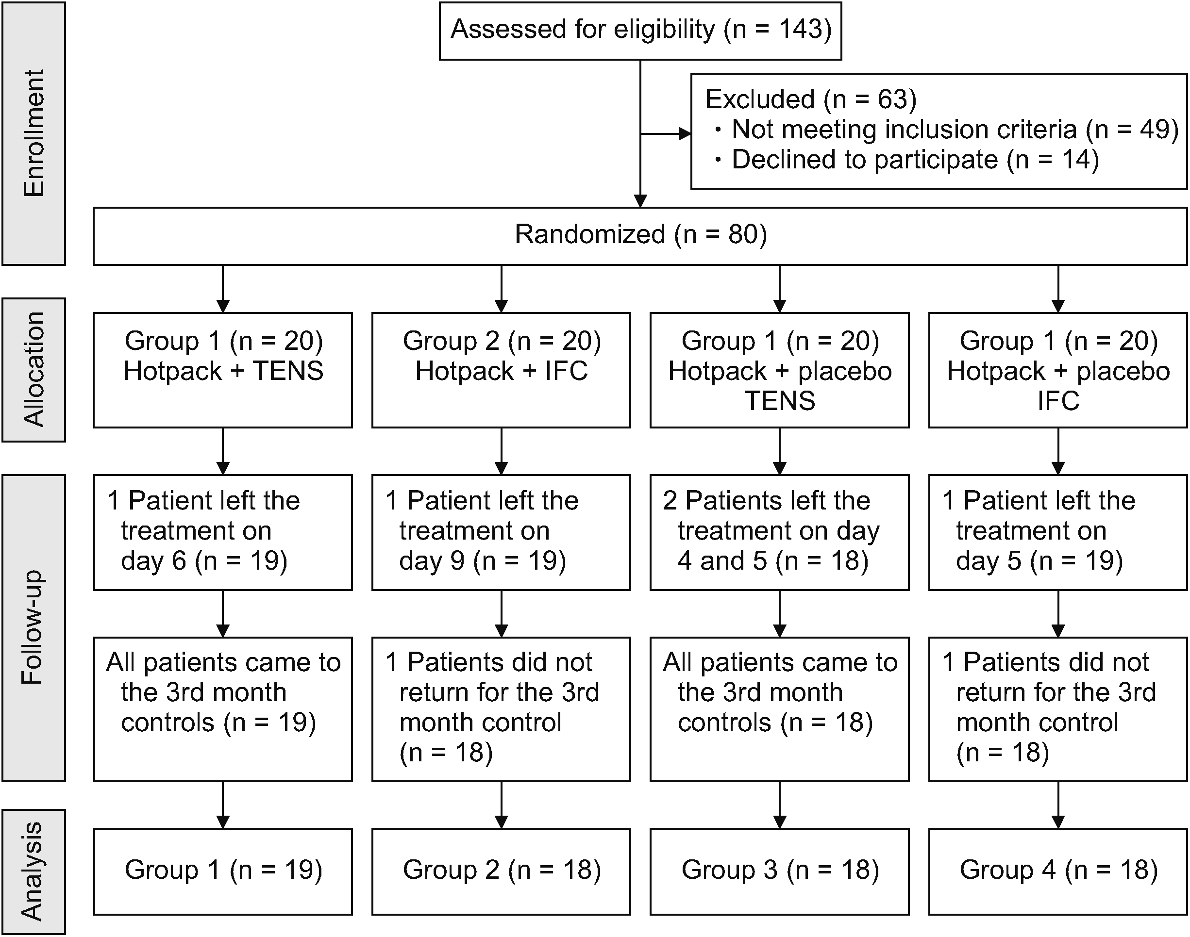
This study was planned as a double-blind, placebo-controlled, randomized, four-arm study. Eligible patients were aged between 40 and 70 years with knee OA according to the American College of Rheumatology (ACR) criteria [19] bilateral stage 2–3 knee OA according to the Kellgren–Lawrence radiological scoring system [20], symptomatic with a score of at least three on the visual analog scale (VAS) in the last 6 months. Patients were excluded if they had received analgesic current previously, had any inflammatory, rheumatological, neurological, lower extremity, vascular, or cognitive diseases, limited knee joint range of motion, a history of knee surgery, history of any contraindication for electrotherapy, or pain in the neck and shoulder area. Patients were also excluded if they had received hyaluronic acid or steroid injections into the knee joint or any conventional or complementary medicine treatment for knee OA within the last 6 months. Ethics committee approval was obtained for the study (Ethics Committee Decision No. E. Kurul-E-18-2044), and the study was registered in the Clinical Trials (No: NCT04153825). Physical examination of the 143 patients who were admitted to Ankara Numune Training and Research Hospital with knee pain between February 1, 2019 and February 1, 2020 and had received posterior–anterior and lateral knee radiography. Of these, 80 patients (160 knees) who agreed to participate in the study and met the inclusion and exclusion criteria were included in the study. The 80 individuals were randomly assigned to the four treatment groups using computer-generated randomization of study numbers. One patient from the TENS and IFC group, two patients from the placebo TENS group, and one patient from the placebo IFC group left the treatment without giving a specific reason, and one patient from the IFC group and placebo IFC group did not return for the third-month follow-up. The CONSORT diagram of the study is shown in Fig. 1.
Firstly, pain severity using a VAS was evaluated separately for both knees in every patient. The knee which showed the highest VAS score was identified as the reference knee. Bilateral therapy was applied to all patients, but the reference knee was used in all evaluations and statistical analyses. Written informed consent was obtained. All participants were asked not to use any analgesics except topical analgesics throughout the study and to inform us in case of any situation that required use of analgesics. The patients were evaluated by a single physician who was blinded to the randomization and had no role in the treatment.

1. Treatment protocol

For the enrolled patients, for 2 weeks, 5 days a week, a program with a total of 10 sessions of physical therapy was applied bilaterally.
To all patients, bilateral hot packs were applied for 20 minutes in a sitting position with their knees extended (Chattanooga Hydrocollator Hotpack, with a surface temperature of approximately 42°C).
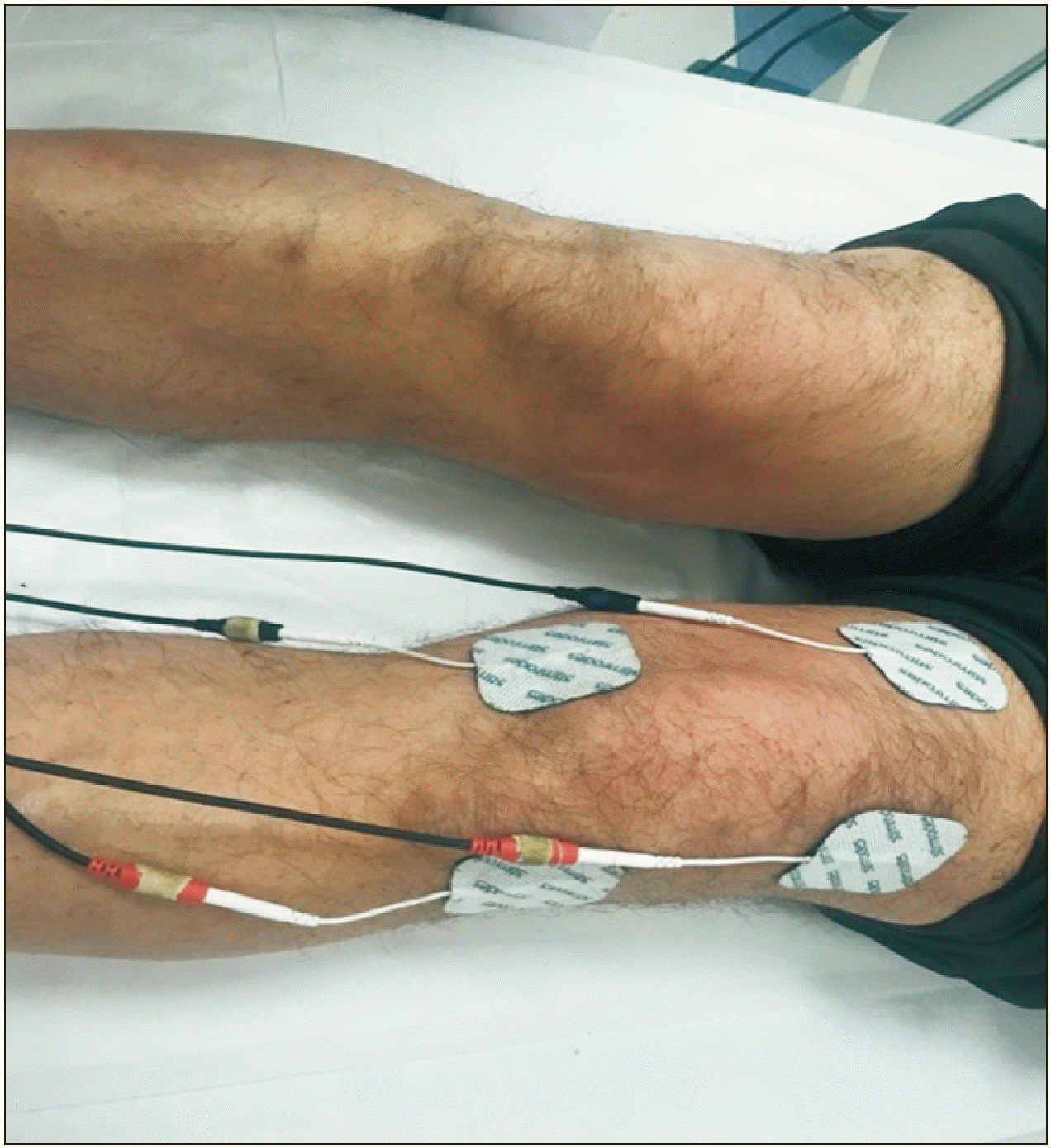
The Intellect Advanced device (Chattanooga Group) with both IFC and TENS features was used in all treatment groups. In the TENS group, electrodes were connected in parallel to the patients. Bilateral conventional TENS, with a frequency of 80 Hz, current transition time of 100 microseconds, and an amplitude density that does not cause contractions or excessive discomfort, but induces a slight tingling sensation, was performed for 20 minutes (Fig. 2) [21].
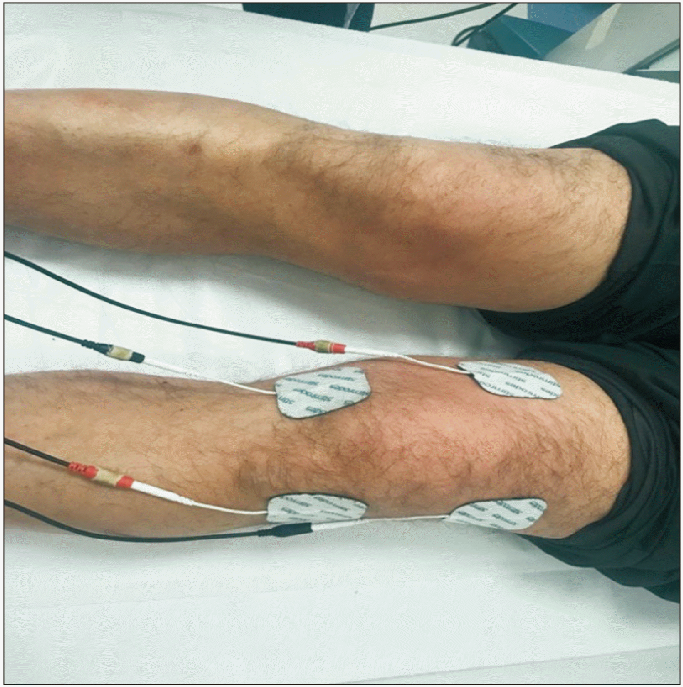
In the IFC group, with a carrier frequency of 4,000 Hz and 100 Hz of amplitude modulated frequencies, four electrodes were placed crosswise, keeping the pain area in the middle, and bilateral IFC was applied for 20 minutes (Fig. 3) [15].
In the placebo groups, electrodes were connected in patients. The device was turned on, but the button that triggered the electric emission was not activated. The lights on the panel of the device were always on, and the electrodes were kept connected for 20 minutes.

2. Evaluation parameters

Demographic characteristics including age, sex, educational status, body mass index, and chronic diseases were recorded. Pain severity was evaluated with a VAS. In this scale, the patient was asked to score the pain level in the range of 0–10 cm (0, no pain; 10, most severe pain) [22]. The Timed Up and Go Test (TUG) was used to evaluate the functional capacity, balance, and fall risk of the patients [23]. The Western Ontario and McMaster Universities Osteoarthritis Index (WOMAC) was used to assess pain and functional status [24]. The degree of the individual’s catastrophic perceptions or thoughts in painful conditions were evaluated using the pain catastrophizing scale (PCS) [25,26]. Depressive symptoms were assessed using the Beck Depression Inventory (BDI) [27]. The Tampa Scale for Kinesiophobia (TSK) was used to assess fear of movement [28,29].
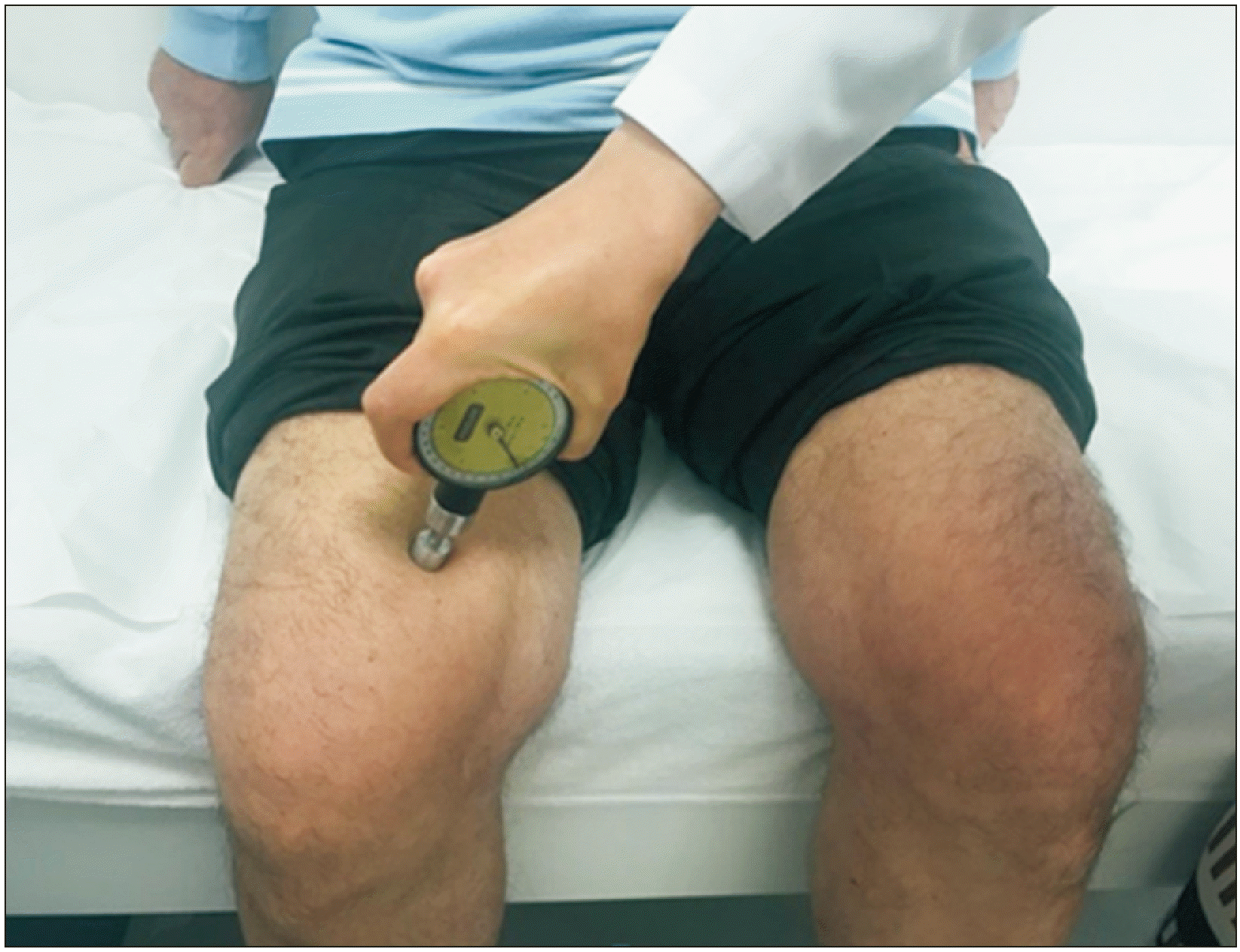
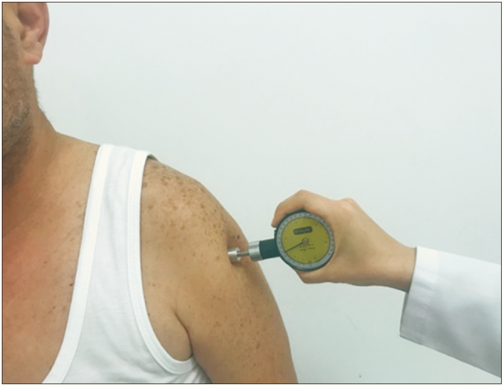
PPT was measured using a hand-held digital algometer (Baseline Dolorimeter). The Baseline Dolorimeter is a device with a 1-cm2 pressure surface and a hand grip, showing the values obtained in kg/cm2. PPTs were evaluated in a quiet environment while the patient was in a sitting position using the m. vastus medialis (3 cm medial to the midpoint of the superior border of the patella) in the reference knee to determine the primary hyperalgesia (Fig. 4). To determine the secondary hyperalgesia, a painless spot away from pathology, the deltoideus muscle (lateral part of the m. deltoideus, 10 cm below the acromion), was used (Fig. 5). The tip of the algometer was placed vertically to the skin on the points to be measured, the pressure was gradually increased until the participant said that only the feeling of pressure turned into a feeling of pressure and pain, and the threshold at the time the pain was noted. From each area, a total of two measurements were taken at intervals of 30 seconds, and the arithmetic mean was recorded [30].

3. Statistical analysis

Study sample size calculation was executed using the G*Power version 3.0.10 software (Heinrich-Heine-Universität Düsseldorf) to ensure a sufficient sample size for one-way analysis of variance (ANOVA): repeated measure between factors test. The total sample size was calculated as 76 for this study under the following conditions: effect size f = 0.4, α (type I error) = 0.05, β (type II error) = 0.10, number of groups = 4, repetitions = 3, and correlation among repeated measures = 0.7.
IBM SPSS Statistics for Windows version 20 (IBM Corp.) was used. Whether the numerical data were normally distributed was measured using the Shapiro–Wilk test. Considering age and sex, the stratified covariant-focused randomization/minimization method was used to balance prognostic factors while assigning patients to treatment groups. General descriptive statistics used included mean, median, standard deviation, and minimum and maximum values for continuous variables, and categorical variables were summarized as number and percentage (%). The chi-square test or Fisher’s exact test was used to examine the distribution of discrete variables among groups. In the four different treatment groups, continuous variables were compared by one-way ANOVA or Kruskal–Wallis analysis. Post hoc tests (Bonferroni) were used to understand which group was different from the other.
Measurement results before and after treatment were evaluated by repeated-measures ANOVA. Following the treatment protocol, multivariate logistic regression analysis was used to determine the initial predictive factors for improvement in the PPT (≥ 15% increase in the third month PPT compared with the initial PPT) [31]. The outcomes were evaluated at a 95% confidence interval, and the significance level was set at P < 0.05.

RESULTS

No statistically significant difference was found between the groups with regards to demographic characteristics or initial scores for all the parameters examined (Table 1). In all groups, significant improvement was found in VAS, WOMAC, PCS, TUG, and TSK scores, when the baseline was compared to 2 weeks and 3 months after treatment (Table 2). However, there was no significant difference in terms of degree of improvement in these above parameters when comparing the groups with each other (Table 3).
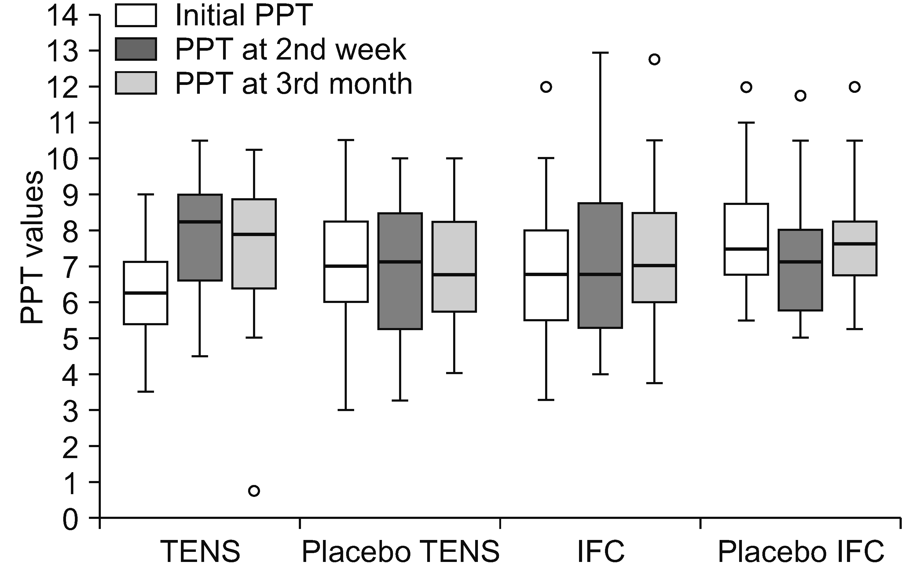
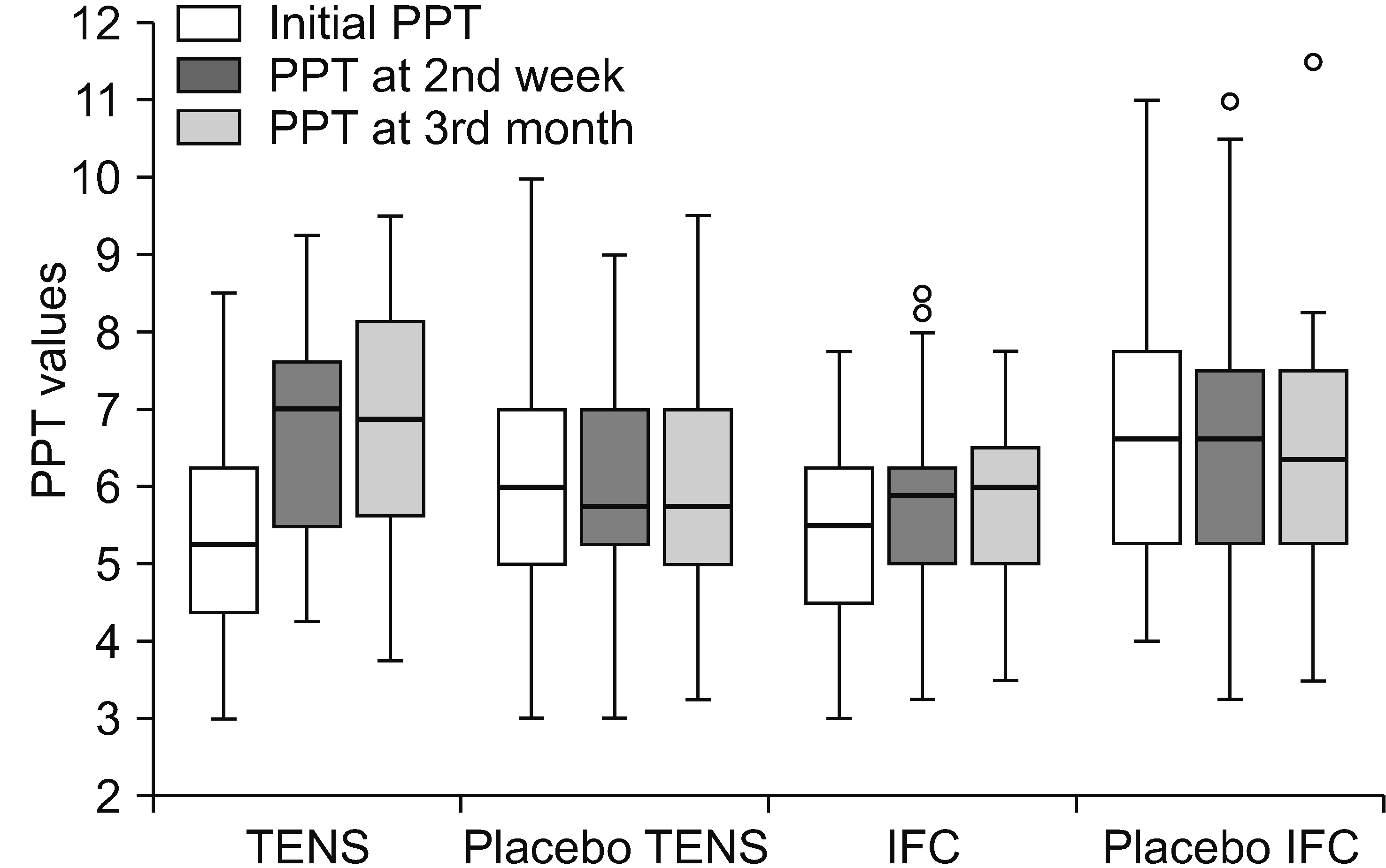
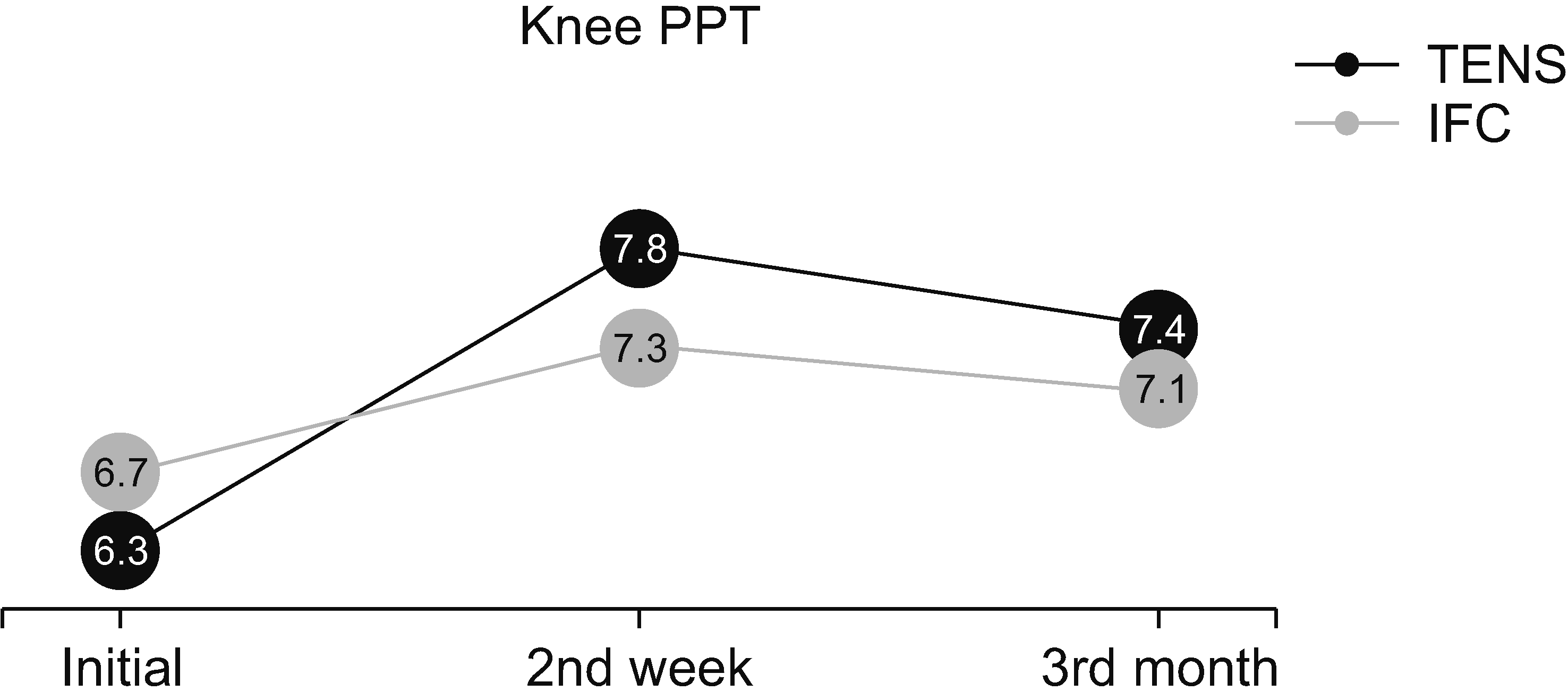
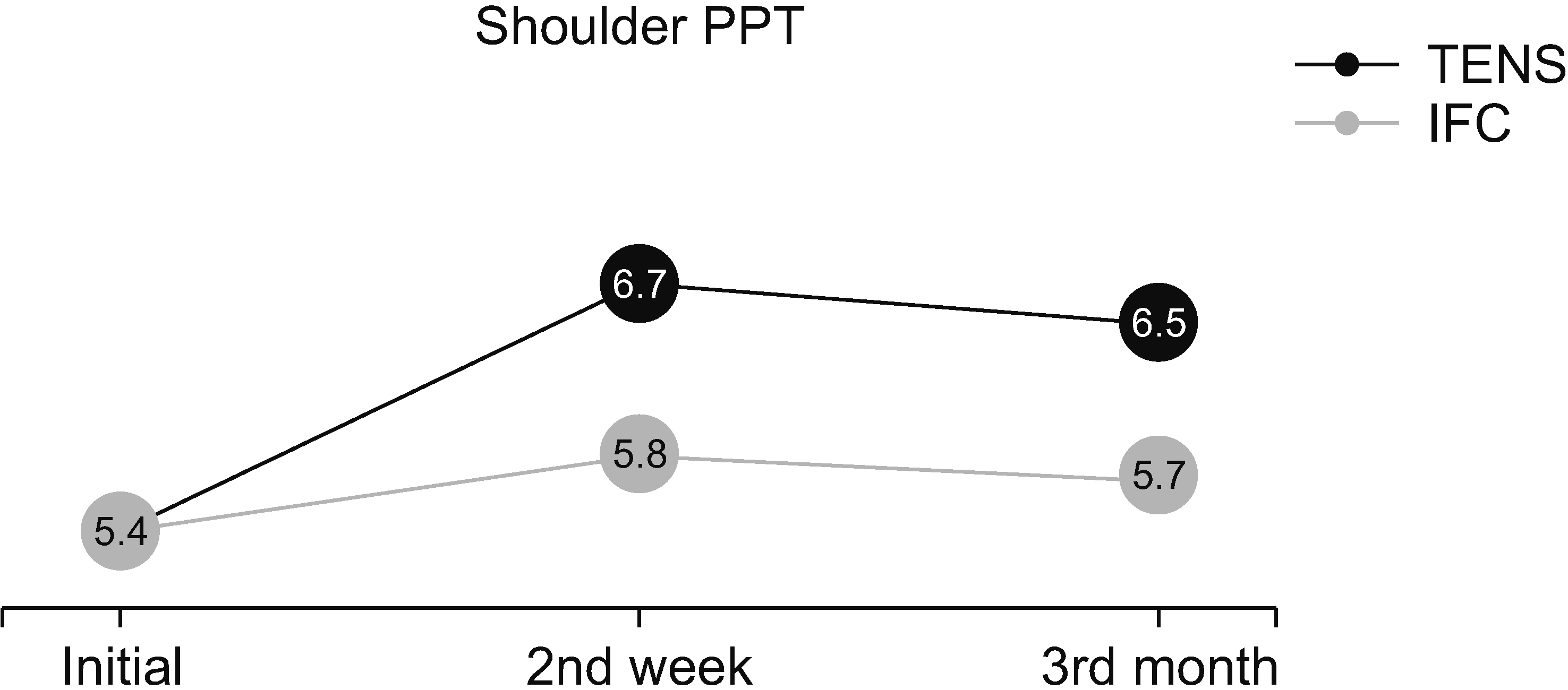
In the TENS and IFC groups, the increase in PPT values in both the painful knee and the shoulder selected as the painless distant point was statistically significant (P < 0.001), and no statistically significant change was found in the placebo TENS and placebo IFC groups compared with the initial value (Table 4, Figs. 6, 7). In the evaluation of knee and shoulder PPT, when the improvements in the TENS and IFC groups were compared, the increase at 2 weeks and 3 months compared with the baseline was statistically significantly higher in the TENS group than in the IFC group (P = 0.023 and P < 0.001) (Figs. 8, 9).
Multivariate logistic regression analysis was performed to find the determinants of improvement in PPT scores at month 3 compare with baseline. The patient’s inclusion in the TENS group and the initial high PPT were found to be the determinants of improvement in knee PPT. The patient’s inclusion in the TENS group, an initially high PPT, and an initially low VAS score were found to be the determinants of shoulder recovery (Tables 5, 6).

DISCUSSION

The present study evaluated the efficacy of TENS and IFC on VAS, WOMAC, PCS, TUG, TSK, and PPT scores of individuals diagnosed with knee OA. Significant improvement was observed compared with the initial values in all parameters, except PPT, in all groups, whereas the primary outcome, PPT scores, significantly improved in the only TENS and IFC groups, not in the two placebo groups. Moreover, the PPT improved more in the TENS group than in the IFC group. In the regression analysis conducted to determine the predictors of this improvement more clearly, in addition to inclusion in the TENS group, the initial low VAS score and initial high PPT were other markers.
In various studies, both TENS and IFC were found to have positive effects on pain and function in patients with knee OA [15,32]. In this study, a statistically significant improvement was observed in all groups; not only the TENS and IFC groups. The improvement of the placebo groups like the real treatment group was consistent with the findings reported in the literature. In a randomized controlled study, Atamaz et al. [18] compared TENS, IFC, and shortwave diathermy in patients with knee OA. In agreement with our study results, VAS and WOMAC scores showed statistically significant improvement in both placebo groups and treatment groups, and no significant difference was found among the groups. Similarly, in another randomized controlled study, no significant difference in improvement was observed between the TENS and placebo TENS groups in patients with knee OA [33]. In the literature, when patients believe they are receiving an actual treatment, a placebo exhibits a morphine-like effect, resulting in an improvement in outcomes like the actual treatment [34]. Thus, patients who had not received analgesic current previously were included in our study to make them think that they were receiving the real treatment. The accordance of the results may show the placebo treatment group believed they received real treatment, and this may strengthen our study. However, all groups were given hot pack treatment. Superficial heat may have improved pain and function through vasodilation, leading to muscle relaxation, and increased tissue perfusion and metabolic activity [35].
Our focus was to evaluate the effect of TENS and IFC on CS. For this, pain catastrophizing and kinesiophobia, which are accepted as subjective indicators of CS, were evaluated [8,9]. However, clear conclusions cannot be drawn with respect to causality between these maladaptive cognitive-emotional factors and CS in knee OA due to data from only cross-sectional studies. Therefore, it may be more appropriate to consider these factors as overlapping factors with CS rather than as clinical indicators of CS [5]. Like our pain and functional evaluation results, a significant improvement was found in all groups, and no difference was found between the groups. In investigating whether kinesiophobia or pain catastrophizing scores improve after the application of any physical therapy agent or surgery, these parameters also improve in parallel with decreased pain intensity and increased function [36,37]. Based on this, the authors can attribute catastrophizing to pain and the improvement in kinesiophobia in all groups to the improvement of pain and function.
In this study, hyperalgesia was evaluated with a more objective method, namely, PPT measurement. Lower PPTs have been described as a risk factor for worsening knee OA symptoms and pain conditions [38]. PPT values in the present study were consistent with the values in previous studies of patients with knee OA [39]. The PPTs increased significantly in the TENS and IFC groups compared with the placebo groups after treatment and the third month compared with the baseline. That is, TENS and IFC reduced both primary and secondary hyperalgesia. The effectiveness of these treatments, especially on secondary hyperalgesia, may indicate that TENS and IFC may have a place in the treatment of CS. When the effects of the two treatments on the PPT were compared, TENS significantly increased the pain threshold compared with IFC. In their study of rats, Hahm et al. [40] experimentally created OA with intra-articular monosodium iodoacetate injection. In that study, it was stated that TENS relieves OA pain by inhibiting active microglia cells in the dorsal horn and decreasing astrocyte expression in the spinal cord and that it improves the paw withdrawal threshold, which is considered the counterpart of the PPT. Similarly, Vance et al. [41] compared the effects of TENS and placebo TENS on pain and pain sensitivity in patients with knee OA and showed that the PPT at both local and painless distal points improved more in the TENS group. TENS shows this effect through its gate-control mechanism by decreasing glial cell activity in the dorsal horn and by increasing endogenous opioid release [40,41]. In the literature, no studies have compared the effects of TENS and IFC treatments on CS in knee OA. In a study of patients with myofascial pain syndrome [42], in contrast to our study, a higher increase in the pain threshold was found in the group receiving IFC treatment compared with the group receiving TENS treatment. However, since different treatment protocols that may affect CS are applied in addition to analgesic current treatments in this study, a clear inference cannot be made. Given the frequency of CS in the knee OA and easy availability of TENS and IFC in clinical practice, additional studies are required to understand the efficacy of these agents on CS.
In the literature, the PPT at the painless distant point, thus secondary hyperalgesia, is a clearer indicator of CS [43]. Therefore, in the present study, regression analysis was performed to clearly determine the predictors of improvement in pain threshold compared with the baseline in the distant pain-free region at the third month. It showed that being in the TENS group, having lower VAS and higher PPT values at baseline were more associated with improvement in pain sensitization. Although no study has reported the predictors of PPT change after current analgesic modalities in patients with knee OA, it had been shown that lower PPTs at baseline predicted nonresponse after total knee replacement [44] and physiotherapy intervention [45] for knee OA. This result shows that when planning treatment for patients with a lower PPT, TENS may be considered a good treatment option. Besides, it should not be forgotten that they may need additional treatments such as anti-depressants, psychological support, and cognitive treatments.
This study has several limitations. First, superficial heat therapy was applied to all groups. The rationale for this was to not leave patients without treatment and ensure participation in our study and continuity by applying real treatment. Second, this study was conducted in a single center, and most of the participants were middle-aged, non-working women, which limits the generalization of our results. Finally, given the high individual differences in response to analgesics, the authors thought that even if the data (dose, duration, type of analgesic) were collected, the use of analgesics would confuse the outcomes in a study comparing analgesic currents. Therefore, to strengthen our results, we specifically instructed all patients not to use any analgesics except topical analgesics throughout the study. However, while focusing extensively on analgesic use for musculoskeletal pain, we may have overlooked patients who unwittingly used analgesics for other reasons such as dental treatment.
In conclusion, TENS and IFC reduced pain sensitivity or increased PPT as compared to placebo groups in patients with knee OA. This effect was more pronounced in the TENS group. Therefore, the present study may recommend the utilization of TENS as an adjunct to treatments of patients with knee OA with CS findings.

ACKNOWLEDGMENTS

We would like to thank the participants in the study.

Notes

DATA AVAILABILITY

The datasets supporting the finding of this study are available from the corresponding author upon reasonable request.

CONFLICT OF INTEREST

No potential conflict of interest relevant to this article was reported.

AUTHOR CONTRIBUTIONS

Şahide Eda Artuç: Writing/manuscript preparation; Aslı Çalışkan Uçkun: Writing/manuscript preparation; Filiz Acar Sivas: Methodology; Fatma Gül Yurdakul: Formal analysis; Hatice Bodur: Writing/manuscript preparation.

REFERENCES

1. Lohmander LS, Roos EM. 2007; Clinical update: treating osteoarthritis. Lancet. 370:2082–4. DOI: 10.1016/S0140-6736(07)61879-0. PMID: 18156017.
crossref
2. Perrot S. 2015; Osteoarthritis pain. Best Pract Res Clin Rheumatol. 29:90–7. DOI: 10.1016/j.berh.2015.04.017. PMID: 26267003.
crossref
3. Fingleton C, Smart K, Moloney N, Fullen BM, Doody C. 2015; Pain sensitization in people with knee osteoarthritis: a systematic review and meta-analysis. Osteoarthritis Cartilage. 23:1043–56. DOI: 10.1016/j.joca.2015.02.163. PMID: 25749012.
crossref
4. Bannuru RR, Osani MC, Vaysbrot EE, Arden NK, Bennell K, Bierma-Zeinstra SMA, et al. 2019; OARSI guidelines for the non-surgical management of knee, hip, and polyarticular osteoarthritis. Osteoarthritis Cartilage. 27:1578–89. DOI: 10.1016/j.joca.2019.06.011. PMID: 31278997.
crossref
5. Lluch E, Nijs J, Courtney CA, Rebbeck T, Wylde V, Baert I, et al. 2018; Clinical descriptors for the recognition of central sensitization pain in patients with knee osteoarthritis. Disabil Rehabil. 40:2836–45. DOI: 10.1080/09638288.2017.1358770. PMID: 28768437.
crossref
6. Moore RL, Clifford AM, Moloney N, Doody C, Smart KM, O'Leary H. 2020; The relationship between clinical and quantitative measures of pain sensitization in knee osteoarthritis. Clin J Pain. 36:336–43. DOI: 10.1097/AJP.0000000000000798. PMID: 31977373.
crossref
7. Arendt-Nielsen L, Skou ST, Nielsen TA, Petersen KK. 2015; Altered central sensitization and pain modulation in the CNS in chronic joint pain. Curr Osteoporos Rep. 13:225–34. DOI: 10.1007/s11914-015-0276-x. PMID: 26026770.
crossref
8. Akin-Akinyosoye K, Frowd N, Marshall L, Stocks J, Fernandes GS, Valdes A, et al. 2018; Traits associated with central pain augmentation in the Knee Pain In the Community (KPIC) cohort. Pain. 159:1035–44. DOI: 10.1097/j.pain.0000000000001183. PMID: 29438225. PMCID: PMC5959005.
crossref
9. Neblett R, Cohen H, Choi Y, Hartzell MM, Williams M, Mayer TG, et al. 2013; The Central Sensitization Inventory (CSI): establishing clinically significant values for identifying central sensitivity syndromes in an outpatient chronic pain sample. J Pain. 14:438–45. DOI: 10.1016/j.jpain.2012.11.012. PMID: 23490634. PMCID: PMC3644381.
crossref
10. Jeong Y, Leem JW, Chung SS, Kim YS, Yoon DM, Nam TS, et al. 2000; Mechanism of hyperalgesia following cutaneous inflammation by complete Freund adjuvant. Korean J Pain. 13:164–74.
11. Cherian JJ, Jauregui JJ, Leichliter AK, Elmallah RK, Bhave A, Mont MA. 2016; The effects of various physical non-operative modalities on the pain in osteoarthritis of the knee. Bone Joint J. 98-B(1 Suppl A):89–94. DOI: 10.1302/0301-620X.98B1.36353. PMID: 26733650.
crossref
12. Tuncer T, Cay FH, Altan L, Gurer G, Kacar C, Ozcakir S, et al. 2018; 2017 update of the Turkish League Against Rheumatism (TLAR) evidence-based recommendations for the management of knee osteoarthritis. Rheumatol Int. 38:1315–31. DOI: 10.1007/s00296-018-4044-y. PMID: 29777340.
crossref
13. Hochberg MC, Altman RD, April KT, Benkhalti M, Guyatt G, McGowan J, et al. American College of Rheumatology. 2012; American College of Rheumatology 2012 recommendations for the use of nonpharmacologic and pharmacologic therapies in osteoarthritis of the hand, hip, and knee. Arthritis Care Res (Hoboken). 64:465–74. DOI: 10.1002/acr.21596. PMID: 22563589.
crossref
14. Vance CGT, Dailey DL, Chimenti RL, Van Gorp BJ, Crofford LJ, Sluka KA. 2022; Using TENS for pain control: update on the state of the evidence. Medicina (Kaunas). 58:1332. DOI: 10.3390/medicina58101332. PMID: 36295493. PMCID: PMC9611192. PMID: 88768f8548d9495083f8f6d4e830f7ab.
crossref
15. Rampazo ÉP, Liebano RE. 2022; Analgesic effects of interferential current therapy: a narrative review. Medicina (Kaunas). 58:141. DOI: 10.3390/medicina58010141. PMID: 35056448. PMCID: PMC8779694. PMID: 35c3ea2b58b94e989fe9dfe97bfb1e00.
crossref
16. Beckwée D, De Hertogh W, Lievens P, Bautmans I, Vaes P. 2012; Effect of tens on pain in relation to central sensitization in patients with osteoarthritis of the knee: study protocol of a randomized controlled trial. Trials. 13:21. DOI: 10.1186/1745-6215-13-21. PMID: 22353582. PMCID: PMC3307484. PMID: 0c2af86a924348e4894f77bcb9b05dd0.
crossref
17. Chen HL, Yang FA, Lee TH, Liou TH, Escorpizo R, Chen HC. 2022; Effectiveness of interferential current therapy in patients with knee osteoarthritis: a systematic review and meta-analysis of randomized controlled trials. Sci Rep. 12:9694. DOI: 10.1038/s41598-022-13478-6. PMID: 35690604. PMCID: PMC9188606. PMID: a679805595db467a9895d88fb902a2cb.
crossref
18. Atamaz FC, Durmaz B, Baydar M, Demircioglu OY, Iyiyapici A, Kuran B, et al. 2012; Comparison of the efficacy of transcutaneous electrical nerve stimulation, interferential currents, and shortwave diathermy in knee osteoarthritis: a double-blind, randomized, controlled, multicenter study. Arch Phys Med Rehabil. 93:748–56. DOI: 10.1016/j.apmr.2011.11.037. PMID: 22459699.
crossref
19. Kawasaki T, Inoue K, Ushiyama T, Fukuda S. 1998; Assessment of the American College of Rheumatology criteria for the classification and reporting of osteoarthritis of the knee. Ryumachi. 38:2–5. Japanese. PMID: 9564771.
20. Kellgren JH, Lawrence JS. 1957; Radiological assessment of osteo-arthrosis. Ann Rheum Dis. 16:494–502. DOI: 10.1136/ard.16.4.494. PMID: 13498604. PMCID: PMC1006995.
crossref
21. Mehendale N, Inamdar M. 2020; A review on transcutaneous electrical nerve stimulation and its applications. SSRN [Priprint]. Available at: https://papers.ssrn.com/sol3/papers.cfm?abstract_id=3602962. cited 2022 Jun. DOI: 10.2139/ssrn.3602962.
crossref
22. Heller GZ, Manuguerra M, Chow R. 2016; How to analyze the visual analogue scale: myths, truths and clinical relevance. Scand J Pain. 13:67–75. DOI: 10.1016/j.sjpain.2016.06.012. PMID: 28850536.
crossref
23. Podsiadlo D, Richardson S. 1991; The timed "Up & Go": a test of basic functional mobility for frail elderly persons. J Am Geriatr Soc. 39:142–8. DOI: 10.1111/j.1532-5415.1991.tb01616.x. PMID: 1991946.
crossref
24. Tüzün EH, Eker L, Aytar A, Daşkapan A, Bayramoğlu M. 2005; Acceptability, reliability, validity and responsiveness of the Turkish version of WOMAC osteoarthritis index. Osteoarthritis Cartilage. 13:28–33. DOI: 10.1016/j.joca.2004.10.010. PMID: 15639634.
crossref
25. İlçin N, Gürpınar B, Bayraktar D, Savcı S, Çetin P, Sarı İ, et al. 2016; Cross-cultural adaptation and validation of the Turkish version of the pain catastrophizing scale among patients with ankylosing spondylitis. J Phys Ther Sci. 28:298–303. DOI: 10.1589/jpts.28.298. PMID: 26957778. PMCID: PMC4756024.
crossref
26. Sullivan MJ, Bishop SR, Pivik J. 1995; The pain catastrophizing scale: development and validation. Psychol Assess. 7:524–32. DOI: 10.1037/1040-3590.7.4.524.
crossref
27. Hisli N. 1989; Use of the Beck Depression Inventory with Turkish university students: reliability, validity and factor analysis. Turk J Psychol. 7:3–13.
28. Tunca Yılmaz Ö, Yakut Y, Uygur F, Uluğ N. 2011; Turkish version of the Tampa Scale for Kinesiophobia and its test-retest reliability. Fizyoter Rehabil. 22:44–49. Turkish.
29. Vlaeyen JW, Kole-Snijders AM, Rotteveel AM, Ruesink R, Heuts PH. 1995; The role of fear of movement/(re)injury in pain disability. J Occup Rehabil. 5:235–52. DOI: 10.1007/BF02109988. PMID: 24234727.
crossref
30. Suokas AK, Walsh DA, McWilliams DF, Condon L, Moreton B, Wylde V, et al. 2012; Quantitative sensory testing in painful osteoarthritis: a systematic review and meta-analysis. Osteoarthritis Cartilage. 20:1075–85. DOI: 10.1016/j.joca.2012.06.009. PMID: 22796624.
crossref
31. Walton DM, Levesque L, Payne M, Schick J. 2014; Clinical pressure pain threshold testing in neck pain: comparing protocols, responsiveness, and association with psychological variables. Phys Ther. 94:827–37. DOI: 10.2522/ptj.20130369. PMID: 24557645. PMCID: PMC4040424.
crossref
32. Wu Y, Zhu F, Chen W, Zhang M. 2022; Effects of transcutaneous electrical nerve stimulation (TENS) in people with knee osteoarthritis: A systematic review and meta-analysis. Clin Rehabil. 36:472–85. DOI: 10.1177/02692155211065636. PMID: 34971318.
crossref
33. Palmer S, Domaille M, Cramp F, Walsh N, Pollock J, Kirwan J, et al. 2014; Transcutaneous electrical nerve stimulation as an adjunct to education and exercise for knee osteoarthritis: a randomized controlled trial. Arthritis Care Res (Hoboken). 66:387–94. DOI: 10.1002/acr.22147. PMID: 23983090.
crossref
34. Rakel BA, Zimmerman BM, Geasland K, Embree J, Clark CR, Noiseux NO, et al. 2014; Transcutaneous electrical nerve stimulation for the control of pain during rehabilitation after total knee arthroplasty: a randomized, blinded, placebo-controlled trial. Pain. 155:2599–611. DOI: 10.1016/j.pain.2014.09.025. PMID: 25270585. PMCID: PMC4250415.
crossref
35. Kim JH, Shin YS. 2020; Effects of localized heating on pain, skin perfusion, and wound healing after lumbar decompression. J Neurosci Nurs. 52:251–6. DOI: 10.1097/JNN.0000000000000529. PMID: 32868698.
crossref
36. Doménech J, Sanchis-Alfonso V, Espejo B. 2014; Changes in catastrophizing and kinesiophobia are predictive of changes in disability and pain after treatment in patients with anterior knee pain. Knee Surg Sports Traumatol Arthrosc. 22:2295–300. DOI: 10.1007/s00167-014-2968-7. PMID: 24691626.
crossref
37. Wade JB, Riddle DL, Thacker LR. 2012; Is pain catastrophizing a stable trait or dynamic state in patients scheduled for knee arthroplasty? Clin J Pain. 28:122–8. DOI: 10.1097/AJP.0b013e318226c3e2. PMID: 22001663.
crossref
38. Carlesso LC, Segal NA, Frey-Law L, Zhang Y, Na L, Nevitt M, et al. 2019; Pain susceptibility phenotypes in those free of knee pain with or at risk of knee osteoarthritis: the multicenter osteoarthritis study. Arthritis Rheumatol. 71:542–9. DOI: 10.1002/art.40752. PMID: 30307131. PMCID: PMC6442725.
crossref
39. Alfieri FM, Vargas E Silva NCO, Dos Santos ACA, Battistella LR. 2020; Cutaneous temperature and pressure pain threshold in individuals with knee osteoarthritis. Reumatologia. 58:272–6. DOI: 10.5114/reum.2020.100195. PMID: 33227096. PMCID: PMC7667939. PMID: 37366b540d4a4495a1435feecca8f439.
crossref
40. Hahm SC, Song E, Jeon H, Yoon YW, Kim J. 2019; Transcutaneous electrical nerve stimulation reduces knee osteoarthritic pain by inhibiting spinal glial cells in rats. Phys Ther. 99:1211–23. DOI: 10.1093/ptj/pzz076. PMID: 31158282.
crossref
41. Vance CG, Rakel BA, Blodgett NP, DeSantana JM, Amendola A, Zimmerman MB, et al. 2012; Effects of transcutaneous electrical nerve stimulation on pain, pain sensitivity, and function in people with knee osteoarthritis: a randomized controlled trial. Phys Ther. 92:898–910. DOI: 10.2522/ptj.20110183. PMID: 22466027. PMCID: PMC3386514.
crossref
42. Hou CR, Tsai LC, Cheng KF, Chung KC, Hong CZ. 2002; Immediate effects of various physical therapeutic modalities on cervical myofascial pain and trigger-point sensitivity. Arch Phys Med Rehabil. 83:1406–14. DOI: 10.1053/apmr.2002.34834. PMID: 12370877.
crossref
43. Moss P, Knight E, Wright A. 2016; Subjects with knee osteoarthritis exhibit widespread hyperalgesia to pressure and cold. PLoS One. 11:e0147526. DOI: 10.1371/journal.pone.0147526. PMID: 26809009. PMCID: PMC4726459. PMID: 62186aa6181f44698b8345ed5c2fa1c5.
crossref
44. Arendt-Nielsen L, Simonsen O, Laursen MB, Roos EM, Rathleff MS, Rasmussen S, et al. 2018; Pain and sensitization after total knee replacement or nonsurgical treatment in patients with knee osteoarthritis: Identifying potential predictors of outcome at 12 months. Eur J Pain. 22:1088–102. DOI: 10.1002/ejp.1193. PMID: 29369450.
crossref
45. O'Leary H, Smart KM, Moloney NA, Blake C, Doody CM. 2018; Pain sensitization associated with nonresponse after physiotherapy in people with knee osteoarthritis. Pain. 159:1877–86. DOI: 10.1097/j.pain.0000000000001288. PMID: 29794610.

Fig. 1
The CONSORT diagram of the study. TENS: transcutaneous electrical nerve stimulation, IFC: interferential current.
kjp-36-3-392-f1.tif
Fig. 2
Transcutaneous electrical nerve stimulation application.
kjp-36-3-392-f2.tif
Fig. 3
Interferential current application.
kjp-36-3-392-f3.tif
Fig. 4
Measurement of pressure pain threshold using a baseline dolorimeter from the knee.
kjp-36-3-392-f4.tif
Fig. 5
Measurement of pressure pain threshold using a baseline dolorimeter from the shoulder.
kjp-36-3-392-f5.tif
Fig. 6
Knee PPT changes (initial, 2nd week, and 3rd month). The line inside the boxes show the median value, the upper and lower borders of the box show the 25% and 75% percentiles, the mustaches 10% and 90% percentiles, the circles show the extreme values. TENS: transcutaneous electrical nerve stimulation, IFC: interferential current, PPT: pressure pain threshold (kg/cm2).
kjp-36-3-392-f6.tif
Fig. 7
Shoulder PPT changes (initial, 2nd week, and 3rd month). The line inside the boxes show the median value, the upper and lower borders of the box show the 25% and 75% percentiles, the mustaches 10% and 90% percentiles, the circles show the extreme values. TENS: transcutaneous electrical nerve stimulation, IFC: interferential current, PPT: pressure pain threshold (kg/cm2).
kjp-36-3-392-f7.tif
Fig. 8
Comparison of knee PPT increase between TENS and IFC groups. TENS: transcutaneous electrical nerve stimulation, IFC: interferential current, PPT: pressure pain threshold (kg/cm2).
kjp-36-3-392-f8.tif
Fig. 9
Comparison of shoulder PPT increase between TENS and IFC groups. TENS: transcutaneous electrical nerve stimulation, IFC: interferential current, PPT: pressure pain threshold (kg/cm2).
kjp-36-3-392-f9.tif
Table 1
Baseline demographic and clinical characteristics of patients
Variables TENS (n = 19) IFC (n = 18) Placebo TENS (n = 18) Placebo IFC (n = 18) P value
Age (yr) 58.50 ± 9.80 61.95 ± 11.78 56.05 ± 9.45 54.00 ± 8.79 0.081
Sex 0.965
Female 85.0 80.0 85.0 85.0
Male 15.0 20.0 15.0 15.0
BMI (kg/m2) 30.33 ± 6.62 31.43 ± 3.51 29.34 ± 4.57 31.20 ± 3.74 0.495
Education status 0.470
Illiterate 40.0 25.0 10.0 15.0
Primaryeducation 25.0 60.0 65.0 75.0
High school 10.0 10.0 10.0 10.0
University 25.0 5.0 15.0 0.0
Comorbidities 65.0 75.0 75.0 65.0 0.813
VAS 7.80 ± 1.39 7.65 ± 1.22 7.90 ± 1.71 7.60 ± 1.60 0.913
WOMAC 58.82 ± 15.26 57.09 ± 9.65 57.09 ± 20.55 52.01 ± 18.09 0.708
PCS 19.15 ± 17.40 20.85 ± 10.83 23.41 ± 6.21 22.10 ± 12.99 0.820
TUG 11.00 ± 3.65 10.47 ± 2.10 10.35 ± 4.01 8.95 ± 1.67 0.181
BDI 10.25 ± 7.95 9.45 ± 6.00 9.40 ± 5.75 9.75 ± 9.79 0.984
TSK 38.30 ± 4.96 32.30 ± 8.47 33.95 ± 8.48 37.85 ± 5.68 0.054
PPT. Knee (kg/cm2) 6.36 ± 1.49 6.83 ± 1.85 6.65 ± 2.19 7.56 ± 1.97 0.233
PPT. Shoulder (kg/cm2) 5.42 ± 1.41 6.20 ± 1.79 5.40 ± 1.34 6.58 ± 2.09 0.073
KL 0.821
KL 2 47.0 55.0 45.0 58.0
KL 3 53.0 45.0 55.0 42.0

Values are presented as % or mean ± standard deviation.

n: number of patients, TENS: transcutaneous electrical nerve stimulation, IFC: interferential current, BMI: body mass index, VAS: visual analog scale (0–10), WOMAC: Western Ontario and McMaster Universities Osteoarthritis Index, PCS: pain catastrophizing scale, TUG: Timed Up and Go test, BDI: Beck Depression Inventory, TSK: Tampa Scale of Kinesiophobia, PPT: pressure pain threshold (kg/cm2), KL: Kellgren–Lawrence radiological scoring system.

Table 2
Intragroup comparison of clinical improvements
Variables TENS (n = 19) IFC (n = 18) Placebo TENS (n = 18) Placebo IFC (n = 18)
Mean ± SD P value Mean ± SD P value Mean ± SD P value Mean ± SD P value
VAS
Initial 7.78 ± 1.43 7.72 ± 1.27 7.83 ± 1.72 7.66 ± 1.53
2nd wk 3.78 ± 1.35 < 0.001a 3.61 ± 1.61 < 0.001a 3.88 ± 2.05 < 0.001a 4.77 ± 1.95 < 0.001a
3rd mo 5.00 ± 1.59 < 0.001b 5.27 ± 1.40 < 0.001b 5.50 ± 2.40 < 0.001b 6.61 ± 1.14 0.015b
WOMAC
Initial 56.97 ± 15.67 57.49 ± 10.00 55.37 ± 19.53 52.59 ± 18.14
2nd wk 32.91 ± 15.35 < 0.001a 29.19 ± 14.07 < 0.001a 32.65 ± 17.97 < 0.001a 35.49 ± 18.76 < 0.001a
3rd mo 39.30 ± 15.09 < 0.001b 42.08 ± 10.41 < 0.001b 38.91 ± 21.02 < 0.001b 44.78 ± 20.39 0.043b
PCS
Initial 19.36 ± 17.92 21.00 ± 11.23 23.11 ± 16.52 20.88 ± 11.80
2nd wk 13.42 ± 17.64 < 0.001a 13.11 ± 13.76 0.003a 16.77 ± 15.90 < 0.001a 14.66 ± 8.84 0.005a
3rd mo 15.42 ± 17.46 < 0.001b 17.88 ± 11.46 0.005b 17.83 ± 16.21 0.039b 18.05 ± 11.07 0.008b
TUG (sec)
Initial 11.12 ± 3.71 10.44 ± 2.15 10.67 ± 4.08 8.57 ± 1.13
2nd wk 9.40 ± 3.43 < 0.001a 9.06 ± 2.83 0.024a 9.44 ± 2.92 0.017a 7.73 ± 1.00 < 0.001a
3rd mo 9.99 ± 3.66 < 0.001b 9.97 ± 2.31 < 0.001b 10.04 ± 3.52 < 0.001b 8.32 ± 1.30 0.045b
TSK
Initial 38.21 ± 5.08 32.26 ± 8.70 33.61 ± 8.73 38.05 ± 5.81
2nd wk 34.52 ± 3.67 < 0.001a 30.63 ± 8.54 0.014a 31.94 ± 8.25 0.005a 35.55 ± 5.70 0.030a
3rd mo 35.26 ± 4.02 < 0.001b 31.47 ± 8.80 0.015b 32.77 ± 8.53 0.048b 36.77 ± 6.00 0.040b

n: number of patients, SD: standard deviation, TENS: transcutaneous electrical nerve stimulation, IFC: interferential current, VAS: visual analog scale (0–10), WOMAC: Western Ontario and McMaster Universities Osteoarthritis Index, PCS: pain catastrophizing scale, TUG: Timed Up and Go test, TSK: Tampa Scale of Kinesiophobia.

aComparison between initial and 2nd week, bComparison between the 2nd week and 3rd month.

Table 3
Comparison of clinical improvements between groups
Variables TENS IFC Placebo TENS Placebo IFC P value
VAS
Initial - 2nd wk 4.00 ± 0.28 4.11 ± 0.36 3.94 ± 0.51 2.88 ± 0.50 0.625
Initial - 3rd mo 2.78 ± 0.33 2.44 ± 0.32 2.33 ± 0.54 1.05 ± 0.39 0.362
WOMAC
Initial - 2nd wk 24.05 ± 2.47 28.30 ± 3.36 22.72 ± 3.88 17.10 ± 3.40 0.981
Initial - 3rd mo 17.66 ± 2.16 15.41 ± 2.32 16.46 ± 2.89 7.81 ± 3.57 0.966
PCS
Initial - 2nd wk 5.94 ± 1.45 7.88 ± 2.32 6.33 ± 1.64 6.22 ± 1.91 0.830
Initial - 3rd mo 3.94 ± 0.97 3.11 ± 0.96 5.27 ± 2.36 2.83 ± 0.93 0.631
TUG
Initial - 2nd wk 1.72 ± 0.19 1.38 ± 0.55 1.23 ± 0.46 0.84 ± 0.12 0.133
Initial- 3rd mo 1.13 ± 0.18 0.46 ± 0.11 0.62 ± 0.20 0.24 ± 0.13 0.122
TSK
Initial - 2nd wk 3.68 ± 0.83 1.63 ± 0.60 1.66 ± 0.51 2.50 ± 0.39 0.085
Initial - 3rd mo 2.94 ± 0.68 0.78 ± 0.53 0.83 ± 0.44 1.27 ± 0.49 0.054

Values are presented as mean difference ± standard deviation.

TENS: transcutaneous electrical nerve stimulation, IFC: interferential current, VAS: visual analog scale (0–10), WOMAC: Western Ontario and McMaster Universities Osteoarthritis Index, PCS: pain catastrophizing scale, TUG: Timed Up and Go test, TSK: Tampa Scale of Kinesiophobia.

Table 4
PPT values in painful knee and the painless distant point shoulder
Variables TENS (n = 19) IFC (n = 18) Placebo TENS (n = 18) Placebo IFC (n = 18)
Mean ± SD P value Mean ± SD P value Mean ± SD P value Mean ± SD P value
PPT; Knee
Initial 6.36 ± 1.49 6.77 ± 2.23 6.97 ± 1.86 7.87 ± 1.82
2nd wk 7.82 ± 1.68 < 0.001a 7.30 ± 2.32 < 0.001a 7.02 ± 1.74 0.881a 7.25 ± 1.91 0.060a
3rd mo 7.42 ± 2.1 < 0.001b 7.16 ± 2.23 < 0.001b 7.06 ± 1.68 0.801b 7.73 ± 1.72 0.326b
PPT; Shoulder
Initial 5.42 ± 1.41 5.41 ± 1.42 6.31 ± 1.85 6.76 ± 2.03
2nd wk 6.72 ± 1.52 < 0.001a 5.84 ± 1.41 < 0.001a 6.08 ± 1.38 0.390a 6.63 ± 1.93 0.717a
3rd mo 6.52 ± 1.45 < 0.001b 5.72 ± 1.26 < 0.001b 5.94 ± 1.47 0.227b 6.44 ± 1.95 0.062b

n: number of patients, SD: standard deviation, TENS: transcutaneous electrical nerve stimulation, IFC: interferential current, PPT: pressure pain threshold.

aComparison between initial and 2nd week, bComparison between the 2nd week and 3rd month.

Table 5
Demonstration of the determinants of improvement of knee PPT by multiple regression analysis
Knee PPT score risea B SE P value 95% CI
Initial PPT 0.864 0.304 0.004 1.308 4.305
Group 2.024 0.580 < 0.001 2.428 23.602
Constant –8.114 2.457 0.001

B: regression coefficient, SE: standard error, CI: confidence interval, PPT: pressure pain threshold (kg/cm2).

a15% increase from baseline in the 3rd month.

Table 6
Demonstration of the determinants of improvement of shoulder PPT by multiple regression analysis
Shoulder PPT score risea B SE P value 95% CI
Group 4.442 1.466 0.003 4.732 1,479.543
Initial VAS –1.198 0.517 0.020 0.110 0.831
Initial PPT 1.712 0.684 0.012 1.450 21.142
Constant –4.926 4.198 0.241

B: regression coefficient, SE: standard error, CI: confidence interval, PPT: pressure pain threshold (kg/cm2), VAS: visual analogue scale (0–10).

a15% increase from baseline in the 3rd month.

TOOLS
Similar articles