

 PDF
PDF Citation
 Print
Print


INTRODUCTION
Inner ear disorders such as hearing loss, tinnitus, and balance disorders affect individuals of all ages and reduce their quality of life. Effective strategies for treating auditory disorders currently rely on the local administration of therapeutic drugs because conventional systemic administration has several limitations, such as the increased risk of overdose, low solubility, unpredictable absorption, and first-pass effect.1 Additionally, this administration method is ineffective mainly because of the blood-perilymph barrier that separates the inner ear from the blood.12 Therefore, there is a need for local delivery systems that can avoid first-pass metabolism associated with systemic administration and ensure the therapeutic drug concentration in the inner ear.
Intratympanic (IT) administration is the most common local delivery method in clinical practice.3 IT administration deposits the drug near the round window (the leading drug absorption site in the inner ear). It facilitates the drug diffusion through the membrane and reach the target site, thereby maximizing drug concentration in the inner ear while minimizing systemic exposure.45 Despite these advantages, the IT-administrated drug solution has a short contact time with the round window membrane because it is easily discharged through the Eustachian tube6; therefore, repeated injections are necessary to obtain the therapeutic effect. To overcome these drawbacks, many studies have been conducted to develop new effective formulations that can improve therapeutic concentration in the inner ear by prolonging drug residence time in the round window membrane of the middle ear.
Thermo-sensitive hydrogels (thermogels) that demonstrate a sol-gel transition behavior in aqueous media are promising biomaterials for designing various drug delivery systems, particularly injectable delivery formulations.78910 They exist as a free-flowing liquid (sol) at room temperature, facilitating drug loading via simple mixing and enabling efficient local administration through direct injection into the target site. After administration, they instantly transform into a solid gel in response to body temperature. Due to their unique thermo-gelling properties, thermo-sensitive hydrogels have been extensively utilized to extend the drug residence time by preventing early clearance of the drug formulation at the administration site.111213 For example, an injectable thermo-sensitive hydrogel that was prepared in an aqueous mixture of chitosan with glycerophosphate has been studied for local delivery to the inner ear.14 Although the hydrogel showed promising results for effective inner ear delivery, it may have limitations in practical applications due to irreversible thermogelation, phase separation, and the excessive presence of glycerophosphate which is potentially toxic to cells.15 On the other hand, owing to its non-toxicity and thermo-reversibility, poloxamer 407 (P407), a tri-block copolymer with ethylene glycol and propylene oxide blocks, has been extensively studied as a representative injectable hydrogel formulation for the delivery of various kinds of drugs,1617 including inner ear disease medication such as steroids and antibiotics.18 In addition to possessing the thermogelation, P407 is amphiphilic with a favorable solubilizing capacity for hydrophobic drugs. P407 can effectively solubilize and encapsulate hydrophobic drugs through micelle formation and deliver them to the target site via diffusion after thermogelation.1920 These properties of IT drug administration could prevent early discharge from the middle ear cavity and increase the contact time with the round window membrane, contributing to better clinical efficacy with enhanced therapeutic drug levels.
Dexamethasone (DEX), a synthetic anti-inflammatory steroid, is mainly used in clinics to alleviate the symptom caused by inner ear disorders.212223 Its poor water solubility has limited its availability as an injectable formulation; therefore, the hydrophilic phosphate form, dexamethasone sodium phosphate (DSP), is commonly used as an alternative despite its limited efficacy. In this study, we prepared and evaluated thermosensitive P407 hydrogel formulations for the solubilization and injectable IT delivery of DEX. We performed a comparative study of injectable P407 formulations of the hydrophilic drug DSP and DEX. Furthermore, drug solubilization, loading capacity, thermogelation, and drug release properties of the DEX-loaded hydrogels were evaluated. In addition, the in vivo animal experiments were performed using guinea pigs to observe cytotoxicity and drug absorption of the P407 formulations after IT injection via histopathological analysis and drug concentration measurement in the inner ear.
METHODS
Materials
DEX (Mw = 392.46 g/mol) was purchased from Farmabios (Gropello Cairoli, Italy) and DSP (Mw = 516.4 g/mol) from Steraloids (Newport, RI, USA), respectively. Kolliphor P407 was purchased from CTC Bio (Hwaseong, Korea). Methanol was supplied from Samchun Chemicals (Seoul, Korea). Dialysis membranes with the molecular weight cut off (MWCO) of 12–14 kDa were obtained from Spectrum Laboratories (San Francisco, CA, USA).
Solubilizing capacity of P407
The solubility of DEX in aqueous media was evaluated as a function of the polymer concentration. An excessive amount of DEX was dissolved in 3 mL of P407 solutions with different concentrations (0–15% w/v, phosphate buffered saline [PBS]). The mixture samples were kept under stirring for 30 minutes and then incubated in a shaking water bath (100 rpm) for 24 hours. Each solution sample was filtered using a syringe filter (0.45 μm) to remove any undissolved residues. The drug concentration in each sample was measured using a ultraviolet (UV)-visible spectrophotometer (V-730; JASCO, Tokyo, Japan). The water-solubility was obtained using a standard calibration curve obtained from different DEX concentrations at 242 nm.
Preparation of DEX-loaded P407 micelles
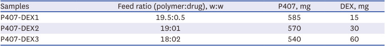
DEX-loaded P407 micelles (P407-DEX) were prepared using the dialysis method as described previously.24 Predetermined amounts of P407 and DEX were dissolved in 30 mL of methanol with stirring for 15 minutes, and diluted with the same amount of deionized water, followed by dialysis (MWCO = 12−14kDa) in water and then lyophilization for 3–4 days. The P407-DEX samples were prepared with three different feed ratios of P407 and DEX (97.5:2.5, 95:5, and 90:10), as show in Table 1. The drug loading content (DLC) of the P407-DEX micelles was measure using UV-visible spectrophotometry. The DLC and drug loading efficiency (DLE) were determined using the following equations:


Characterization of DEX-loaded P407 micelles
The size and size distribution of the P407-DEX samples were determined using dynamic light scattering (DLS) (ELSZ series; Otsuka Electronics, Osaka, Japan). The samples were dissolved in PBS at a concentration of 1% w/v. The thermal characteristics of DEX, P407, and P407-DEX samples were investigated and compared using a differential scanning calorimeter (DSC 4000 PerkinElmer; PerkinElmer, Waltham, MA, USA). Approximately 3 mg of each sample was equilibrated to 30°C for 2 minutes and then heated from 30°C to 300°C (10°C/min) using a perforated aluminium pan and nitrogen as the purge gas (20 mL/min). An empty aluminium pan was used as the reference. The powder crystallinity of DEX, P407, and P407-DEX samples were assessed at room temperature by X-ray diffraction measurements (XRD; Bruker AXS D8 Advance diffractometer, Bruker, Billerica, MA, USA) with Cu Kα radiation source (40 kV and 40 mA), 1°/min of scan speed, 0.02 scan step and 0−80°(2θ) range.
Thermosensitive sol-gel transition properties
The thermosensitive sol-gel transition properties of P407 hydrogel formulations with or without the drug (DSP or DEX) were compared. Aqueous solutions of P407 were prepared at a concentration of 20% w/v in PBS (pH 7.4). The DSP-loaded P407 formulation was prepared by dissolving DSP at the same concentration (3% w/v) as DEX in the P407-DEX hydrogel. The P407-DEX hydrogel was prepared by directly dissolving the P407-DEX powder in 1 mL of cold PBS (pH 7.4) at a P407 concentration of 20% w/v. All samples were kept at 4°C before use.25 The sol-gel transition behavior was observed macroscopically with a gradual temperature increase (1°C/min). The gelation temperature (Tgel) was determined by the tube tilting method. All the experiments were repeated in triplicate and the average value was used for comparison.
Rheological analysis
Rheological properties of the P407 hydrogel formulations were observed using an HR10 rheometer (TA Instruments, New Castle, DE, USA). Aqueous solutions of P407, P407-DSP, and P407-DEX were placed between two titanium parallel plates with a diameter of 60 mm and a gap of 1 mm. Under the constant stress of 20 Pa, the temperature-dependent rheological behaviors were analyzed using the frequency sweep method with a frequency of 1 Hz at a heating rate of 3°C/min from 10 to 40°C.
In vitro drug release
The drug release kinetics of the P407 hydrogel formulations were observed in PBS (pH 7.4). The hydrogel formulations, P407-DSP and P407-DEX, with a drug concentration of 3% w/v were inserted into dialysis membrane tubes (MWCO = 12−14 kDa) and immersed in 50 mL of PBS. The test samples were incubated in a shaking water bath at 37°C and 100 rpm, and 10 mL of release medium samples were taken at predetermined time points with replenishment of the same volume of fresh medium. The released drug amount was characterized by the UV-Vis absorbance measurements at 242 nm.
Animals
In this study, we used 6-week-old male albino guinea pigs, each weighing about 200–250 g. A total of 72 animals were used for perilymph sampling after IT administration of the DSP, P407-DSP, and P407-DEX formulations into the tympanic cavity and histopathological analysis. Two animals were used as normal controls for histopathological studies.
IT administration
IT injection was performed according to our previous study.26 The animals were anesthetized and kept at 37°C using a heating pad. A retro-auricular incision was made, and the temporal bone was opened to visualize the round window membrane. Aqueous formulations of P407-DSP and P407-DEX (sample volume = 80 μL and the drug concentration = 6 mg/mL) were prepared and an aqueous solution of DSP with the same drug concentration was used as a control because the free form of DEX was not suitable for injectable formulations due to its limited solubility and dispersibility. They were administered by IT injection with a 26 gauge needle (BD, Seoul, Korea), followed by application of dental cement (Durelon™ Carboxylate Luting Cement; 3M, St. Paul, MN, USA). The skin incision was then closed. All guinea pigs were observed to recover and show normal activities.
Histopathological analysis after IT injection
Histopathologic analysis was performed to investigate the adverse effects of the IT-administered P407 formulations on the middle and inner ear. Tissue samples were prepared according to the previous reports.27 The middle ear samples were harvested 7 days after IT administration with P407-DSP and P407-DEX formulations to evaluate the inflammation of the middle ear mucosa. The prepared sections were stained with hematoxylin and eosin (H&E). Cochleae samples were harvested 7 days after IT injection for whole-mount immunostaining to observe whether cochlear hair cells were damaged. Tissues were fixed in 4% paraformaldehyde (PFA) in PBS for 30 minutes and then rinsed in PBS for 30 minutes. After blocking, the primary antibody (myosin VIIa) was incubated at 4°C overnight, then washed 6 times with PBS. The Alexa Fluor 594 goat anti-mouse secondary antibody and Alexa Fluor 488 phalloidin antibody were incubated for 2 hours and washed with PBS. To observe the absorption of DEX in the cochlea, cochleae samples were collected at 30 minutes, 90 minutes, and 1 day after IT injection, infusion of 4% PFA at 4°C for 30 minutes, 10% ethylenediaminetetraacetic acid in PBS for 3–4 weeks of decalcification, paraffin-embedded, and 4 μm thickness sections for immunohistochemistry.
Drug concentration in the cochlear perilymph
The DEX concentration in the cochlear perilymph were assessed at 30 minutes, 90 minutes, 3 hours, 1 day, 3 days, and 7 days after IT administration. The sampling and analytical methods were carried out in the same manner as previously reported.26 Small cochleostomy was performed at the tip of the cochlea, and then the capillary tubes (Sigma-Aldrich, St. Louis, MO, USA) were used to collect the perilymph. These operations were performed under a surgical microscope. Finally, all the perilymph and standard samples were diluted and analyzed using liquid chromatography-tandem mass spectrometry (LC-MS/MS) method equipped with an Agilent 1290 Infinity II LC system (Agilent Technologies, Santa Clara, CA, USA) and a QTRAP ® 6500 LC-MS/MS System (SCIEX, Framingham, MA, USA). The date scan was performed using multiple reaction monitoring.
Statistical analysis
All statistical analysis and data graphing were performed using GraphPad Prism 6 (GraphPad Software, San Diego, CA, USA). All measurements were made on different samples. Adobe Photoshop (version 7.0; Adobe Systems Inc., San Jose, CA, USA) was used to perform colorization of monochrome fluorescence images, adjustment of image contrast and superimposition of images. The DEX concentration measurements were used two-way analysis of variance. All data were expressed as the mean ± standard error of mean, and statistical significance was set at P < 0.05.
RESULTS
Preparation and characterization of DEX-loaded P407 micelles
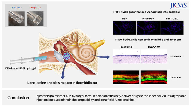
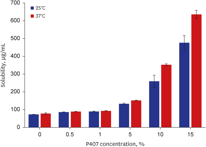
An injectable hydrogel formulation was prepared using thermosensitive P407 for efficient delivery of the hydrophobic drug DEX to the inner ear (Fig. 1A). This hydrogel formulation exhibited temperature-induced gelation behaviour after IT administration, which is expected to contribute to effective drug absorption in the inner ear by reducing premature discharge of the drug from the middle ear cavity, thereby increasing the contact time with the round window membrane (Fig. 1B). The solubilization effect of P407 on DEX was investigated in current study (Fig. 2). The water-solubility of DEX was observed to be 72.5 μg/mL and 77.48 μg/mL at 25°C and 37°C, respectively, which increased up to 475.7 μg/mL and 635.09 μg/mL as the P407 concentration increased from 0 to 15% w/v, respectively. The increase in the DEX solubility was more prominent at 37°C compared to 25°C. The results showed that P407 is effective in improving the aqueous solubility of hydrophobic DEX.
Fig. 1
Schematic illustration of (A) preparation and (B) intratympanic injection of DEX-loaded P407 hydrogel formulation.
DEX = dexamethasone, P407 = poloxamer 407, PEO = poly(ethylene oxide), PPO = poly(propylene oxide).
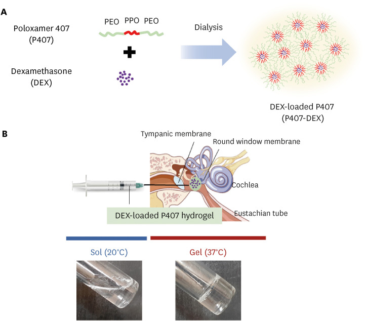
DEX-loaded P407 formulations were prepared using the dialysis method (Fig. 1A). To evaluate the drug-loading capacity of P407 formulations for DEX, various P407-DEX formulations with different polymer to drug feed ratios were analyzed for DLC and DLE (Table 2). The P407-DEX1 (drug feed ratio of 2.5) exhibited a high DLE but a low DLC (1.7%). P407-DEX3 (highest drug feed ratio of 10) exhibited the highest DLC but the lowest DLE. Moderate DLC and DLE were demonstrated by P407-DEX2 at a drug feed ratio of 5. The change in particle size was confirmed via DLS before and after drug loading into P407 micelles. The particle size of P407 micelles before drug loading was 27 nm (Fig. 3A). After drug loading, a significant increase in particle size was observed for all the P407 formulations. In particular, P407-DEX3, with the highest drug content, presented a particle size close to 2,000 nm, while other formulations presented relatively uniform and smaller particle sizes of approximately 450 nm. We decided to use P407-DEX2 for further experiments, as it displayed appropriate drug content and particle sizes in the drug loading and DLS results.
Table 2
Characteristics of drug-loaded P407 micelles prepared by the dialysis method
Fig. 3
Physical status characterization of DEX-loaded P407. Particle size distribution of (A) P407 and (B) P407-DEX at concentration of 1% w/v in phosphate buffered saline (pH 7.4), measured by dynamic light scattering. (C) Differential scanning calorimeter thermograms and (D) powder X-ray diffraction patterns of DEX, P407, and P407-DEX.
DEX = dexamethasone, P407 = poloxamer 407.
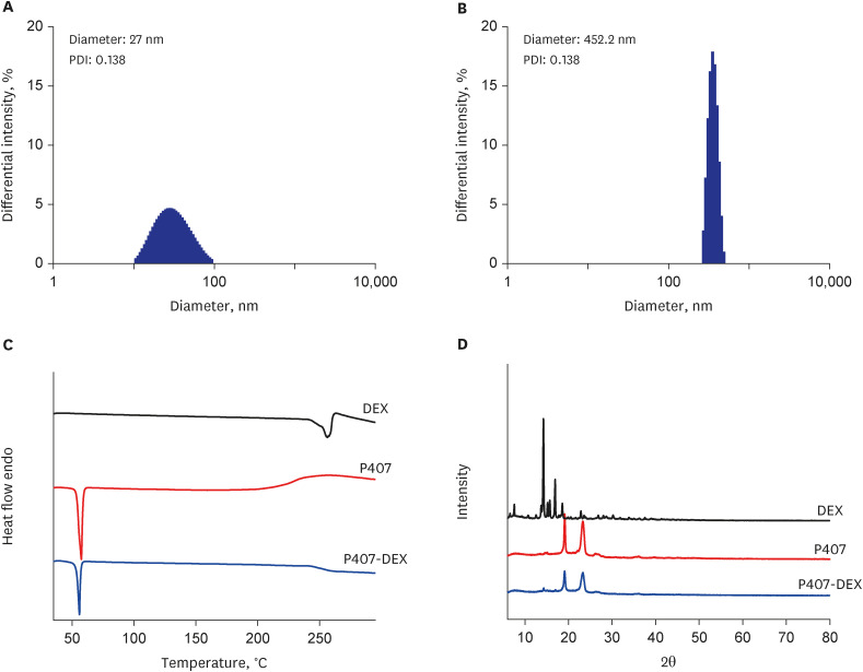
Next, the thermal behaviors of DEX, P407, and P407-DEX were evaluated (Fig. 3C). In pure DEX, a single endothermic peak is observed at approximately 260°C, which corresponds to its melting point. P407 exhibited a sharp melting endotherm at 57.1°C, which is consistent with a previous report.28 However, P407-DEX did not show the characteristic DEX melting peak but showed only the melting peak of P407. This indicated that the hydrophobic DEX was homogenously dispersed within P407 without the formation of crystalline domains. The XRD patterns of the DEX, P407, and P407-DEX powders were evaluated (Fig. 3D). The DEX powder exhibited typical sharp patterns in the range of 5 to 40° of 2, which is attributed to the characteristics of a crystalline state. However, the diffractogram of P407-DEX revealed that the typical crystalline peaks of DEX disappeared, and only P407 characteristic peaks remained at 19.1° and 23.09°. These results suggested that DEX molecules were homogeneously present in P407, similar to the DSC results.
Thermo-sensitive sol-gel transition
The thermosensitive sol-gel transition properties of P407, P407-DSP, and P407-DEX were investigated and compared by the tube tilting method and rheological analysis (Fig. 4). It is known that a 20% P407 solution exhibits a typical phase transition from a flowing sol state to a non-flowing gel state at 24°C. The P407-DSP and P407-DEX samples both showed effective thermogelation near 24°C, which was not significantly different from that of P407 alone (Table 3).
Fig. 4
The sol-gel transition behavior. (A) Sol-gel transition images of P407, P407-DSP, and P407-DEX at 20°C and 37°C, respectively. Temperature-dependent rheological behavior of (B) P407, (C) P407-DSP, and (D) P407-DEX.
DSP = dexamethasone sodium phosphate, DEX = dexamethasone, P407 = poloxamer 407.
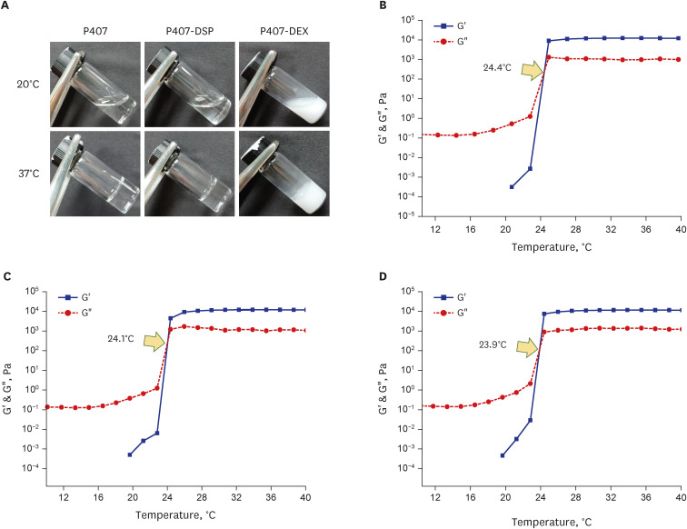
Table 3
Chemical compositions and gelation temperatures of hydrogel formulations
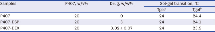
| Samples | P407, w/v% | Drug, w/w% | Sol-gel transition, °C | |
|---|---|---|---|---|
| Tgela | Tgelb | |||
| P407 | 20 | 0 | 24 | 24.4 |
| P407-DSP | 20 | 3 | 24 | 24.1 |
| P407-DEX | 20 | 3.02 ± 0.07 | 24 | 23.9 |
Rheological analysis was performed to determine the viscoelastic properties of P407, P407-DSP, and P407-DEX as a function of temperature. The Tgel is defined as the crossover point of the elastic (G’), and loss moduli (G”). The G’ value of P407 was continuously lower than G” at low temperatures; however, the G’ value increased dramatically over G” value from the Tgel point of 24.4°C (Fig. 4B). The G’ and G” values of P407-DSP and P407-DEX crossed at the Tgel point of 24.1°C, and 23.9°C, respectively (Fig. 4C and D). The presence of DEX did not significantly affect the thermogelling properties of P407.
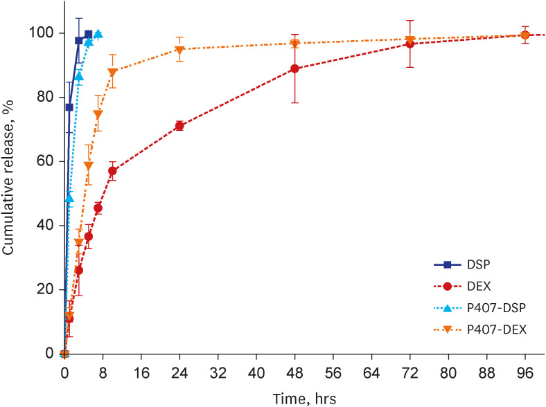
In vitro drug release kinetics
The in vitro drug release kinetics of P407-DSP and P407-DEX were compared to the hydrophilic and hydrophobic forms of DEX (Fig. 5). The hydrophilic DSP formulations with and without P407 showed fast release patterns, and their drug release was completed within a few hours. The hydrophobic DEX formulation showed a slow-release behavior owing to its limited solubility. On the other hand, P407-DEX showed a more sustained drug release than the hydrophilic DSP formulations due to the hydrophobic interaction between DEX and the P407 micelles, but an enhanced release behavior compared to the DEX alone. Such enhanced release property of P407-DEX could be attributed to the solubilization effect of P407.
Evaluation of in vivo drug uptake
Effectiveness of the P407 hydrogel formulations for inner ear drug delivery was evaluated by observing the drug concentration and distribution in the cochlear perilymph and tissue. Following IT-injection of DSP, P407-DSP, and P407-DEX formulations, the intracochlear perilymph was collected from the cochlea after 30 minutes, 90 minutes, 3 hours, 1 day, 3 days, and 7 days. The DEX concentration was measured using LC-MS/MS. Significant DEX concentrations were detected in the perilymph fluid 30 minutes after local IT administration of all the formulations (Fig. 6A). The aqueous DSP formulation exhibited a high concentration after 30 minutes. However, the drug concentration decreased after 90 minutes and was no longer detectable after 3 hours. In contrast, both P407-DSP and P407-DEX maintained high drug concentrations over a longer period. Even in the case of the P407-DSP formulation, DEX concentration peaked after 90 minutes of treatment, maintaining a higher drug concentration for longer than that of the DSP control. The P407-DEX formulation exhibited a drug concentration peak after 3 hours and maintained a significantly high drug concentration after 1 day.
Fig. 6
Evaluation of in vivo drug uptake. (A) DEX concentration in the intracochlear perilymph of DSP, P407-DSP and P407-DEX group after IT injection at 30 minutes, 90 minutes, 3 hours, 1 day, 3 days and 7 days after treatment. (B) Distribution of DEX in the cochlear tissue in DSP, P407-DSP and P407-DEX. Tissues were stained for DEX (red) and cell nucleus (blue) for assessing DEX uptake. Scale bar = 500 µm. (C) Quantitative analysis of DEX uptake in DSP, P407-DSP and P407-DEX at 30 minutes; 90 minutes and 1 day after IT injection.
DEX = dexamethasone, DSP = dexamethasone sodium phosphate, P407 = poloxamer 407.
*P < 0.05.
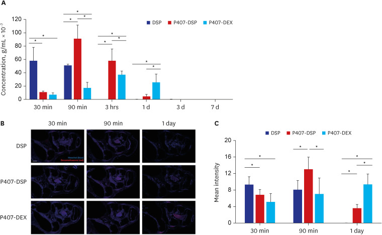
Immunohistochemical staining showed the presence and distribution of DEX in the cochlear tissues (Fig. 6B). Intensity analysis of these tissues showed that DPS resulted in a high drug uptake in the cochlear tissue after 30 minutes of treatment, but uptake decreased from the 90 minutes time point and was undetectable after one day. In contrast, P407-DSP continued to increase in drug uptake after 90 minutes, and the effective concentration was maintained after one day. The P407-DEX group, compared with the DSP only and P407-DSP groups, showed an increased cochlear tissue drug concentration even after 1 day with continuous drug uptake (Fig. 6C).
In vivo safety evaluation of the P407 hydrogel formulations
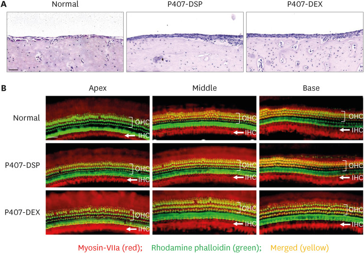
The possible negative effects of IT administration of the hydrogel formulations were evaluated by observing the survival of auditory hair cells using histopathological analyses. Histological evaluation of the middle and inner ears was performed seven days after IT injection. H&E staining was performed to detect mucosal inflammation in the middle ear. The results revealed that no significant inflammatory responses, such as edema and fibrosis, were present after P407-DSP or P407-DEX administration when compared with those in the untreated middle ear mucosa (Fig. 7A). Additionally, whole mount staining of inner hair cells and outer hair cells showed that no cellular loss or death was observed in any turns of the cochlea compared with the normal group (Fig. 7B).
Fig. 7
Evaluation of in vivo safety. (A) Sectional histopathologic findings of the middle ear mucosa in normal (left), 7 days after P407-DSP injection (middle) and 7 days after P407-DEX injection (right). Scale bar =50 μm. (B) Whole-mount of auditory epithelium in normal, 7 days after P407-DSP injection and P407-DEX injection. Tissues were stained for myosin-VIIa (red), rhodamine Phalloidin (green) to visualize hair cells and actin and then merged with the myosin-VIIa and rhodamine phalloidin. Scale bar = 50 μm.
DSP = dexamethasone sodium phosphate, DEX = dexamethasone, P407 = poloxamer 407, OHC = outer hair cell, IHC = inner hair cell.
DISCUSSION
The development of inner ear drug delivery therapy has become a rapidly expanding field of otolaryngology. In recent years, different materials have been developed to extend the retention time of drugs in the middle ear after IT administration. Thermo-sensitive hydrogels that can undergo sol-to-gel transition have been extensively utilized for injectable drug delivery systems, including IT administration for inner ear treatment strategy. Our previous study demonstrates that the hexanoyl glycol chitosan thermogel formulations could maintain a high drug concentration in the inner ear for a long time due to good residual stability by thermogelation.26 In the present study, we developed P407 as one of the representative thermo-sensitive hydrogels as an injectable formulation for the IT administration of DEX.
P407 is an amphiphilic block copolymer consisting of a hydrophilic poly (ethylene glycol) block and a hydrophobic poly (propylene glycol) block. P407 can enhance the water-solubility of hydrophobic drugs by self-assembly in aqueous media to form micellar structures.29 The results of the solubility tests confirmed that P407 could effectively solubilize the hydrophobic drug DEX. The enhanced DEX solubility may facilitate DEX's release from P407 formulations. P407 was also helpful in preparing DEX-loaded P407 formulations due to its micelle-forming ability. A hydrogel formulation prepared by simple physical mixing of P407 with DEX is not preferable because the non-uniform drug distribution or phase separation may occur. The DEX-loaded P407 micelles may help prepare more homogeneous hydrogel formulations without phase separation. The DSC thermogram and powder X-ray diffraction pattern of P407-DEX revealed the disappearance of the intrinsic crystalline characteristics of DEX after encapsulation with P407, indicating that the hydrophobic DEX is miscible with P407 and exists in a homogenously dispersed state. The thermo-sensitive sol-gel transition temperature of P407-DEX hydrogels was observed around 24°C, indicating that the hydrogels exist as an injectable liquid at room temperature but solidify in the body by thermogelation. All these unique physicochemical and thermogelling properties of P407 make it useful as a promising injectable hydrogel formulation for IT administration of DEX.
In in vivo experiments, we first evaluated the delivery efficiency of P407 formulations to the inner ear. In the case of the DSP solution, the drug concentration decreased so rapidly and could not be measured even 3 hours after IT injection because most of the drug in aqueous solution are likely to be rapidly removed from the tympanic cavity through the Eustachian tube and lymphatic channels before passing through the RWM. In contrast, both P407 formulations, P407-DSP and P407-DEX, demonstrated a significantly higher drug uptake due to their thermogelling properties that could enhance the retention time of the drug by preventing premature drug elimination. On the other hand, P407-DSP exhibited a higher drug concentration at the initial time, but the drug concentration quickly decreased, while P407-DEX showed that significant drug concentrations could be maintained for a longer time due to the relatively slow release characteristics of hydrophobic DEX without any initial burst release. For the P407-DEX formulation, a significant DEX concentration was detected even after 1 day, significantly longer than the half-life of DEX in the perilymph of animal models, which is about 18 to 130 minutes,3031 and this phenomenon was further confirmed by immunohistochemical staining. Therefore, the P407 thermogel formulations have the advantage of remaining in a gel state for a longer period in the cavity, which can provide more opportunities and time to contact with RWM, thereby increasing the efficiency of delivery to the inner ear. Although the P407 thermogel formulations offer a high potential to improve the delivery efficiency of the inner ear by increasing the contact time with RWM, its safety was a concern. After IT injection of P407 thermogel formulations for 7 days, histopathological analysis revealed no cytotoxicity or inflammation responses in the mucosa of the middle ear and whole-mount staining showed no significant hair cell loss in all turns of the cochlear. Therefore, P407 thermogel formulation is safe, and it has no adverse effects on the middle and inner ear.
In this study, we prepared and evaluated injectable P407 hydrogel formulations to efficiently deliver DEX to the inner ear. The P407 polymer could effectively solubilize the hydrophobic DEX and be used as an injectable hydrogel formulation with thermosensitive sol-gel transition and sustained release properties. In vivo studies, using a guinea pig model, showed that the hydrogel formulations delivered considerably high drug concentrations to the inner ear via IT injection and demonstrated a favorable safety profile without any apparent cytotoxicity or inflammation in the middle and inner ear. These findings indicate that injectable P407 hydrogel formulations can efficiently deliver drugs to the inner ear via IT injection because of their biocompatibility and beneficial functionalities, such as drug solubilization capacity, thermogelation, and controlled release. Further studies using an animal model of hearing loss will be conducted to observe how effective the injectable P407 formulation is for treatment and recovery.
 XML Download