

 PDF
PDF Citation
 Print
Print


INTRODUCTION
Primary gastrointestinal lymphoma accounts for approximately one-third of extranodal non-Hodgkin’s lymphomas and a small proportion of gastrointestinal malignancies. The most common subtypes of primary gastrointestinal lymphoma are diffuse large B-cell lymphoma and mucosa-associated lymphoid tissue (MALT) lymphoma.1-3 Primary MALT lymphoma is a rare distinct subtype of non-Hodgkin’s lymphomas, occurring in approximately 8% of all non-Hodgkin lymphomas.4-6 Most primary gastrointestinal MALT lymphomas occur in the stomach, and duodenal involvement is extremely rare.7,8 Therefore, the clinical manifestations, treatment, and prognosis of primary duodenal MALT lymphoma have not been validated because of its rarity.
This paper reports a case of a 40-year-old male with MALT lymphoma arising in the duodenum that was treated successfully with radiation therapy alone, along with a review of the relevant literature.
CASE REPORT
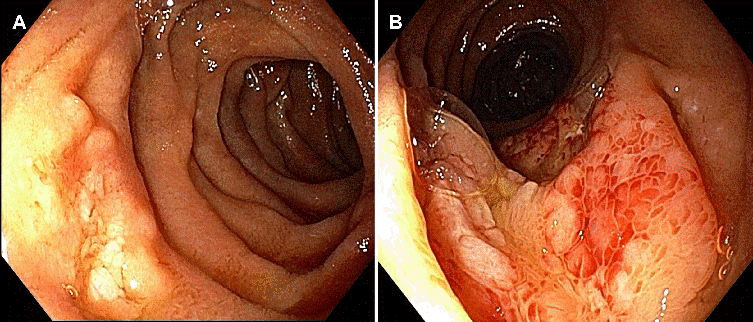
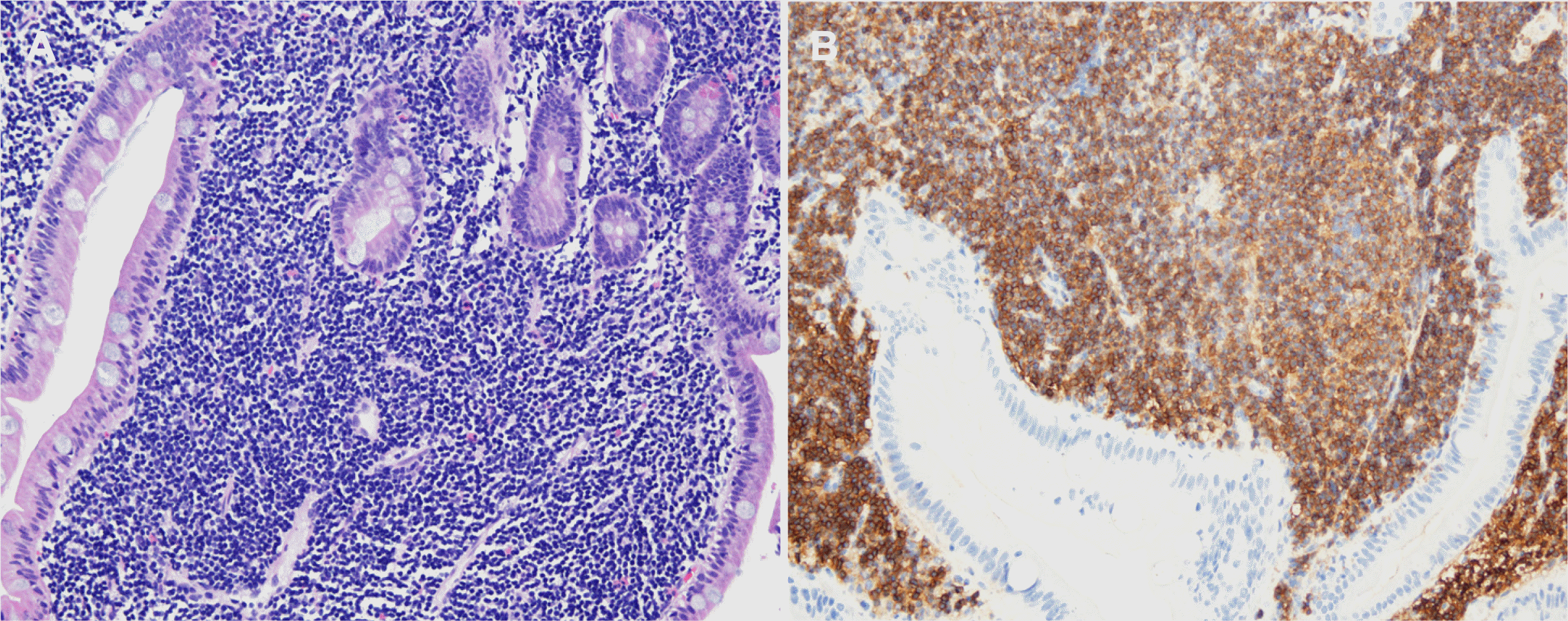
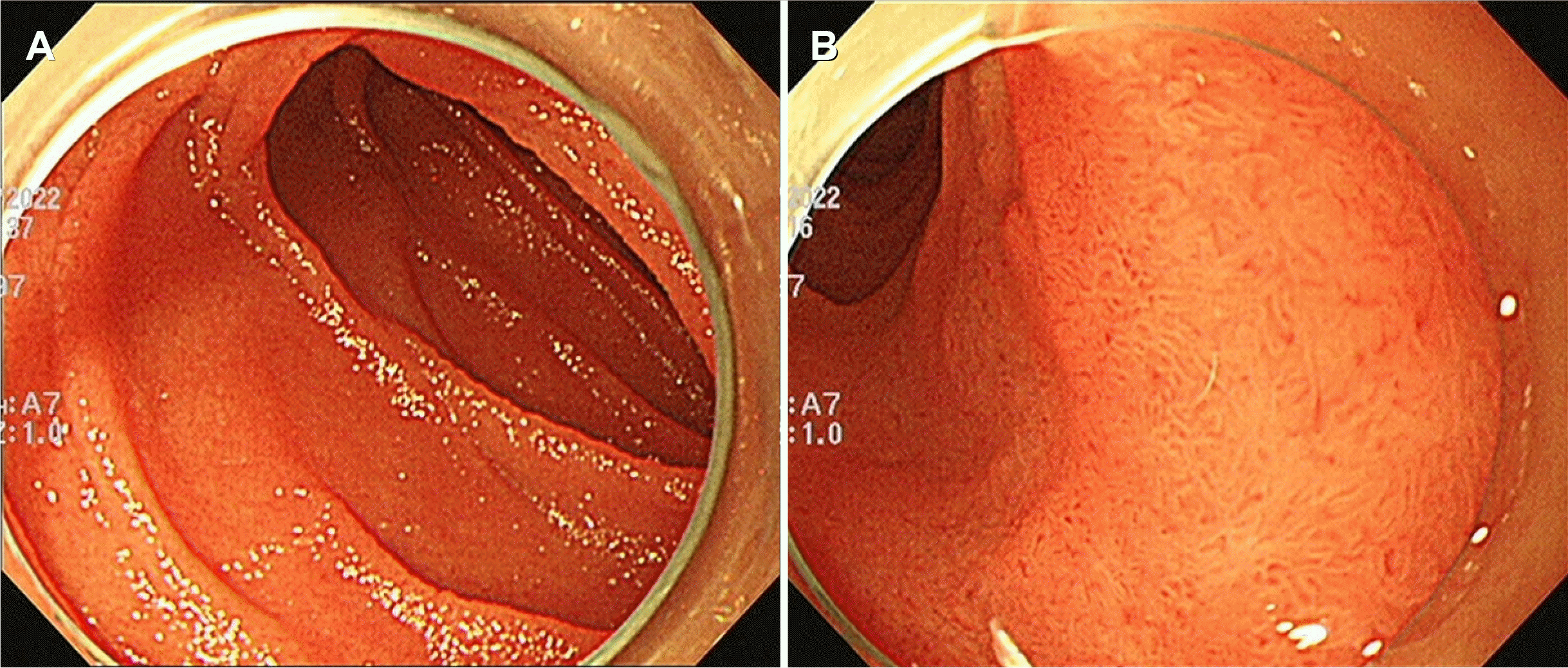
A 40-year-old male visited the authors’ clinic for a medical check-up. He had no past medical history. He denied any gastrointestinal and systemic B symptoms or signs, such as abdominal pain, nausea, vomiting, fever, night sweats, and weight loss. He was afebrile, and his blood pressure and pulse rates were normal. His abdominal examination was unremarkable. The findings included a soft and non-tender abdomen, no hepatosplenomegaly, and no palpable superficial lymph nodes. The laboratory examinations revealed the following findings: white blood cell count 5,400/mm3 (normal range: 6,000–10,000/mm3), hemoglobin 15.7 g/dL (normal range: 12–16 g/dL), platelet count 161,000/mm3 (normal range: 130,000–450,000/mm3), blood urea nitrogen 12.6 mg/dL (normal range: 8–23 mg/dL), creatinine 0.85 mg/dL (normal range: 0.5–1.3 mg/dL), serum albumin 4.5 g/dL (normal range: 3.0–5.0 g/dL), aspartate aminotransferase 26 U/L (normal range: 5–37 U/L), alanine aminotransferase 21 U/L (normal range: 5–40 U/L), alkaline phosphatase 57 U/L (normal range: 39–117 U/L), or lactate dehydrogenase 335 IU/L (normal range: 218–472 IU/L). Total bilirubin was 0.89 mg/dL with a direct fraction of 0.21 mg/dL (normal range: 0.2–1.2/0.05–0.3 mg/dL). Serum carcinoembryonic antigen was within normal limits. Esophagogastroduodenoscopy showed whitish multi-nodular mucosal lesions in the second and third portions of the duodenum. The polymerase chain reaction test for Helicobacter pylori (H. pylori) DNA showed negative (Fig. 1A, B). The histological examinations, including hematoxylin–eosin staining, showed dense infiltration of atypical lymphoid cells with lymphoepithelial lesions in the duodenal mucosa (Fig. 2A). The immunohistochemical examination showed that tumor cells were positive for the B-cell marker CD20 (Fig. 2B) and negative for CD3 and Bcl-6. The biopsy specimens indicated MALT lymphoma of the duodenum. Contrast-enhanced neck, chest, abdomen, and pelvis CT, PET-CT, and bone marrow aspiration revealed negativity for lymphoma involvement. The tumor was diagnosed as primary duodenal MALT lymphoma and classified as stage IE according to the Ann Arbor staging classification. A discussion with the multidisciplinary medical team, including a medical oncologist, radiation oncologist, gastroenterologist, and surgeon, led to the decision to perform radiation therapy. The patient received a total dose of 3,000 cGy in 15 fractions with involved site external beam radiation therapy for three weeks. Three months after the completion of radiation therapy, the esophagogastroduodenoscopy and pathologic examination revealed complete resolution of the duodenal lesions (Fig. 3A, B). The patient has been on a regular follow-up schedule at the outpatient clinic. The serial follow-up exams with a biopsy at three months and 12 months after radiation therapy revealed no evidence of tumor recurrence. Informed consent was obtained from the patient for publication.
DISCUSSION
MALT lymphoma is an extranodal marginal zone B cell lymphoma arising from MALT in various organs, including the gastrointestinal tract, salivary gland, parotid gland, lung, thyroid gland, thymus, skin, bladder, mammary gland, and lacrimal gland.4-6 The stomach is the most common site of gastrointestinal MALT lymphoma involvement, but duodenal involvement is extremely rare.7,8 Therefore, little is known about the clinical data, presenting manifestations, treatment, or prognosis of primary duodenal MALT lymphoma owing to its rarity.
Two reports from USA and Korea revealed the mean age at diagnosis of duodenal MALT lymphoma to be 56.2 years (range 20–78) in USA9 and 49.7 years (range 20–72) in Korea.10 The gender ratio was 11 males:15 females in USA,9 and seven males: six females in Korea.10 Duodenal MALT lymphoma can be asymptomatic in the early stages, but it may induce abdominal discomfort or pain, vomiting, hematemesis, melena, and occasionally outlet obstruction according to the lesion type, location, size, and degree of obstruction.9,10 The most common site was the bulb, followed by the second portion and multiple sites. The main endoscopic feature was a nodular or polypoid lesion, followed by ulceration, granular lesion, and mass.9,10 In the present case, a 40-year-old man with asymptomatic MALT lymphoma arising from the second and third portions of the duodenum, which presented as whitish multi-nodular mucosal lesions were found incidentally on health screening endoscopy.
Previously, duodenal MALT lymphomas were treated with various modalities, including single or combination of H. pylori eradication, surgery, chemotherapy, or radiation therapy.9,10 Despite this, the complete remission rates in patients with duodenal MALT lymphoma have been disappointing compared to gastric MALT lymphoma patients.10 The treatment and outcome of duodenal MALT lymphoma are not yet well established because of its rarity.
Gastric MALT lymphoma is associated strongly with H. pylori infection, and H. pylori eradication is a well-known standardized therapy for gastric MALT lymphoma.7,8 Although some reports have shown that H. pylori eradication alone was effective for treating duodenal MALT lymphomas,11-14 the association between duodenal MALT lymphoma and H. pylori infection is unclear. A comparison report of 13 duodenal MALT lymphomas and 130 gastric MALT lymphomas showed that the H. pylori infection rates were less than half in duodenal MALT lymphomas.10 In the present case, duodenal MALT lymphoma was not associated with a H. pylori infection. Some reports have shown that a duodenal MALT lymphoma was resistant to H. pylori eradication therapy.15,16
Radiation therapy is associated with better survival and lower mortality in patients with early-stage or localized MALT lymphomas arising in the gastrointestinal tract and other organs, compared to other treatment options and modalities, such as surgical resection or chemotherapy.4-8,17 Therefore, radiation therapy may be a feasible and safe treatment, allowing organ preservation and avoiding toxicity, morbidity, and mortality, which are associated with aggressive surgical resection or chemotherapy in early and localized duodenal MALT lymphoma. In the present case, definitive treatment with involved site radiation therapy was planned by a discussion with the multidisciplinary medical team because the lesion was localized to the duodenum alone and was detected early. Early detection was achieved by closely examining the duodenum 2nd to 3rd portions, which is easily missed, especially during a routine medical examination. Three months after radiation therapy, endoscopic examination with biopsy for pathologic examination revealed complete resolution of the duodenal lesions. Twelve months after radiation therapy, the patient had not developed any tumor recurrence. On the other hand, it was difficult to accurately target the lesion in radiation therapy because the small intestine is movable and not fixed in the abdominal cavity. Further study will be necessary to determine the long-term efficacy and probability of normal tissue complications of this treatment approach.
 XML Download