

 PDF
PDF Citation
 Print
Print


INTRODUCTION
Anchorage in orthodontic treatment is a crucial consideration when correcting different types of malocclusions. Orthodontists have created many types of intra- and extra-oral appliances to enhance anchorage. The function of different types of anchorage is to facilitate planned tooth movement and avoid side effects in other parts of the dentition.
The first animal study on basal bone anchorage for orthodontic treatment failed to maintain anchorage for more than 1 month because of the lack of osseointegration.1 However, after many years of research and development, the concept of osseointegration using titanium implants was established and successfully applied for restorative purposes by Dr. Brånemark in the 1970’s.2 Creekmore and Eklund3 succeeded in applying the first temporary anchorage device (TAD) at the anterior nasal spine for deep bite correction.
TADs facilitate different types of orthodontic tooth movement, including molar intrusion, which is considered a challenging tooth movement. After many clinical trials in the application of TADs, controlling the posterior dentoalveolar height by intruding the molars into the supporting bone became achievable. Molar intrusion with TADs is now a predictable technique for correcting over-erupted molars and flattening the occlusal plane4,5 or reducing posterior dentoalveolar height6-11 for counterclockwise rotation of the mandible.
The envelope of intrusive movement with TADs affects treatment planning.4-11 The mean amount of molar intrusion with TADs has been clarified and ranges from 2.1 mm to 4 mm.4-6,10,12,13 For extreme supra-erupted second molars, intrusion can reach 8 mm.14
Alveolar bone and gingiva remodeling follows orthodontic tooth movement. The tissue response after molar intrusion has been discussed in many studies,12,15-20 including clinical and radiographic evaluation of periodontal conditions and histological analysis in animal experiments. Nevertheless, no histological findings have been reported on patients after molar intrusion because of ethical concerns.
Therefore, we report the case of a patient treated with five TADs to reduce protrusion and gummy smile with a high mandibular plane angle (MPA: SN-MP). While retracting the upper and lower anterior teeth, we intruded the upper molars with bilateral infra-zygomatic crest (IZC) mini-screws and maintained the vertical height of the lower molars with mini-screws at the buccal shelf. After 5 months of molar intrusion, shortened clinical crowns of the bilateral upper molars were noticed, concomitant with the difficulty of further orthodontic tooth movement. After full discussion with the patient, osseous resective surgeries were performed in the bilateral upper posterior region to prevent redundant alveolar bone from causing physical hindrance. During the surgery, the hard and soft tissue surrounding the crown portion of the intruded molars was carefully removed to obtain biological width and a specimen was sent for biopsy. The surgery resulted in a healthy periodontium with normal architecture and increased the ease of maintaining oral hygiene.
DIAGNOSIS AND ETIOLOGY
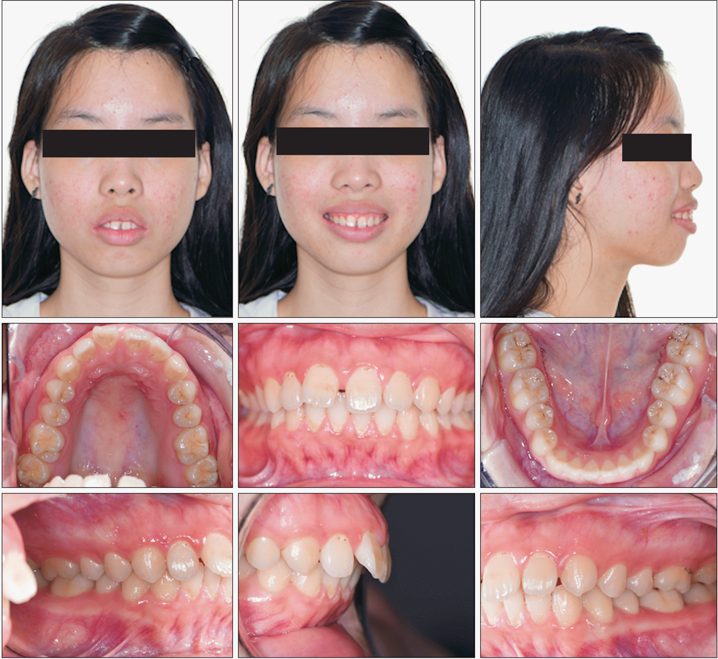
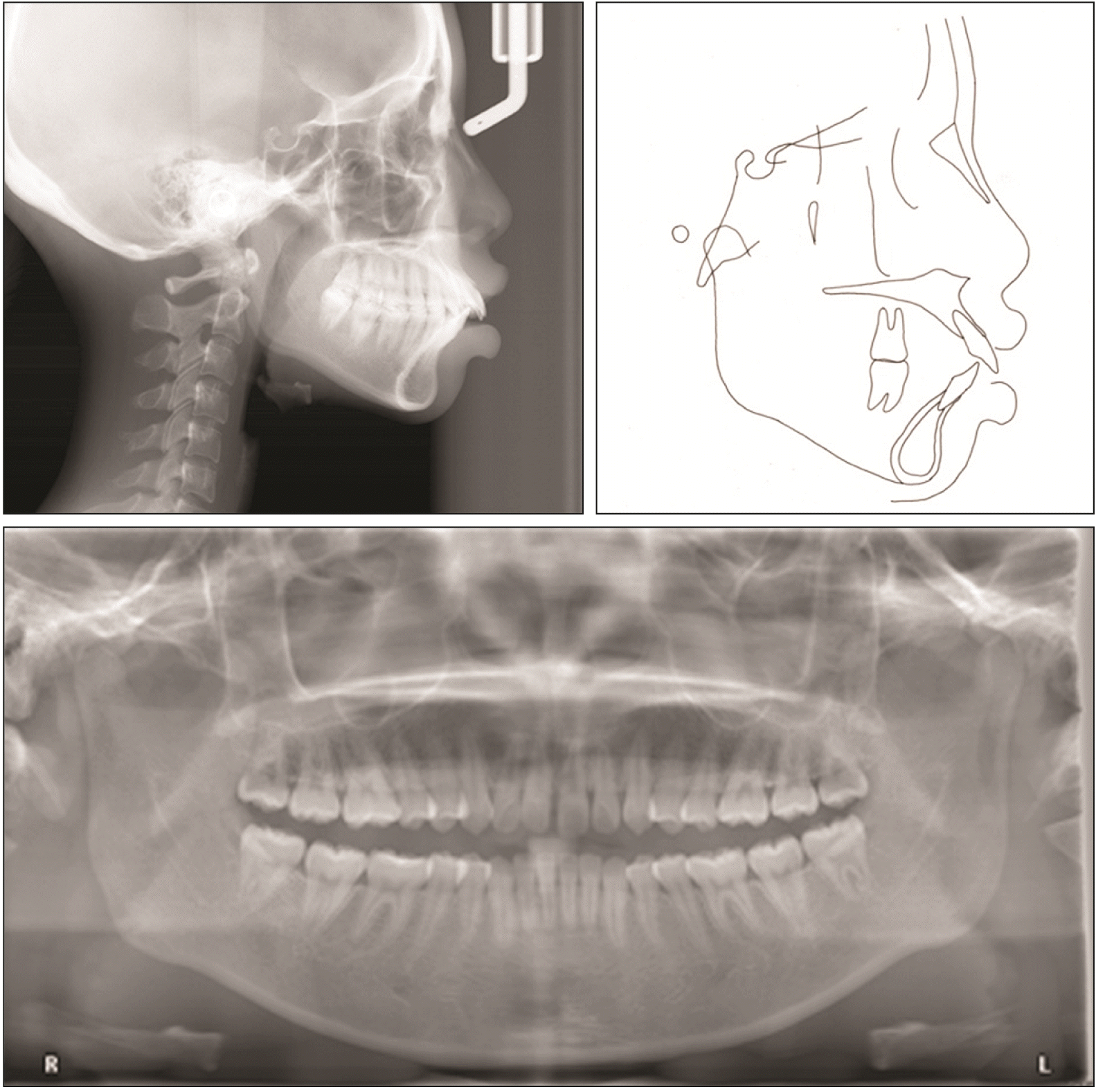
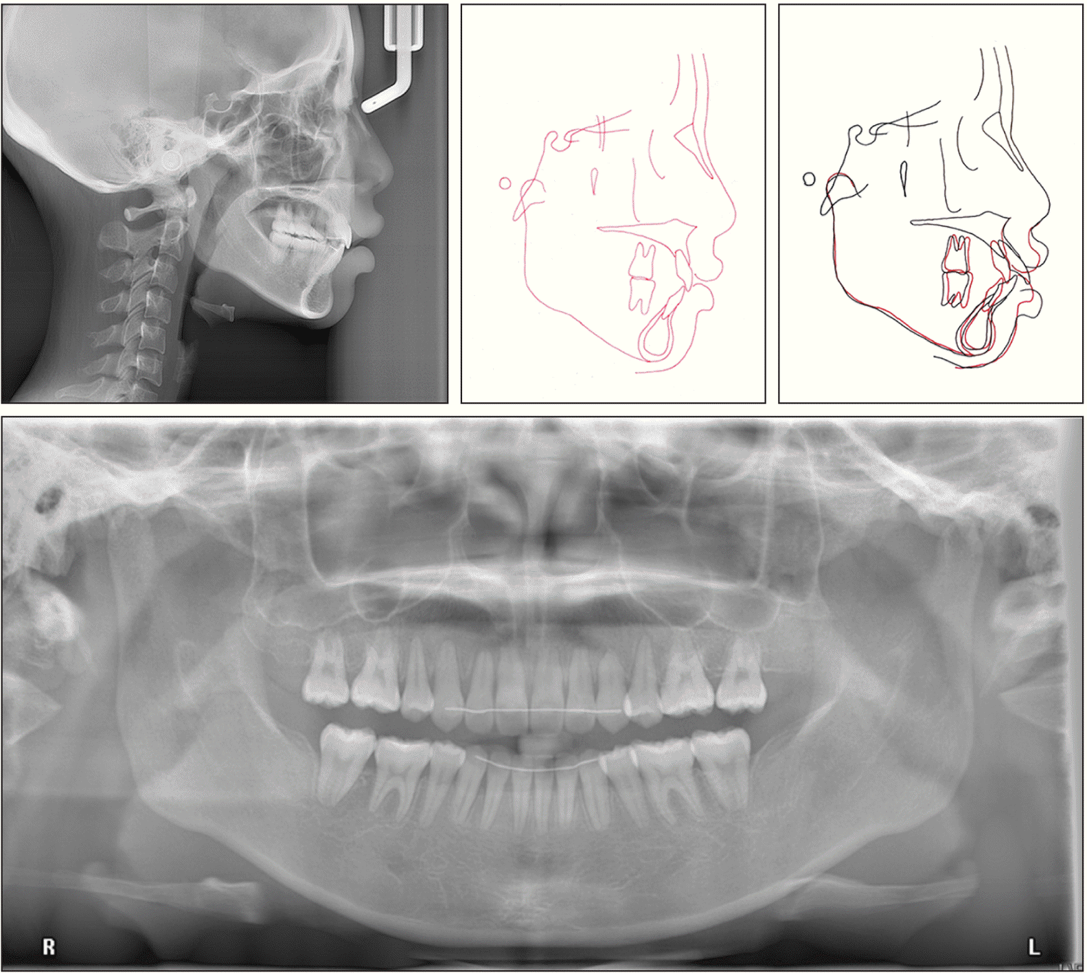
A 17-year-old girl complained of protrusive front teeth and gummy smile. This patient had a convex facial profile with protrusive upper and lower lips. Facial proportions were within normal range with a 4-mm gummy smile. A 2-mm space between the upper central incisors, as well as a 1-mm space at the distal side of both the upper central incisors were noticed. The lower midline deviated to the left side by 1 mm. She had bilateral Class I molar and Class II canine relationships (Figure 1). Panoramic radiography showed that this patient had no missing teeth and none of the third molars were impacted (Figure 2). Cephalometric analysis (Table 1) revealed an orthognathic maxilla (SNA 79.5°), mild mandibular retrognathism (SNB 75°), and a high MPA (37°). This patient had excessive upper anterior and upper posterior dental heights (UADH 34 mm, UPDH 29 mm), and proclined upper and lower incisors (U1-SN 113°, L1-MP 105°). The upper and lower lips were protrusive to the E-line (approximately 6 mm and 11 mm, respectively).
TREATMENT OBJECTIVE
Four first premolar extractions with maximal anchorage for retraction and intrusion were planned to reduce protrusion and eliminate gummy smile. Two mini-screws at the IZC were applied to intrude and retract the upper teeth simultaneously for more balanced lateral profile with good chin projection, pleasing smiling, and solid Class I occlusion.
TREATMENT ALTERNATIVES
We provided two treatment alternatives:
1. Improvement of the patient’s facial profile using full fixed orthodontic appliances combined with orthographic surgery. Le Fort I three-piece osteotomy would be performed with the extraction of two upper first premolars and anterior sub-apical osteotomy would be performed at the mandible with the extraction of two lower first premolars. This option could efficiently correct her gummy smile and protrusion.
2. Improvement of the patient’s facial profile using full fixed orthodontic appliances with TADs and four first bicuspid extractions. TADs would be required in the four quadrants and the anterior maxilla for maximal retraction of the anterior teeth and vertical control of the upper posterior teeth. Upper molar and incisal intrusion would be performed to reduce the posterior vertical dimension for mandibular rotation to further improve the patient’s lateral profile and her gummy smile. Esthetic crown lengthening surgery would be necessary if gingival remodeling following orthodontic intrusion was lacking. The patient chose this option to avoid the risks associated with orthognathic surgery.
Written informed consent was obtained from the patients and their parents.
TREATMENT PROGRESS
Pre-adjusted, fixed, orthodontic brackets (Victory Series Brackets, 0.018 slot; 3M Unitek, Monrovia, CA, USA) were bonded after the four third molars and four first bicuspids were extracted. Two 0.014-inch nickel-titanium arch wires were used initially at the upper and lower arches. Two months after banding and bonding, we inserted bilateral mini-screws (Bio-Ray A1-P mini screw, 2.0 mm in diameter, 17 mm in length) in the IZC areas. Two mini-screws (Bio-Ray A1-P mini screw, 2.0 mm in diameter, 10 mm in length) at the bilateral buccal shelf were inserted 3 months after the orthodontic treatment was initiated.
For the mandibular arch, ligature wire fixation from the mini-screws to the first molars provided absolute anchorage of the posterior teeth. Two nickel-titanium coil springs were then used between the first molars and canines for retraction of the lower anterior teeth.
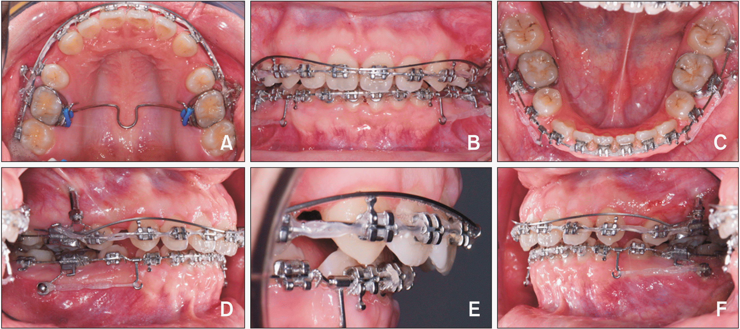
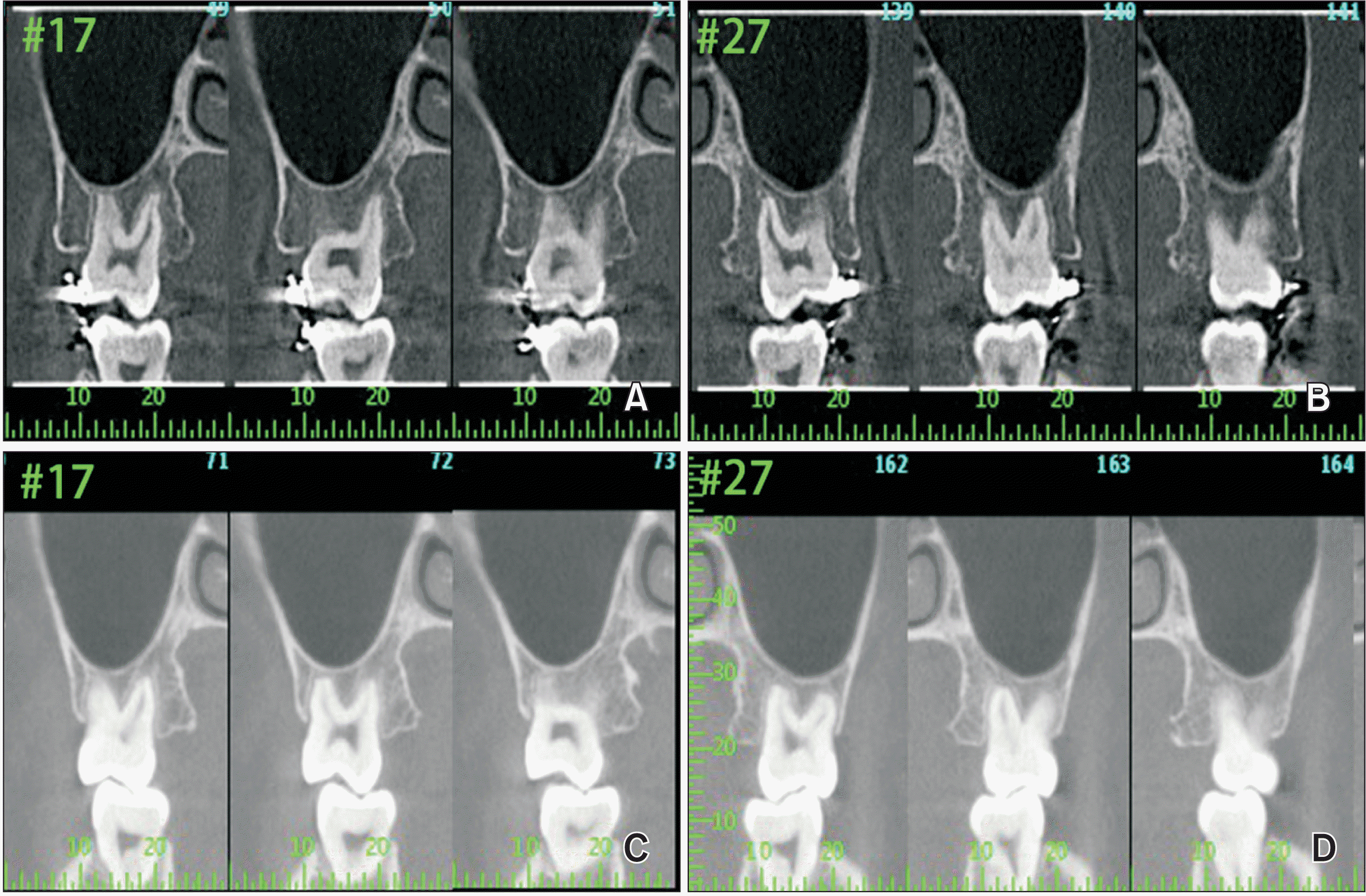
In the maxillary arch, major efforts were made for retraction and intrusion. When inserting 0.016 × 0.022-inch stainless steel archwire, the archwire was bent into a compensating curve, and also an intrusive archwire was delivered for anterior vertical control. At the same time, during the anterior retraction process, nickel-titanium coil springs were inserted between the bilateral upper canines and the mini-screws. During en masse retraction, power chains with 300 grams of force, were used between the bilateral IZC mini-screws and the main archwire for upper molar intrusion. Ten months into treatment, a trans-palatal arch (TPA) was used for torque control of the upper molars during the molar intrusion process. At the same appointment, a mini-screw (Bio-Ray A1-P mini screw, 2.0 mm in diameter, 10 mm in length) was inserted in the sub-apical area, between the two upper central incisors, for anterior vertical control. After 5 months of molar intrusion, the orthodontist noticed that the molar brackets were impinging on the gingiva, hindering both orthodontic tooth movement and oral hygiene care (Figure 3).
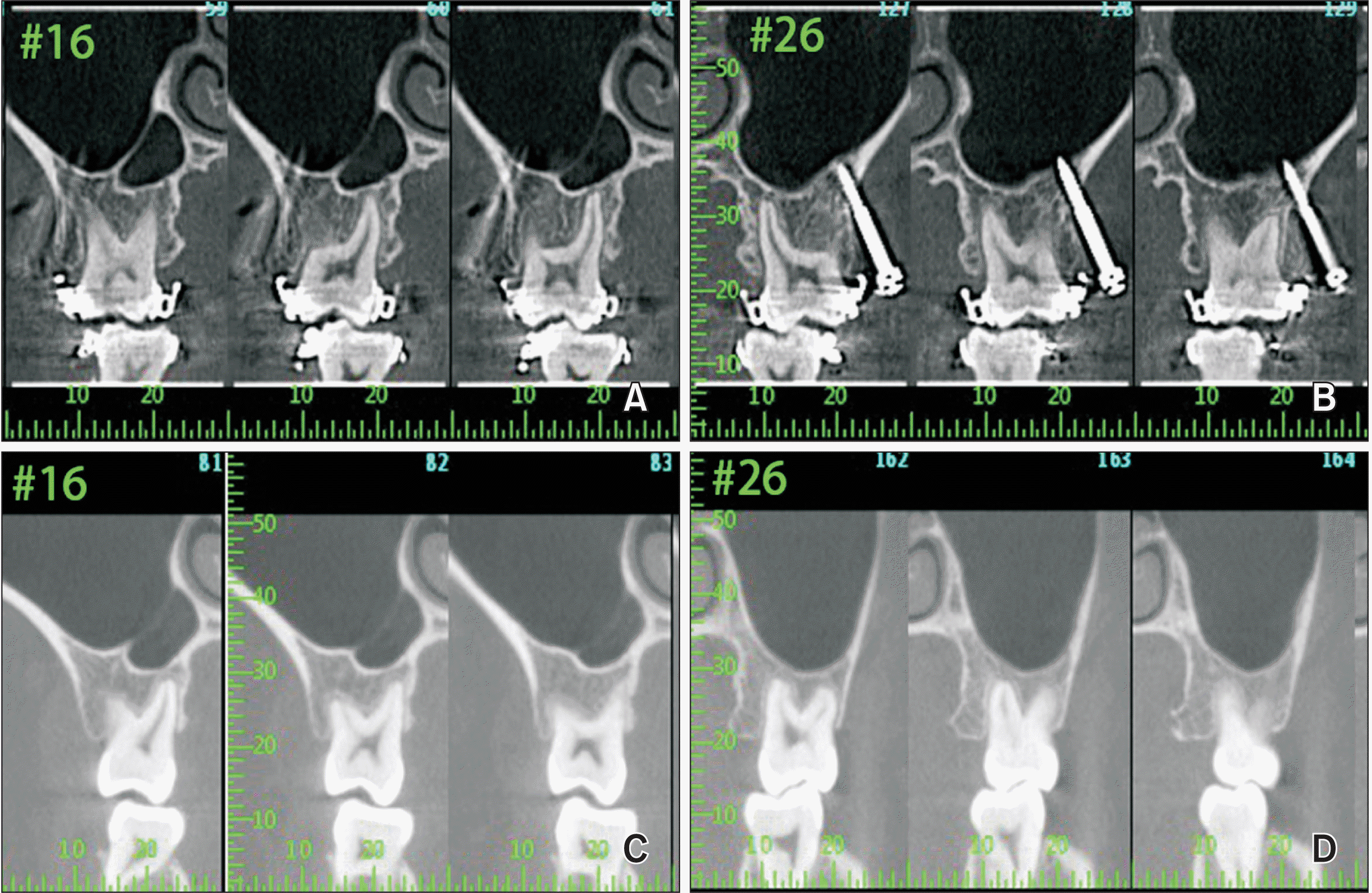
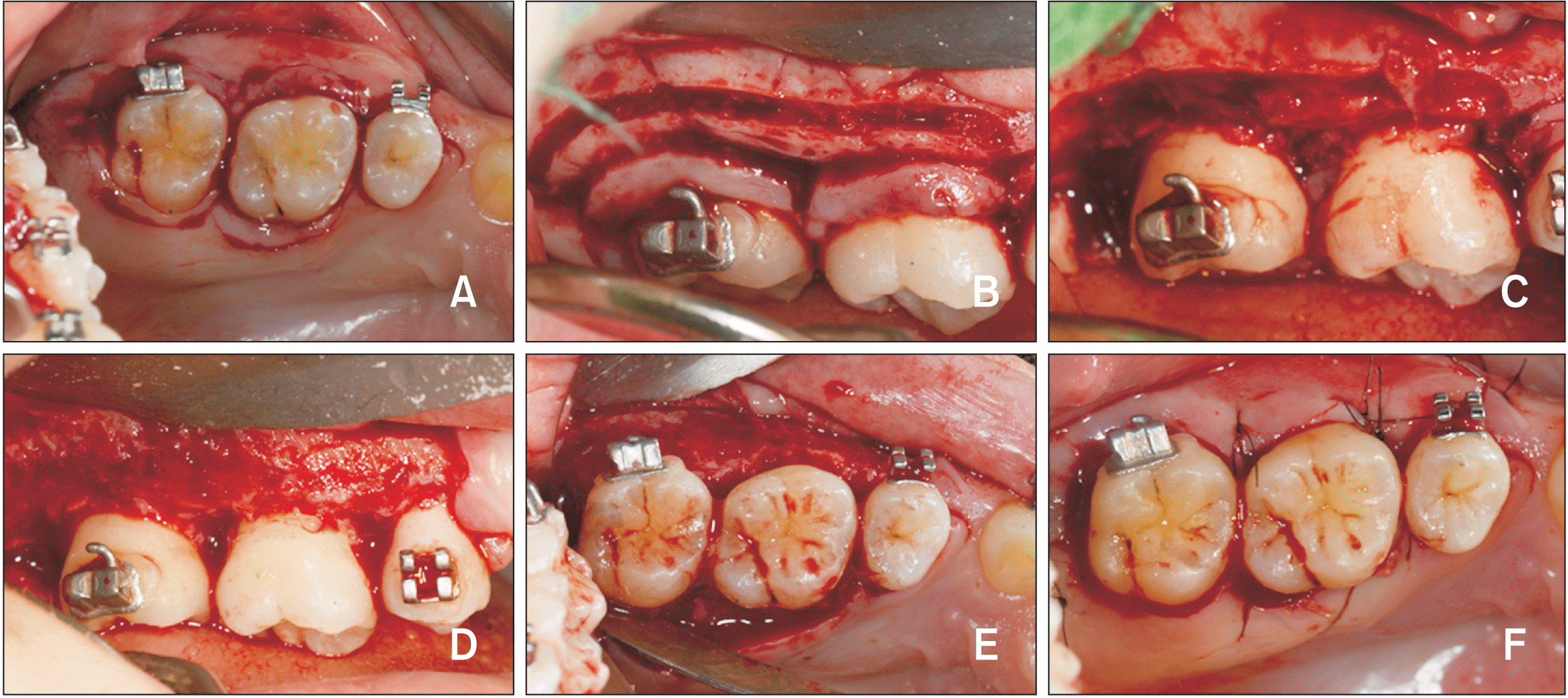
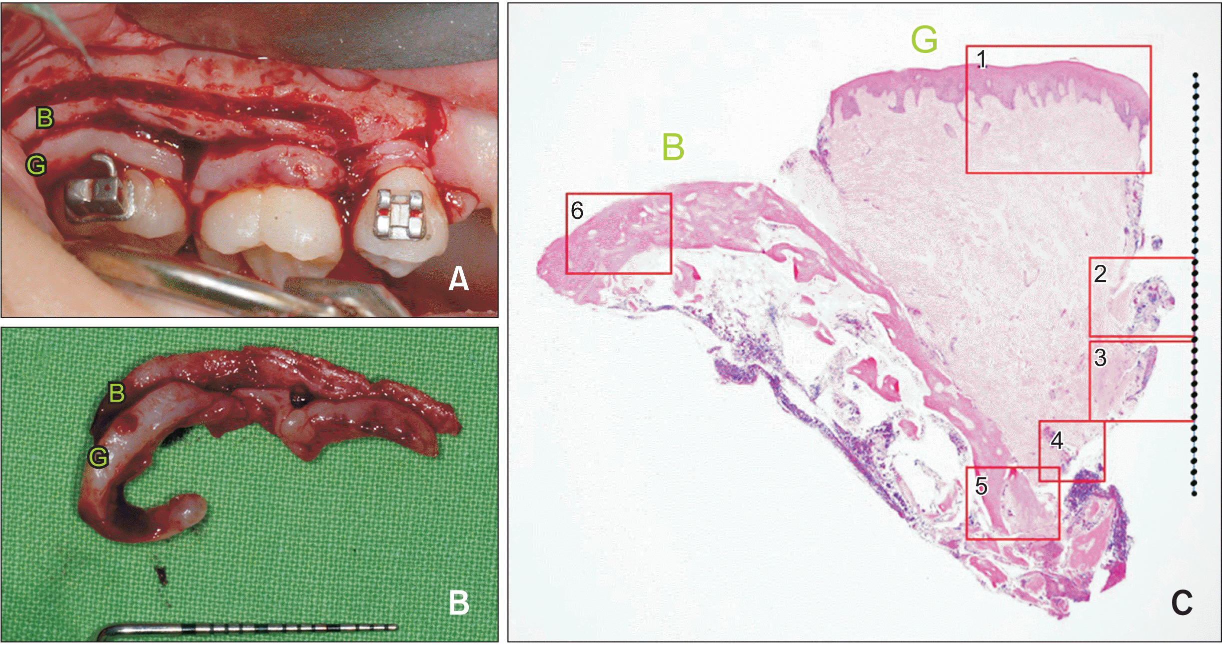
Cone-beam computed tomography (CBCT) images, which were taken at 11 months into treatment, revealed the sites of impingement. The clinical attachment levels of the upper molars were below the crest of the bulging alveolar bone, and the hooks of the molar brackets were impinging on the alveolar bone (Figures 4 and 5). Osseous resective surgery was scheduled by the periodontist after a thorough discussion with the patient and her family. The purpose of the periodontal surgery was to re-contour the osseous tissue and create natural bony architecture for effective hygiene. Resective periodontal surgeries on the upper posterior region were delayed till 18 months into the treatment because of issues related to the COVID-19 pandemic. During the first surgery, intra-sulcular incisions were made around the molars and a vertical incision was made at the disto-buccal line angle of the upper left second premolar. After a full-thickness flap was reflected, 3-mm infra-bony defects were noticed on the buccal side of the molars. Alveolar bone reshaping was performed using a high-speed carbide bur and a back action chisel (Figures 6 and 7). The second resective periodontal surgery was performed 2 months later on the upper right posterior molars. With awareness of the amount of redundant tissue from the first surgery, this time, submarginal incisions were made and a horizontal bony cut was made 2 mm lower using a high-speed carbide bur at the upper right molars. The bulging alveolar bone and gingiva around the molars was harvested for biopsy using a back action chisel. After natural bony architecture was created around the molars, the flap was then primarily closed (Figure 7). Bilateral IZC screws were removed during the surgeries for better access.
The orthodontic treatment for further retraction was continued 1 week after the completion of periodontal surgeries. The lower main archwire was shaped into a reverse curve of Spee, and Class II elastics were used for retracting upper anterior teeth. The TADs at the bilateral buccal shelf provided absolute anchorage for the lower arch and prevented lower molar extrusion. Upper archwire with a compensating curve was used along with an intrusive archwire for controlling the overbite. The Class II elastics were used again, this time without the lower molars being tied to TADs to enable free movement, thus achieving a Class I molar relationship in the final 6 months of treatment.
Treatment was completed after 2 years and 10 months when normal Class I occlusion was achieved. Lingual fixed retainers and Essix retainers for the upper and lower arches were provided for retention.
RESULTS
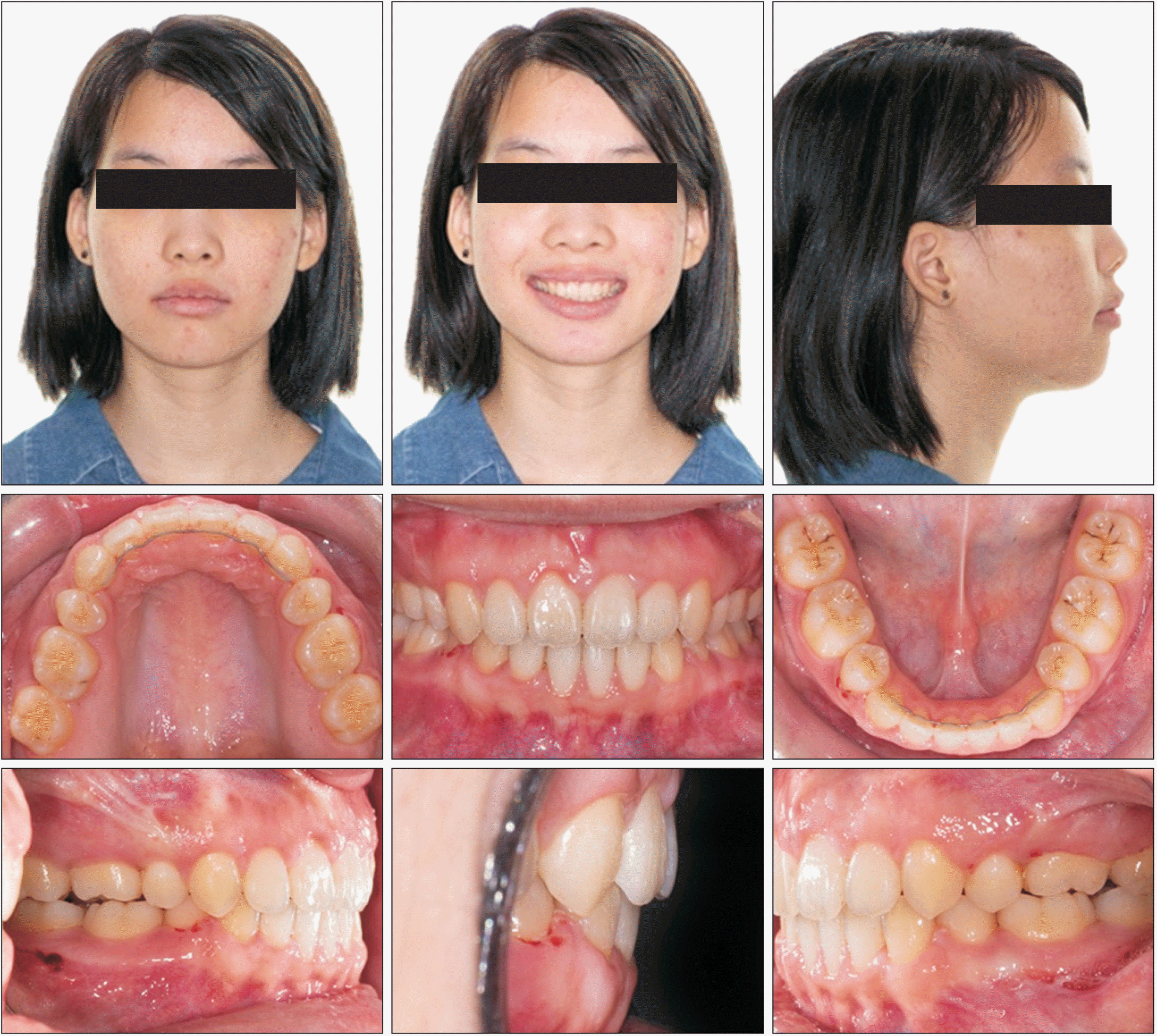
The orthodontic treatment achieved an acceptable overjet, overbite, bilateral Class I molar and canine relationships, and centered midlines (Figure 8). The superimposition of lateral cephalograms showed that MPA had decreased by 1.5° (from 37° to 35.5°). The UPDH decreased from 29 mm to 27.5 mm. This may be contributed by the use of TADs at the bilateral IZC for molar intrusion. The upper and lower incisors were upright after the orthodontic treatment (U1-SN from 113° to 97°, L1-MP from 105° to 97°). The distance from the upper lip to the E-line was 2 mm and the distance from the lower lip to the E-line was 4 mm. The cephalometric analysis data are listed in Table 1.
The chief complaints in this case, dental protrusion and gummy smile, were successfully addressed with four first bicuspid extractions with maximal anchorage using TADs. Furthermore, vertical control to reduce anterior and posterior dental height with TADs, facilitated counterclockwise rotation of the mandible and better chin projection, and further improved the convex lateral facial profile (Figure 9).
HISTOLOGICAL FINDINGS
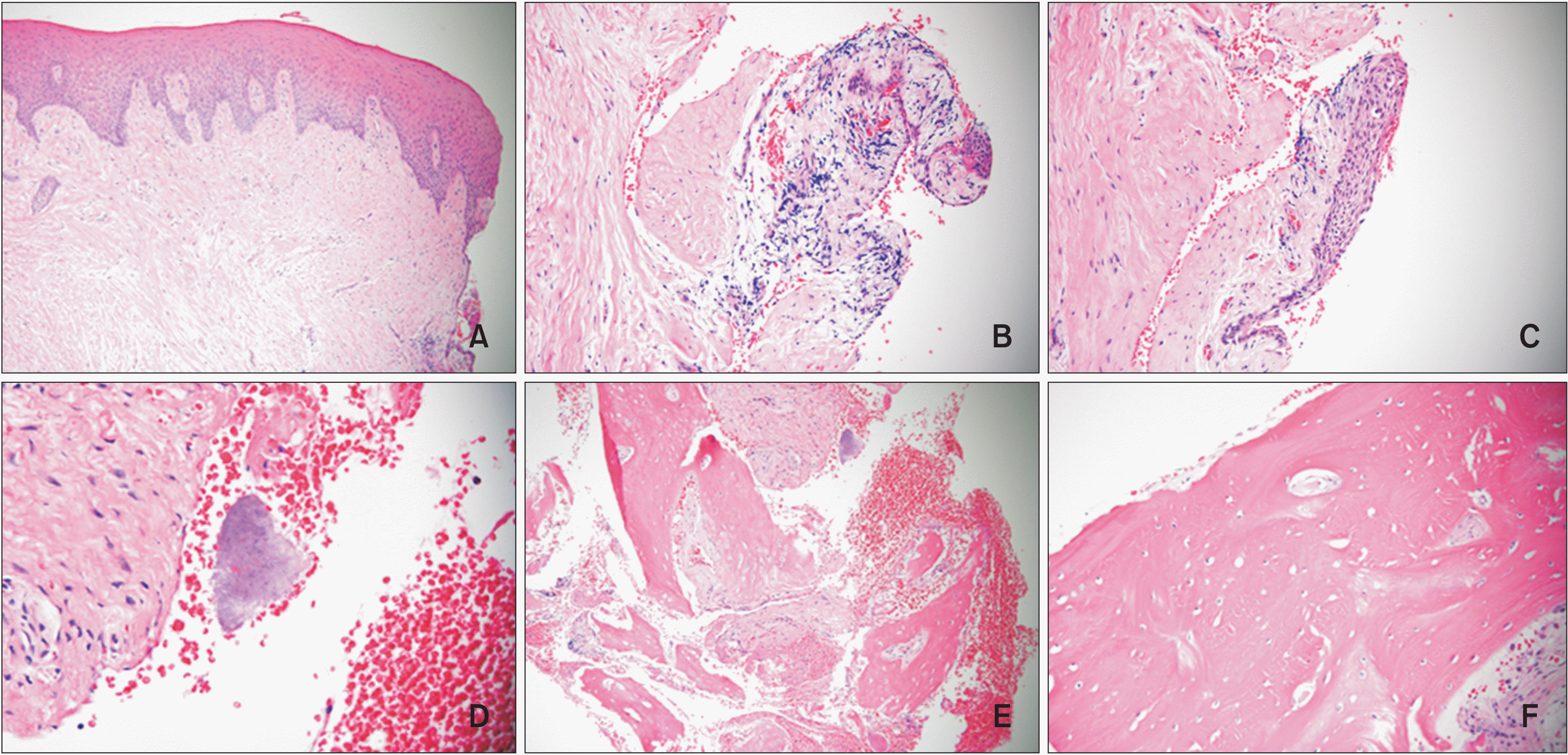
A specimen that demonstrated the relationship between the soft and hard tissue was preserved during resective surgery on the buccal side of the right second molar (Figure 10). Dimensions were measured using a histological section: the width of the soft tissue sulcus was 2.7 mm at top; the length of tissue facing the tooth was approximately 3.0 mm from the top of the gingiva to the bottom where the bone with inflammatory cells infiltrated.
On the buccal side, the gingiva was covered by parakeratinized squamous epithelium and supported by dense fibrous lamina propria without inflammatory cell infiltration. Along the surface of the gingival sulcus facing the clinical crown (Figure 11), few sulcular epithelium and bacterial colonies were present. Some chronic inflammatory cells that infiltrated below the non-keratinized sulcular epithelium were noted, with abundant capillaries filled with red blood cells (Figure 11E). The bone facing the sulcular region at the bottom showed bone remodeling and woven bone formation with plump osteocytes in the lacunae. The bone facing the buccal side without covering soft tissue, which was reflected and preserved for clinical suturing, was composed of dense and mature laminated bone with small osteocytes in the lacunae and fibrovascular tissue in the Haversian canals, indicating a relatively slow turnover of bone here (Figure 11F).
DISCUSSION
To present date, no human histological study of molar intrusion has been reported due to ethical issues. In our clinical case, an unexpected delay in tissue remodeling was noticed after 5 months of the active intrusion. The dentoalveolar remodeling had not followed the tooth movement at a distance from the periodontal ligament (PDL). The redundant tissue had to be corrected via osseous resective surgeries to restore periodontal health and resume orthodontic tooth movement. Two surgeries on this patient allowed us to obtain biopsy tissue with accuracy. The amount of redundant gingiva-alveolar bone tissue around the intruded molars was estimated from the first surgery, we then could cautiously detach unwanted tissues as a whole piece during the second surgery and send for biopsy (Figure 10). This well-preserved piece around intruded molar provided a valuable histological study as described in Figures 10 and 11.
The goal of the osseous resective surgery was to reshape the bulging marginal bone to better simulate physiological architecture, thus achieving a proper tooth–alveolar bone relationship.21 It is widely accepted that alveolar bone remodeling is expected to follow orthodontic treatment, but in some cases, uneven bony architecture around the teeth after orthodontic tooth movement is noticed. In our molar intrusion case, the alveolar bone and the gingiva did not follow the tooth movement. Few reports are available on clinical periodontal changes after molar intrusion,12,18,20 including histological findings from animal experiments.15,16,19,22 According to Kanzaki et al.,16 during molar intrusion with skeletal anchorage devices, pressure from supra-alveolar fibers induces alveolar bone resorption and remodeling. In the case of a large amount of intrusion,19 dento-periosteal fibers and dento-gingival fibers gradually separate from the cementum. Bone crest resorption then decreases and unfavorable bony architecture forms.
No studies have discussed the range of alveolar bone remodeling, nor have they discussed the time needed for periodontium remodeling. Influence of the initial alveolar bone thickness on the alveolar bone remodeling was discussed by Hong et al.20 It was concluded that many factors potentially influence the treatment and affect the result. According to Chung et al.,23 the alveolar bone remodeling with orthodontic tooth movement is dependent on the PDL component, the stress/strain distribution, in the periodontium. Bone surrounding the tooth has more PDL insertion, however over a particular distance from the root surface, the mechanical stress mediated from PDL decreases and the biological reaction of the tissue changes. In our clinical case, after 5 months of active upper molar intrusion into the wide alveolar ridge, the orthodontist noticed the molar bracket hooks were impinging on the gingiva, and therefore surgery was needed. Due to the issues with COVID-19 however, the osseous resective surgeries could not be performed until 13 months after the intrusion, and even till then, the tooth-bone relationship and the morphology of the alveolar bone had not changed. Further investigations on the extent and duration needed for alveolar bone remodeling around moving roots will justify the clinical timing for resective surgery.
Root resorption induced by orthodontic treatment as found in this clinical case after 5 months of active upper molar intrusion on the CBCT images, is a common side effect, which is undesirable and compromises tooth structure and our treatment goals. Upon detection, active intrusion was discontinued to prevent further root resorption in our case. Although Burstone suggested 50 grams of intrusive force for the upper central incisors, and 200 grams for the six upper anterior teeth, an optimal intrusive force for premolars and molars has not yet been established. An intrusive force ranging from 90 to 300 grams for posterior teeth is commonly used.24,25 Many investigations have tried to identify factors which relate to orthodontic induced root resorption.25,26 It was concluded that continuous heavy force causes greater root resorption than continuous light force, and increased force levels can increase the risk of root resorption.25,26 Therefore, optimal light continuous force is crucial to avoid excessive root resorption. During orthodontic treatment, monitoring the root resorption condition is also essential. CBCT examination is an accurate method to evaluate the root length and morphological changes in three dimensions. With reasonable low radiation exposure, CBCT could be widely applied for mid-treatment examination for susceptible patients.
Intrusion of the upper molars leads to counterclockwise rotation of the mandible and changes the vertical dimension and the chin projection. Using TPA or TAD on the palatal side from the initial stage of the molar intrusion process would be beneficial to prevent the buccal tipping of the molars in posterior intrusion cases since the torquing of the wire alone is generally insufficient.
In this patient, the MPA decreased from 37° to 35.5° and the UPDH decreased from 29 mm to 27.5 mm with molar intrusion using TADs. Cephalometric superimposition revealed that the UPDH had only decreased by 1 mm at the end of treatment despite 3 mm of intrusion before periodontal surgeries. Relapse of intrusion might have resulted from the removal of IZC mini-screws and the use of Class II elastics at the later stage without the lower molars tied to the bilateral buccal shelf screws, which allowed free movement of the lower molars. Further, adaptation of oral facial muscle activity to the intruded molar position together with use of intrusive archwire without the presence of skeletal anchorage during the final stage of treatment may have led to partial relapse of intruded upper molars. Options to prevent relapse of molar intrusion may include keeping TADs in place until 1 year after treatment, or use of a high pull headgear. In our case, adequate duration was devoted to observe the change in vertical dimension in relation to patient-specific physiology during the later stage of treatment without the presence of TADs. Thus, its long term stability should be anticipated as relapse is often seen within 1 year after treatment.13,27-30 Nonetheless, factors related to long-term stability of orthodontically changed vertical dimension, remain to be determined.
CONCLUSIONS
In this clinical case, unfavorable alveolar bone remodeling during molar intrusion became an obstacle to orthodontic treatment. The histological findings of redundant tissue removed during osseous resective surgery revealed bacterial colonies present below the sulcus. This indicates that unfavorable tissue architecture endangers the periodontal health of the patient. Patients should always be informed about the necessity of osseous resective surgery before extensive intrusion. Delivering optimal light continuous force for orthodontic intrusion, and monitoring the root resorption condition, are essential during orthodontic treatment. Some relapse after upper molar intrusion with TADs should also be taken into consideration.

 XML Download