

 PDF
PDF Citation
 Print
Print


INTRODUCTION
Myocardial infarction (MI) remains a major public health issue with extremely high morbidity and mortality rate worldwide. MI is caused by reduced blood flow associated with coronary artery occlusion, resulting in insufficient oxygen supply to the heart tissue [1]. The resulting cardiac ischemia leads to the occurrence of various cardiac pathological responses, including a large number of cardiomyocytes death, which ultimately results in irreversible structural destruction of the heart [2,3]. Although thrombolytic anticoagulation and percutaneous coronary intervention can largely restore reperfusion of occluded coronary arteries after MI, they have failed to repair the structural destruction of the heart caused by coronary occlusion. Therefore, effective therapeutic approaches for cardiac repair after MI still need to be explored.
MI also leads to an acute inflammatory response, which plays a pivotal role in the determination of the infarction size and the outcome of the repair [4]. The inflammatory response triggered by myocardial ischemic injury is predominantly composed of a time-dependent infiltration of macrophages [5]. Several studies have shown a significant increase in the release of damage-associated molecular patterns (DAMPs) from necrotic cardiomyocytes during myocardial ischemic injury [6]. These DAMPs can be recognized by Toll-like receptors (TLRs) which are mainly expressed on macrophages [7,8]. Recognition of DAMPs by TLRs activates the downstream NF-κB and MAPK signaling pathways, leading to the production of inflammatory factors in macrophages [9]. Nevertheless, excessive inflammatory response and cytokine production lead to exacerbating cardiomyocytes death and cardiac injury [10]. Therefore, tight control of the inflammatory response after MI is essential for the protection of the heart from adverse cardiac remodeling and progressive dysfunction.
Recent evidence indicates that a portion of transcription factors have a decisive role in the outcome of MI. In particular, we identified a transcription factor KLF9 that is upregulated in MI. KLF9 is involved in the regulation of a wide range of biological processes including neural development, B-cell differentiation, proliferation, and apoptosis [11]. In addition, KLF9 is also participating in the regulation of oxidative stress responses caused by a variety of pathological conditions [12-14]. Nevertheless, the role of KLF9 in inflammatory responses triggered by MI remains unknown. Therefore, the underlying mechanisms of cardiac injury after MI, in particular the regulatory role of KLF9, still need to be thoroughly investigated.
In this study, we found an increased expression of KLF9 in macrophages after MI. KLF9 deficiency mice showed improved cardiac function and reduced infarct size. Loss of KLF9 also contributed to the decreased production of inflammatory cytokines and impaired activation of NF-κB and MAPK signaling pathways in macrophages. Mechanistically, KLF9 directly promoted the expression of TLR2 via a transcription factor regulation pattern. Our study identified a crucial regulatory role for KLF9 in the pathogenesis of aberrant inflammatory responses induced by MI.
METHODS
Animal model
Wild type 7–8 weeks old C57BL/6 mice were purchased from Beijing Vital River Laboratory Animal Technology Co., Ltd. KLF9-deficient mice were originally obtained from Jackson Laboratory. All mice were held in a specific pathogen-free environment in the central animal facility. The mouse model of MI was established by permanent ligation of the left anterior descending according to previous protocols [15]. Briefly, mice were anesthetized with sodium pentobarbital at 60 mg/kg and ventilated with a rodent ventilator through endotracheal intubation. The left anterior descending coronary artery was exposed and ligated with 8-0 prolene sutures after left thoracotomy at the fourth intercostal space. Sham-operated mice underwent the same procedure but without ligation. Two-dimensional echocardiography (Vevo 2100; VisualSonics) with an M-mode and 30-MHz frequency probe was used to measure the cardiac function of anesthetized mice in a blinded approach at prearranged time points.
Histological analysis
Hearts were harvested and fixed with 4% paraformaldehyde solution, then paraffin-embedded, and cut into 4 μm thick sections. Paraffin-embedded mouse myocardium sections were dewaxed and rehydrated according to standard procedures. Masson trichromatic staining was performed to evaluate the fibrotic scar area according to the manufacturer’s instructions. Hematoxylin and eosin (H&E) staining were used to assess infiltrating immune cells. Immunofluorescence staining was performed as described previously to detect KLF9 expression in macrophages. Finally, the stained sections were photographed and documented with a microscope.
ELISA detection of inflammatory cytokines
Heart tissues were extracted, homogenized in normal saline, and the supernatant after centrifugation was used to measure inflammatory cytokine production. According to the manufacturer’s instructions, the concentrations of tumor necrosis factor-α (TNF-α), interleukin (IL)-1β, and IL-6 production in macrophage supernatants and tissue homogenates were detected with ELISA kits.
Cell culture
Primary bone marrow-derived macrophages were prepared from bone marrow progenitor cells by culture in endotoxin-free DMEM containing 10% FBS and 25 ng/ml recombinant M-CSF at 37°C in 5% CO2. On day 6, the adherent clusters of bone marrow derived macrophage (BMDM) were maintained for further experiments. For cardiac macrophage isolation, mouse hearts were removed from mice and cut into small pieces, which were then digested into single cell suspension. F4/80+ macrophages were isolated by positive selection using F4/80 microbeads from mononuclear cells with magnetic cell sorting separators.
Quantitative real-time PCR
Total RNA was extracted from infarcted heart tissues or macrophages using Trizol reagent (Invitrogen) according to the manufacturer’s protocol subjected to reverse-transcription reaction using the First Strand cDNA Synthesis kit (Toyobo). The concentration of total RNA was measured and then subject to the reverse-transcription reaction by the Primescript RT Reagent Kit (RR037A; Takara). The cDNA was subject to quantitative polymerase chain reaction by using ABI7500 real-time qPCR system (Thermo Fisher Scientific) and SYBR Green Real-Time PCR Master Mix (Toyobo). Data were analyzed by the 2−ΔΔCt method and normalized to GAPDH.
ChIP assay
The ChIP experiments were performed with a ChIP Assay Kit (Millipore) according to the manufacturer’s protocol. Briefly, approximately 1 × 10⁷ cells were fixed with 1% formaldehyde for 10 min at 37°C and neutralized with 125 mM glycine. The fixed cells were lysed and chromatin was fragmented by sonication. Immunoprecipitation was performed with 2–4 μg of KLF9 antibodies (Invitrogen) overnight at 4°C. The cross-linking of bound DNA fragments was then reversed and purified for real-time PCR. The primers used for Tlr2 promoter detection were as follows: 5’-CAGTCAGTGCGACATAGGGT-3’(sense) and 5’-TTGCCCTTGGGATCAGCTAC-3’(antisense).
Western blotting
Total proteins from infarcted heart tissue or macrophages were extracted with cell lysis buffer (Cell Signaling Technology) containing a protease inhibitor mixture (Calbiochem). The concentrations of proteins were quantified with the BCA Protein Assay Reagent Kit (Thermo Fisher Scientific). Immunoblotting of equal amounts of proteins from each sample was performed using the SDS-PAGE electrophoresis system according to standard operating procedures.
Statistical analysis
Statistical analysis was performed by using GraphPad Prism software version 5.0 (GraphPad Software Inc.). Data are represented as mean ± SD. For statistical significance analysis, unpaired t-tests were used for two groups and one-way analysis of variance (ANOVA) was used for more than two groups. p < 0.05 was considered statistically significant.
RESULTS
KLF9 expression is enhanced in the heart after MI
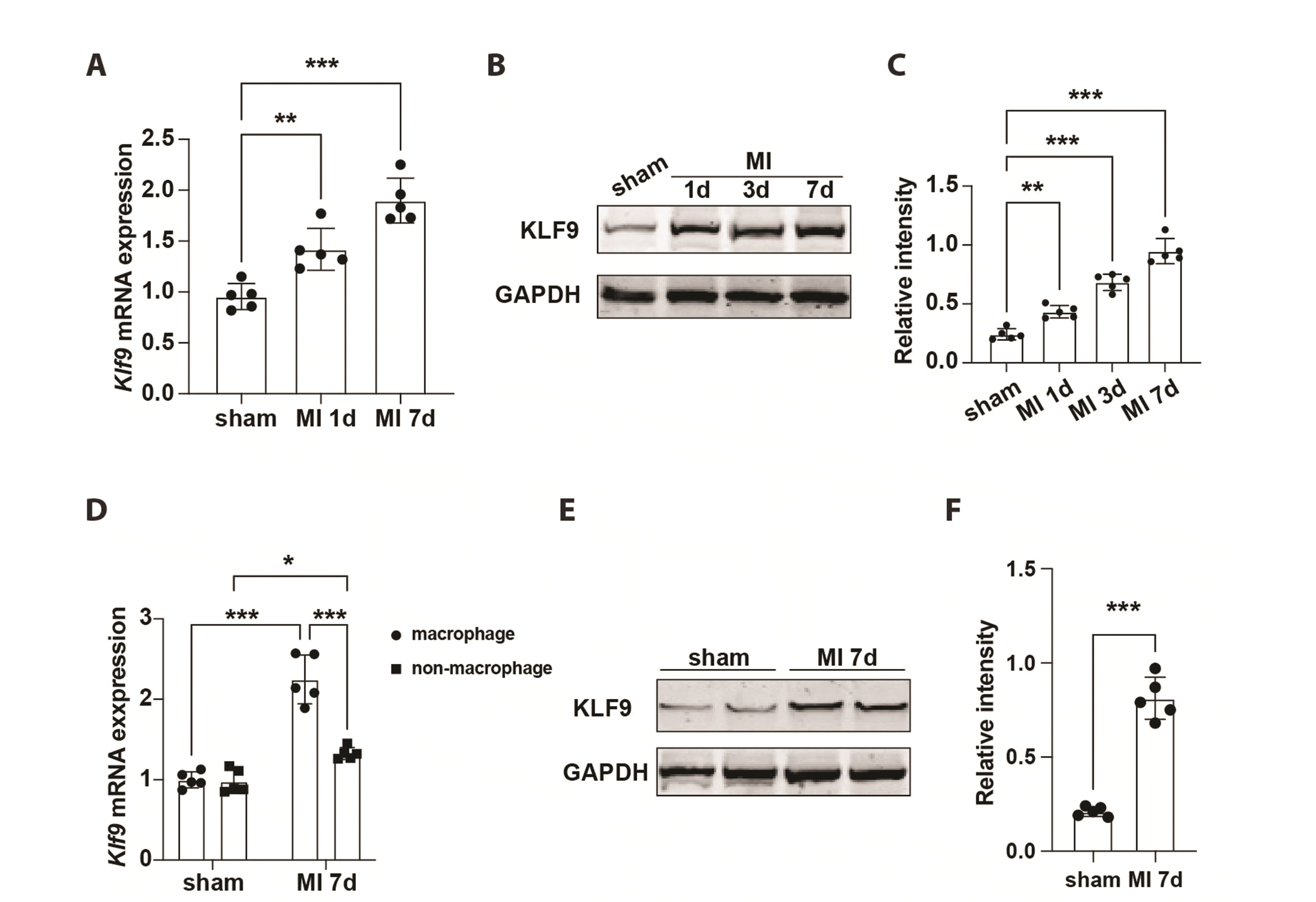
To explore the biological function of KLF9 in myocardial injury, we first evaluated the expression changes of KLF9 in heart tissue after MI. As shown in Fig. 1A–C, both mRNA and protein levels of KLF9 were elevated in mice hearts following MI injury. Furthermore, F4/80 positive macrophages were isolated from MI or sham-operated heart tissues, and the results showed that the mRNA and protein expression levels of KLF9 were significantly increased in macrophages from heart tissues after MI (Fig. 1D–F). In addition, we found a slightly increased expression of KLF9 in non-macrophages after MI. The expression of KLF9 in macrophages was significantly higher than in non-macrophages (Fig. 1D). These results indicate increased levels of KLF9 in infiltrated macrophages from ischemic heart tissue and predict an important role of KLF9 in MI injury.
KLF9 deficiency prevents cardiac dysfunction and adverse cardiac remodeling
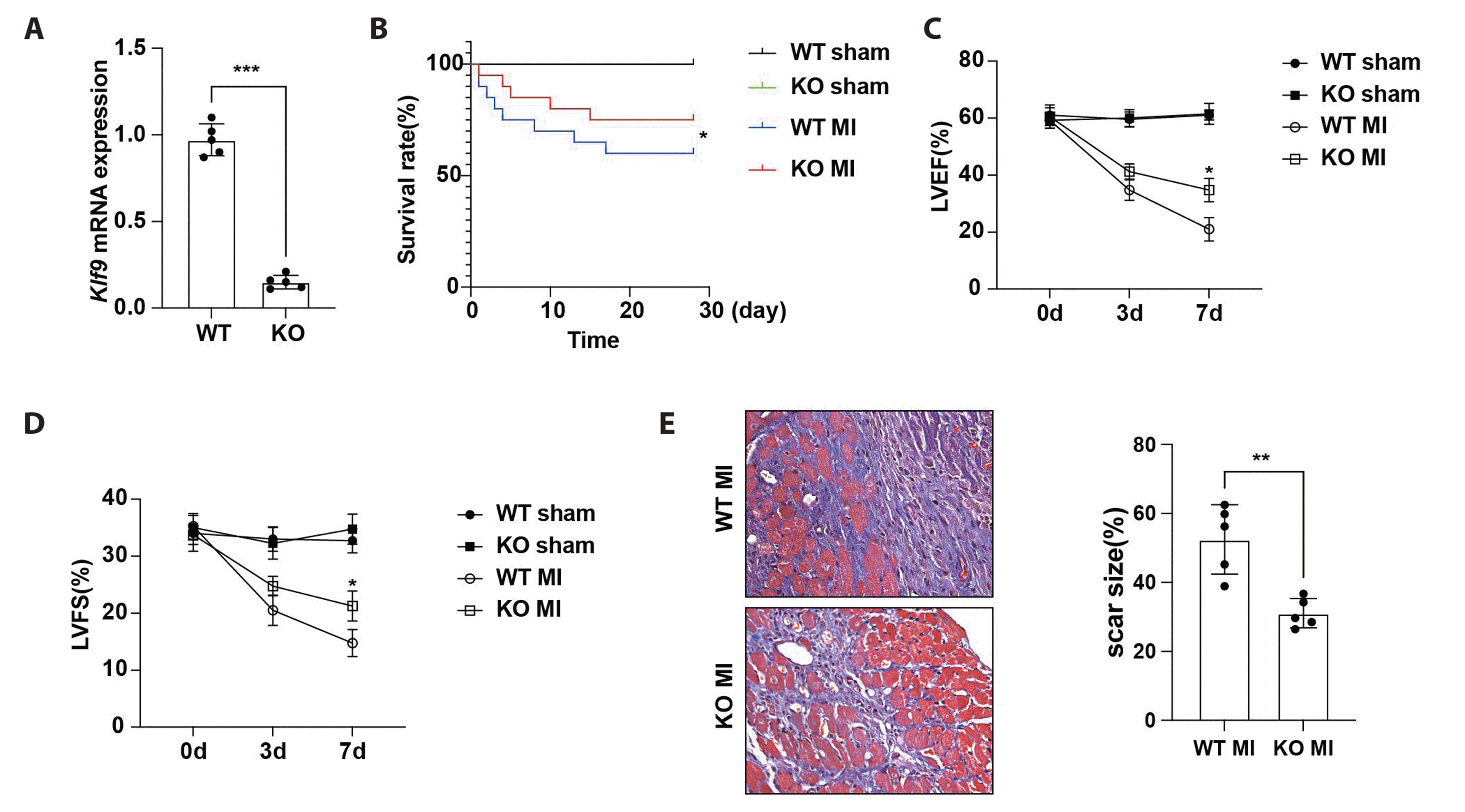
To further verify the regulatory role of KLF9 in myocardial ischemic injury, we constructed a MI model by utilizing KLF9-deficient mice. KLF9-deficient mice showed reduced expression of KLF9 at the mRNA level (Fig. 2A). The survival rate of infarcted mice was markedly improved in KLF9-deficient mice compared with littermate wild-type mice (WT) (Fig. 2B). Loss of KLF9 showed increased percentages of left ventricular ejection fraction and left ventricular fractional shortening compared with WT mice after MI (Fig. 2C and D; Supplementary Fig. 1A). Masson’s trichrome staining was performed to detect the adverse cardiac remodeling induced by MI. As shown in Fig. 2E and Supplementary Fig. 1B, KLF9 deficiency mice showed markedly decreased ECM deposition in border zone of myocardium, suggesting that KLF9 deficiency significantly prevented adverse cardiac remodeling post-MI. We further explored the role of KLF9 in myocardial hypertrophy under physiological and pathological conditions. As shown in Supplementary Fig. 1C and D, we did not observe myocardial hypertrophy in normal hearts from WT or knockout (KO) mice. In addition, KLF9 deficiency did not affect myocardial hypertrophy after MI, suggesting that KLF9 did not contribute to the cardiomyocyte-mediated pathological responses following MI (Supplementary Fig. 1E and F). These results suggest that KLF9 aggravates the cardiac injury, leading to worsened cardiac dysfunction and adverse cardiac remodeling after MI.
KLF9 deficiency suppresses cardiac inflammatory response triggered by MI
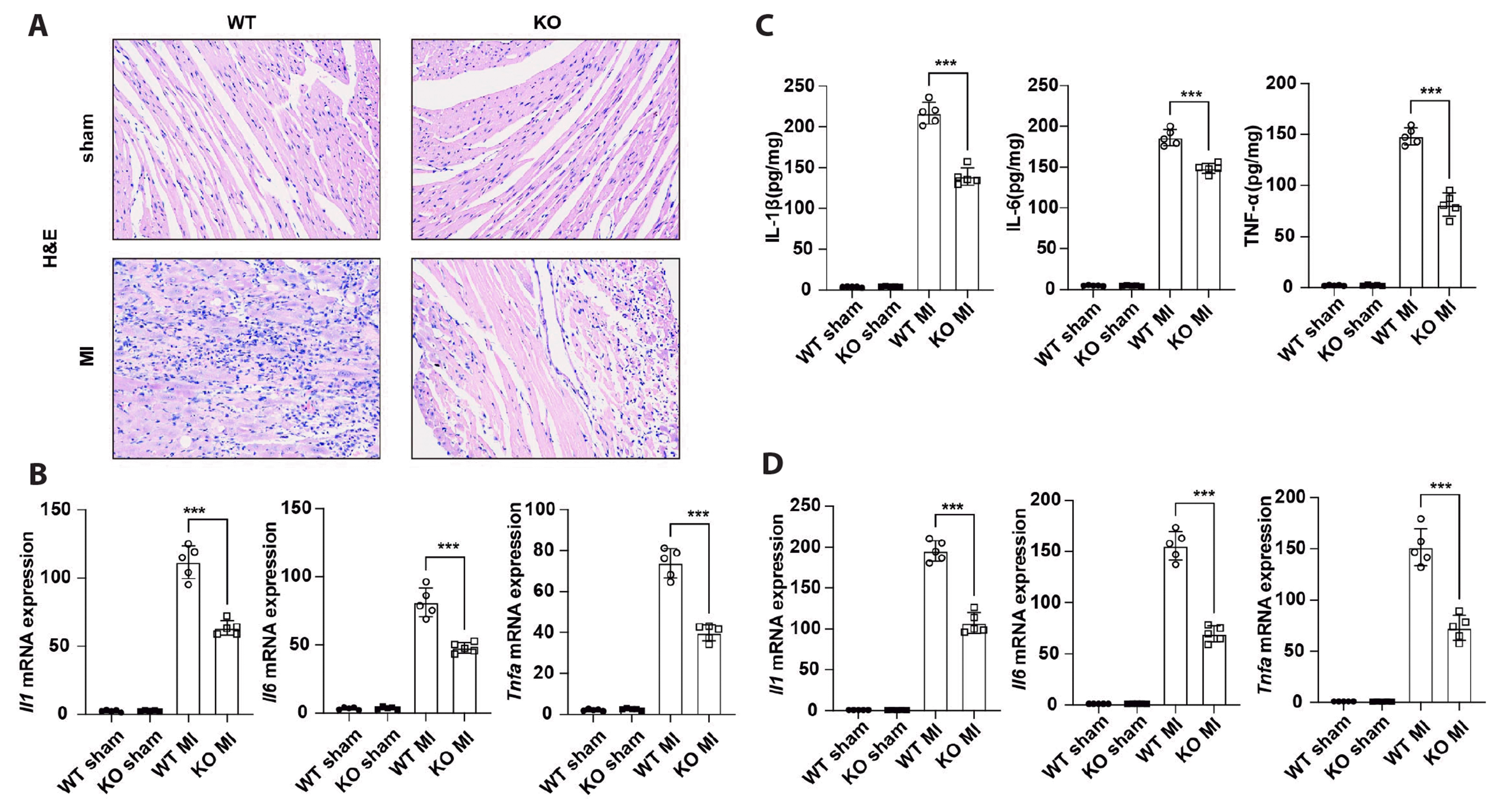
The inflammatory response is a major contributor to the deterioration of cardiac function and adverse cardiac remodeling after MI [16]. We then assessed the modulatory effects of KLF9 knockdown on the inflammatory response induced by MI. H&E staining showed markedly decreased inflammatory cell infiltration in the peri-infarct area at 7 days post-MI in the KLF9 deficiency group compared with the WT group (Fig. 3A and Supplementary Fig. 2). The mRNA levels of the inflammatory cytokines Il1, Il6, and Tnfa were much lower in KLF9-KO mouse hearts than in WT hearts post-MI (Fig. 3B). KLF9 knockdown was also associated with a significant reduction in the production of inflammatory cytokine proteins such as IL-1β, IL-6, and TNF-α (Fig. 3C). F4/80 positive macrophages were isolated from KLF9 KO or WT MI heart tissues, and the results showed that the mRNA expression levels of inflammatory cytokines Il1b, Il6, and Tnfa were significantly decreased after KLF9 deficiency (Fig. 3D). These results indicate that KLF9 enhances the expression of pro-inflammatory cytokines in cardiac macrophages and exacerbates the inflammatory responses of the heart post-MI.
Loss of KLF9 attenuates the inflammatory response of macrophages triggered by DAMPs
MI leads to the release of large amounts of DAMPs from necrotic cardiomyocytes, which can be recognized by macrophage surface pattern recognition receptors (PRRs) to activate downstream inflammatory signaling pathways. High-mobility group box 1 (HMGB1) belongs to a kind of DAMPs, and we subsequently utilized HMGB1 for in vitro functional validation experiments of KLF9. BMDMs were isolated from KLF9 KO or WT mice and subsequently treated with HMGB1 for 12 h. As shown in Fig. 4A, the production of inflammatory cytokines IL-1β, IL-6 and TNF-α was significantly decreased in KLF9 KO BMDMs following HMGB1 treatment. We further observed the effect of KLF9 silencing with specific siRNAs on inflammatory cytokine production in BMDMs. Consistently, the results showed that KLF9 silencing markedly suppressed the production of inflammatory cytokines IL-1β, IL-6 and TNF-α induced by HMGB1 (Fig. 4B). Next, we aim to explore the activation of MAPK and NF-κB signaling pathways triggered by DAMPs in macrophages. The phosphorylation levels of p38, ERK and p65 were decreased in KLF9 KO macrophages after HMGB1 treatment (Fig. 4C). Furthermore, KLF9 silencing repressed the phosphorylation levels of p38, ERK and p65 in macrophages after HMGB1 treatment (Fig. 4D). Together, these findings suggest that KLF9 serves as a positive regulator of the inflammatory responses in response to DAMPs.
KLF9 directly binds to the TLR2 promoter region and promotes its expression in macrophages
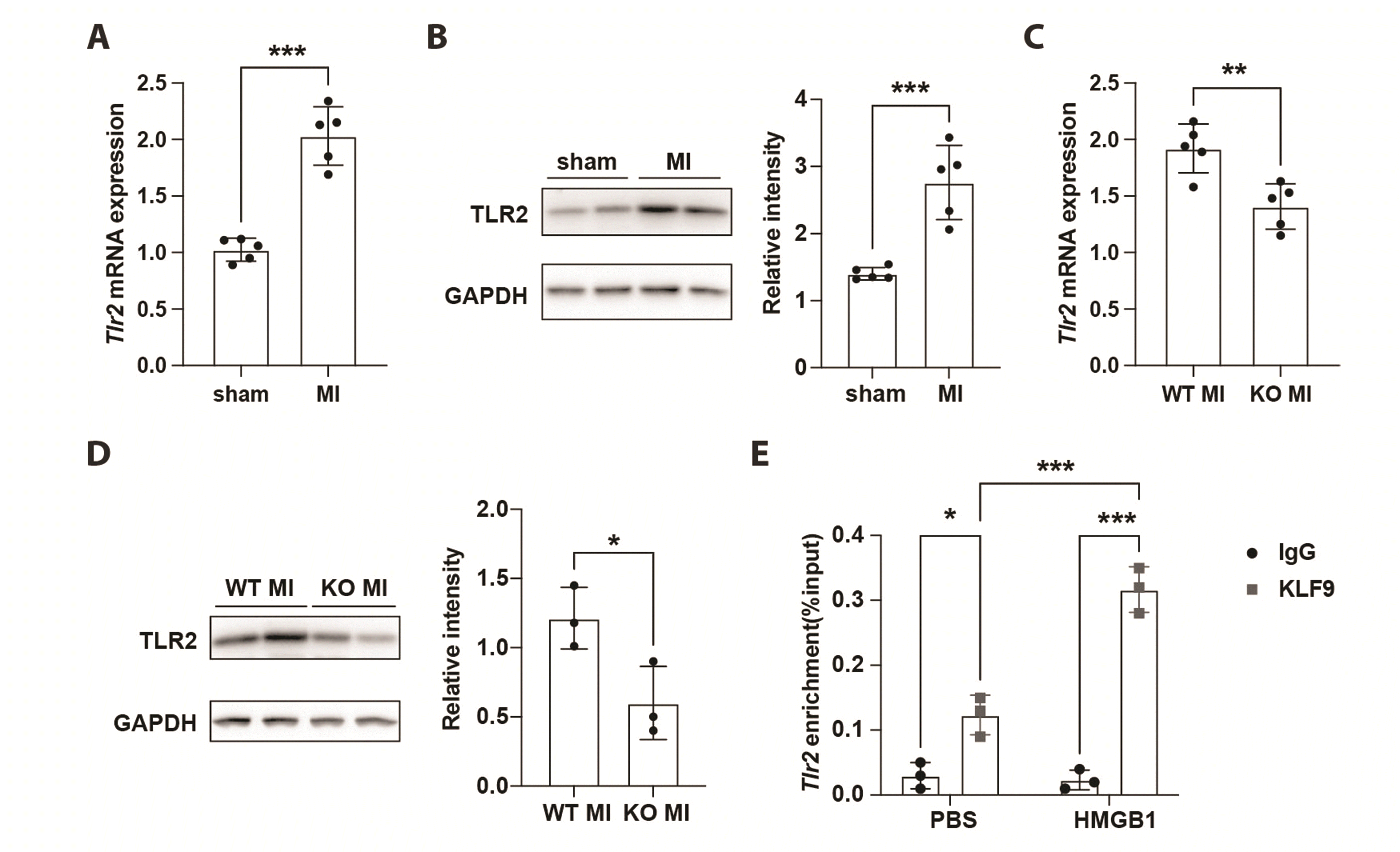
The activation of MAPK and NF-κB signaling pathways is reliant on the recognition of signaling triggered by DAMPs through macrophage PRRs after MI. We found that the mRNA and protein levels of TLR2 were significantly increased during myocardial ischemic injury (Fig. 5A, B). We then investigated the effect of the KLF9 knockdown on TLR2 expression. We found that KLF9 knockdown significantly inhibited TLR2 mRNA and protein expression in the heart after MI (Fig. 5C, D). Given that KLF9 is a transcription factor that is involved in the transcriptional regulation of target genes, we then assessed whether KLF9 directly affects the transcriptional regulation of TLR2. The result showed that the recruitment of KLF9 to the promoter of TLR2 was significantly increased in macrophages following HMGB1 treatment (Fig. 5E). These results indicate that KLF9 directly binds to the TLR2 promoter and enhances the expression of TLR2 in macrophages during ischemic stimulation.
DISCUSSION
In this study, we demonstrated the involvement of KLF9 in exacerbating cardiac dysfunction and deteriorating cardiac remodeling after MI. We found the activated expression of KLF9 aggravates the inflammatory responses induced by MI. Loss-of-function experiments indicated that KLF9 deficiency significantly prevented cardiac dysfunction and adverse cardiac remodeling triggered by MI. In addition, KLF9 deficiency also showed the inhibitory ability on activation of MAPK and NF-kB signaling induced by HMGB1 in macrophages. We subsequently indicated that KLF9 binds directly to the promoter of Tlr2, accelerating TLR2 transcription, promoting an inflammatory response, and deteriorating cardiac function after MI.
The inflammatory response is thought to be involved in the pathogenesis of MI, coordinating beneficial homeostatic responses in the heart to promote cardiac repair after MI [17]. The innate immune cells which are recruited into myocardial tissue mediated the clearance of necrotic cardiomyocytes through phagocytosis after MI [18]. However, DAMPs and ischemia triggered an excessive inflammatory response of infiltrative inflammatory cells, leading to increased myocardial damage. Anti-inflammatory therapy has been shown to reduce the extent of infarction and prevent cardiac remodeling in the treatment of MI [19]. Macrophages are one of the most abundant cardiac inflammatory cell clusters and are responsible for the predominant role in the inflammatory response of the heart after MI [20]. A number of PRRs, such as TLR2, are located on the surface of macrophages [21]. During the acute inflammatory phase of MI, apoptosis and necrosis of cardiomyocytes due to ischemia can produce a variety of endogenous ligands that are also known as DAMPs, mainly including HMGB1 [22]. HMGB1 can be recognized by TLR2, leading to the activation of downstream MAPK and NF-kB signaling pathways as well as the increased production of cytokines [23,24]. Previous studies have demonstrated that TLR2 deficiency show improved cardiac function, prevented cardiac remodeling and inflammatory responses after MI [25,26]. Therefore, how precisely manipulating the deleterious inflammatory response of macrophages is a valuable strategy to prevent pathological remodeling after MI. Herein, we demonstrate that KLF9 deficiency suppresses the expression of TLR2 in macrophages after HMGB1 stimulation. The downregulated of TLR2 impairs the activation of MAPK and NF-kB signaling pathways, thereby preventing cardiac dysfunction and adverse cardiac remodeling post-MI. Our findings revealed that KLF9 serves as a novel positive regulator of cardiac injury and shows a potential therapeutic effect on inflammatory responses triggered by MI.
Multiple mechanisms have been identified to be involved in regulating the pathological process after MI, including the transcriptional control of transcription factors. Krüppel-like factors (KLFs) belong to the zinc-finger transcription factor family and contribute to diverse biological processes such as embryonic development, regeneration, and human disease development [27]. The DNA binding capability of KLFs is reliant on the conserved carboxy-terminal zinc finger domain and that allows KLFs to bind CACCC- or GC-rich sites in promoter and enhancer regions [28]. Particularly, alterations in KLFs function have been implicated in the pathophysiology of many human diseases, including cardiovascular disease. Previous studies have shown that KLFs are critical mediators of cardiac development, hypertrophy, remodeling, injury repair, and electrical activity [29]. KLF9 is part of the KLFs family of members and has been reported to be associated with the progression of various cancers [30]. Additional studies have demonstrated multiple regulatory roles of KLF9 in the development of pathology. KLF9 can promote the expression of Gasdermin-D (GSDMD) via binding with its promoter, which in turn exacerbates the inflammatory injury triggered by acute lung injury [31]. KLF9 specifically binds to the promoter region of matrix metalloproteinase 28 and inhibits its transcriptional, thereby suppressing the invasion and metastasis of gastric cancer cells [32]. KLF9 directly binds to the PADI4 gene promoter to promote PAD4 overexpression in colorectal cancer cells, thereby enhancing cell growth and migration [33]. A recent study also indicated that KLF9 is associated with the deterioration of cardiac injury by impairing the clearance of reactive oxygen species after MI [34]. Although, the more in-depth mechanism of KLF9 in the pathogenesis of MI remains to be further explored. Considering the critical role of KLF9 in cardiovascular diseases, our results reveal that KLF9 aggravates cardiac injury via mediating excessive inflammatory responses of macrophages after MI. KLF9 directly promotes the expression of TLR2 dependent on transcriptional regulatory capacity. Based on the identified role of KLF9 in ischemic diseases, this could provide valuable insights for the establishment of a possible clinical treatment for MI.
In summary, our data indicated that the upregulation of KLF9 exhibits a positive correlation with excessive inflammatory responses and exacerbated cardiac dysfunction after MI. KLF9 directly promotes the expression of TLRs and enhances the activation of MAPK and NF-kB signaling, which subsequently contributes to excessive inflammatory responses triggered by MI. Our results reveal a potential molecular foundation for the regulation of KLF9 in inflammatory responses, and it may become a novel therapeutical target for MI.
SUPPLEMENTARY MATERIALS
Supplementary data including two figures can be found with this article online at https://doi.org/10.4196/kjpp.2023.27.2.177.

 XML Download