Abstract
Fixation of radial head fracture with minimally invasive posterior approach remains a significant challenge. The aim of this study was to determine the feasibility of trans-anconeus posterior elbow approach and to observe lateral ulnar collateral ligament (LUCL) in extended elbows. This cadaveric study was performed in twenty upper limbs of fresh fixed adult male cadavers. An oblique incision was made in the middle segment of anconeus until the lateral ligament complex and the joint capsule had been revealed. A deep dissection was explored to observe the anatomical relationship of the LUCL to the anconeus. Measurements of the LUCL were recorded while the elbow was fully extended. The mean distance between the edge of the radial head and the proximal insertion of the LUCL was 13.3 mm (11.5–16.2 mm); the mean distance between the edge of the radial head and the distal insertion of the LUCL was 20.9 mm (19.2–23.4 mm); the distance between the edge of the radial head and the distal edge of the annular ligament was 11.2 mm (8.22–11.7 mm). By estimate correlation of the previous measurements, the direct and accessible way to expose the posterolateral articular capsule of the elbow joint was through a window in medial 2/3 of the middle segment of anconeus muscle. These trans-anconeus approach is useful. It provides good visualization, facilitates applying the implants, and lessens the risk of radial nerve injury. Awareness of the anatomy is mandatory to avoid injury of LUCL.
Isolated radial head fractures account for about 2%–4% of all fractures, however; it comprises more than 30% of total elbow fractures [1, 2]. The most common pattern of radial head fracture involves the anterolateral part, while the posteromedial part is much rarer. Usually, isolated posteromedial radial head fractures are not associated with either elbow dislocation or ligamentous injury [3]. The scarcity of the literature on posteromedial radial head fracture in addition to our novel approach used to access the fracture have urged us to propose this cadaveric study.
Controversy remains regarding the anatomy of the lateral collateral ligament complex of the elbow and their relation to the surrounding forearm extensor muscles. The lateral ulnar collateral ligament (LUCL) has been described as an important stabilizer against posterolateral rotatory forces in several studies [4-6]. Hackl et al. [7] considered the LUCL is coalescing ligament with the annular ligament (AL) rather than representing a distinct ligament. This cadaveric study was conducted to examine some anatomical features related to the approach, most importantly, is the relationship of the ulnar lateral collateral ligament to the incision in the anconeus muscle. The aim of our study was to address the exact location of the trans-anconeus approach through cadaveric study. Furthermore, to apply the modified approach in fixing radial head fracture in patients undergoing surgery as a controversy exists regarding the visualization of both anterior and posterior parts of the radial head fracture in addition of making the reduction of the fracture easier.
Twenty upper limbs of fresh fixed adult male cadavers were dissected, without distinction of ethnical group or daily activities. The cadaveric specimens were provided by anatomy department, Ain Shams University following the ethical guidelines. Each upper limb was placed in pronated position on the dissection table. The anconeus was identified and traced till its insertion. In order to define aspects of the anconeus, we used the nomenclature proposed by Coriolano et al. [8]: superior border (from the lateral epicondyle [LE] to the ulna adjacent to the olecranon), lateral border (from the LE to the union of the proximal and middle thirds of the ulna) and base (insertion of the anconeus along the ulna). The anconeus was divided into 4 parts: proximal transverse fibers (upper part): those arising from the proximal third of the tendon, adjacent to the superior border of the muscle; middle part: those arising from the middle third of the tendon; distal part: those arising from the distal third of the tendon; and terminal fibers: those originating from the tip of the tendon. The obliquity of the muscle fibers of the middle part of the muscle was calculated. The deep side of the anconeus was dissected to expose the elbow joint capsule. An oblique incision was made in the middle segment of the anconeus until the lateral ligament complex and the joint capsule had been revealed proximally, along with the supinator muscle distally. Measurements of the LUCL were recorded while the elbow was in full limit of extension.
Descriptive statistics were generated from the cadaver specimens to determine values for 1) the length of the borders (superior, lateral and base), 2) the length of the anconeus muscle fibers of upper and lower parts. 3) The angle of the muscle fibers angle of the middle segment with the sagittal plane of ulna, 4) the LUCL length; the distance from the proximal and distal attachments of the LUCL to the proximal edge of the cartilage of the radial head and 5) the distance from the proximal edge of the cartilage of the radial head to the distal edge of the AL. For the measurements we used a drawing compass and millimetered ruler and photographs were taken with a digital camera. The cadaver dissection measurement is pictorially represented in Fig. 1. The statistical analyses were performed using IBM SPSS Statistics for Windows, Version 20.0 (IBM Co.). Descriptive statistics (mean and standard deviation) were used to describe anconeus muscle.
Ten patients undergoing posterior elbow surgery were selected for the present study. All patients were subjected to standard elbow radiographs (Fig. 2) as well as computed tomography (CT) scan (Fig. 3) with all procedures conducted in the orthopedic department of El-Demerdash hospital, Ain Shams University (prospective study). The study was approved by the University’s ethics committee (approval no. FMASU R 191/2022) and informed consent was signed by each patient.
Patients with displaced Mason type II isolated posteromedial radial head fracture with no neurovascular deficit were included in the study. On clinical examination, the patients had tenderness over the radial head and limitation of flexion-extension arc. The elbows were blocked in pronation with no active or passive supination. Exclusion criteria were: patients under the age of 18 years and revision surgery.
The anconeus muscle was observed to have a sharp, tendinous expansion, started just inferiorly to the LE of the humerus and extended along the lateral border of the muscle (Fig. 4). The anconeus muscle fibers arise obliquely from the tendinous expansion. From the end of the tendinous expansion, the terminal muscle fibers arise. Some cadavers (twelve specimens) showed connection between the medial head of the triceps and the anconeus near its origin. The anconeus nerve branched from the radial nerve of the medial head of triceps and run among or at the bottom of the superior margin of anconeus muscle. The measures of the anconeus borders, the tendinous expansion, the length of the anconeus muscle fibers of upper and lower parts and anconeus muscle fiber angle of the lower part are tabulated in (Table 1).
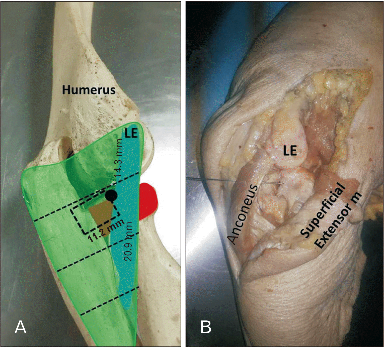
An oblique anconeus muscle incision was made along the middle segment from the tendinous expansion to the proximal ulna, and the LUCL was identified (Fig. 4). The anconeus muscle fibers had parallel orientation to the LUCL fibers. LUCL could be identified as they coalesce with the AL at the head of radius. The mean distance between the edge of the radial head and the proximal insertion of the LUCL was 13.3 mm (range, 11.5–16.2 mm); the mean distance between the edge of the radial head and the distal insertion of the LUCL was 20.9 mm (range, 19.2–23.4 mm); the distance between the edge of the radial head and the distal edge of the AL was 11.2 mm (range, 8.22–11.7 mm).
By estimate correlation of the previous measurements, the direct and easier accessible way to expose the posterolateral articular capsule of the elbow joint was through a window in medial 2/3 of the middle segment of anconeus muscle (Fig. 5).
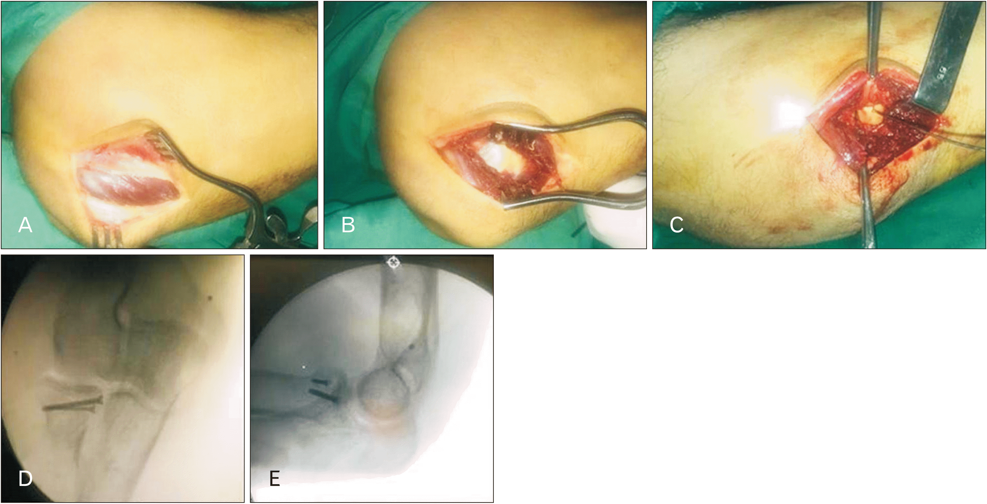
Elbow surgery to fix the fracture by screws was performed under general anesthesia in elbow flexion 90 degrees position. It was our speculation that using trans-anconeus approach would facilitate the visualization of both anterior and posterior edges of the fracture. Moreover, it will enable us to insert the implants (mini screws) comfortably. The trans-anconeus approach can be stereotyped as a midway approach between both Kocher and Boyd approaches. With the elbow flexed 90 degrees, the LE and the head radius were identified then a lateral skin incision was made about 6–7 cm toward the posterior border of the proximal ulna. The anconeus muscle was exposed after incising the overlying fascia (Fig. 6). Through a muscle split approach (midway and parallel to its fibers) the capsule of the joint was exposed. The capsule was incised just posterior to the LUCL along its extension between the LE and the footprint of the LUCL at the supinator crest of the ulna approximately 2 cm distal to the head radius. A preliminary anatomical reduction of the fracture was achieved by means of K-wire. Anterior and posterior edges of the fragment were visualized by performing supination -pronation to check reduction. Finally, definitive fixation was done by two mini screws perpendicular to the fracture line (Fig. 6). The tourniquet was released to achieve good hemostasis and the capsule was tightly sutured followed by wound closure.
Postoperatively, an above elbow slab was applied in 90 degrees of elbow flexion for one week then it was replaced by a hinged elbow brace for a month with gradual increase in elbow extension. The patients were referred to physiotherapy department one month postoperatively for range of motion and strengthening exercises. At three months, patients were able to obtain full flexion-extension arc with fully united fracture radiologically and clinical.
Posteromedial radial head fractures are not common and mostly are not associated with elbow dislocation or ligamentous injury [3, 9]. In our cases the mechanism of injury was the result of a fall on flexed elbow with the forearm supinated.
In most previous studies, a very high percentage of fractures occurred in the forearm bones [10]. Mason [11] classified the radial head fractures into 3 types: non-displaced, displaced and comminuted then a fourth type fracture-dislocation was added by Broberg and Morrey [12] however; it was first described by Johnston [13]. Regrettably, Mason classification was based only on the displacement and the comminution of the fracture but did not mention its location. The posteromedial fracture was described in CT classification and we considered the CT classification in interpretation of the current results. Capo et al. [3] conducted a study on 25 patients with partial articular radial head fracture based on the CT scan and they divided the radial head into four quadrants guided by the radial tuberosity that was assigned as 6 o’clock, 3 o’clock anterior and 9 o’clock posterior. Fractures between 12 o’clock and 3 o’clock were categorized as anterolateral fragments, between 3 o’clock and 6 o’clock as posterolateral fragments, between 6 o’clock and 9 o’clock as posteromedial fragments, and between 9 o’clock and 12 o’clock as anteromedial fragments.
The tarns-anconeus approach was first (only) described by Iwasaki et al. [14] when treating patients with osteochondritis dissecans of the capitellum by mosaicplasty. However; the distal limit of their approach was the proximal edge of the AL. We made a modification on this approach by extending the capsular incision 2 cm distal to the head radius heading to the supinator crest just proximal to the footprint of the LUCL, and by leaving about 3 mm of the AL posteriorly for later repair (Fig. 6).
The LUCL runs parallel and deep to the aponeurosis of the anconeus muscle with a footprint of about 1 cm originating from the tubercle of the supinator crest. The anconeus muscle is a fan shaped triangular muscle that originates from the posterosuperior aspect of the LE to be inserted in the posterolateral surface of the proximal ulna [4, 15]. The triceps–anconeus muscle innervated mainly by the radial nerve, which is liable to be damaged during posterior elbow approaches [16].
The cadaveric study shed some light to increase the safety and the utility of this approach; 1- It is better to leave 2 cm at least from superior margin during anconeus incision as the present cadaveric study (in twelve specimens) showed connection between the medial head of the triceps and anconeus. Also, it could be at risk in cutting the nerve to anconeus because it branches from the radial nerve of the medial head of triceps at the anconeus superior margin [17]. 2- It is better to plan splitting of the anconeus muscle in the medial 2/3 of the middle segment. The fibers are angled about 40° and this is analogous to the obliquity of the LUCL. 3- The approach allows incising a window in the posterolateral part of the capsule measuring about 11 mm which can be increased few mm more by further retracting the ULCL. 4- LUCL insertion in the ulna could be injured by extending the incision of the capsule more than 2 cm distal to the head radius.
The cadaveric study may be subject to some limitations that merit discussion. First, the anatomic dimensions of the structures examined in this study may vary significantly based on patient sex, hand dominance, activity level, and size, as evidenced by the large ranges for some of the variables. Further studies examining these anatomic differences may help guide during elbow surgery based on patient characteristics. Secondly, additional dimension measurements during different anatomical positions are needed to avoid potential bias to flexed or extended positions.
We believe this novel approach was beneficial in isolated posteromedial radial head fractures cases. It allowed us to reach the fracture directly without much dissection. We were able to judge the reduction easily by direct visualization of both edges of the fragment. Insertion of the screws was straight forward with consideration of the mean maximal capacity of elbow joint was 40.5 ml [18]. Meanwhile, through meticulous dissection we were capable to preserve LUCL. Bartoníček et al. [19] stated that the Kocher incision between the anconeus and extensor carpi ulnaris (Kocher interval) is relatively more posterior and thus risks injuring the lateral collateral ligament complex. Furthermore, we are skeptical that either Kocher or Boyd approach could allow us to assess the whole circumference of the fragment. They probably permit visualization of only anterior (in Kocher approach) or posterior edges of the fracture (in Boyd approach; plane between anconeus and proximal ulna). In addition, we think it would have been more difficult to insert the screws from the more anterior Kocher approach. Boyd approach may be more extensive and entails separation of the anconeus from its bony insertion. Finally, the risk of injury to posterior interosseous nerve is very low. Nevertheless, the trans-anconeus approach has its limitations; it is only suitable to selected types of head radius fractures. It is basically not an extensile approach. However, it has the potential to extend the visualization capability by opening another window anterior to LUCL by incising the AL.
In conclusion, trans-anconeus approach is useful in cases of isolated posteromedial radial head fractures. It provides good visualization, facilitates applying the implants, and lessens the risk of posterior interosseous nerve injury. Awareness of the anatomy is mandatory to avoid injury of LUCL.
Acknowledgements
The authors would like to extend their appreciation to Technical Staff of the Center of Donors of Cadavers and dissecting Rooms of anatomy department, Faculty of medicine, Ain Shams University for supporting this research. Also, we would like to thank the participants, the study investigators and the staff at the clinical centers for generating the image data sets used in this research.
Notes
References
1. Duckworth AD, McQueen MM, Ring D. 2013; Fractures of the radial head. Bone Joint J. 95-B:151–9. DOI: 10.1302/0301-620X.95B2.29877. PMID: 23365021.

2. Gawande J, Jain S, Santoshi JA. 2017; Neglected bilateral radial head fracture with a rare presentation: a case report. Chin J Traumatol. 20:246–8. DOI: 10.1016/j.cjtee.2016.07.003. PMID: 28684037. PMCID: PMC5555247.

3. Capo JT, Shamian B, Francisco R, Tan V, Preston JS, Uko L, Yoon RS, Liporace FA. 2015; Fracture pattern characteristics and associated injuries of high-energy, large fragment, partial articular radial head fractures: a preliminary imaging analysis. J Orthop Traumatol. 16:125–31. DOI: 10.1007/s10195-014-0331-x. PMID: 25542062. PMCID: PMC4441642.

4. Moritomo H, Murase T, Arimitsu S, Oka K, Yoshikawa H, Sugamoto K. 2007; The in vivo isometric point of the lateral ligament of the elbow. J Bone Joint Surg Am. 89:2011–7. DOI: 10.2106/00004623-200709000-00017. PMID: 17768199.
5. Stipp WN, Ribeiro FR, Tenor Junior AC, Filardi Filho CS, Molin DCD, Petros RSB, Brasil Filho R. 2013; Anatomical parameters in the lateral ulnar collateral ligament reconstruction: a cadaver study. Rev Bras Ortop. 48:52–6. DOI: 10.1016/j.rbo.2012.05.005. PMID: 31304111. PMCID: PMC6565991.

6. Wegmann K, Burkhart KJ, Zimmermann J, Dargel J, Nijs S, Konerding MA, Müller LP. 2014; The interference of distal humeral plating with the medial and lateral collateral ligaments of the elbow. Arch Orthop Trauma Surg. 134:501–7. DOI: 10.1007/s00402-014-1952-5. PMID: 24531976.

7. Hackl M, Bercher M, Wegmann K, Müller LP, Dargel J. 2016; Functional anatomy of the lateral collateral ligament of the elbow. Arch Orthop Trauma Surg. 136:1031–7. DOI: 10.1007/s00402-016-2479-8. PMID: 27245451.

8. Coriolano M, Lins OG, Amorim MJ, Amorim AA. 2009; Anatomy and functional architecture of the anconeus muscle. Int J Morphol. 27:1009–12. DOI: 10.4067/S0717-95022009000400008.

9. Mellema JJ, Eygendaal D, van Dijk CN, Ring D, Doornberg JN. 2016; Fracture mapping of displaced partial articular fractures of the radial head. J Shoulder Elbow Surg. 25:1509–16. DOI: 10.1016/j.jse.2016.01.030. PMID: 27052270.

10. Kim DK, Kim MJ, Kim YS, Oh CS, Lee SS, Lim SB, Ki HC, Shin DH. 2013; Long bone fractures identified in the Joseon Dynasty human skeletons of Korea. Anat Cell Biol. 46:203–9. DOI: 10.5115/acb.2013.46.3.203. PMID: 24179696. PMCID: PMC3811853.

11. Mason ML. 1954; Some observations on fractures of the head of the radius with a review of one hundred cases. Br J Surg. 42:123–32. DOI: 10.1002/bjs.18004217203. PMID: 13209035.

12. Broberg MA, Morrey BF. 1987; Results of treatment of fracture-dislocations of the elbow. Clin Orthop Relat Res. (216):109–19. DOI: 10.1097/00003086-198703000-00017. PMID: 3102139.

13. Johnston GW. 1962; A follow-up of one hundred cases of fracture of the head of the radius with a review of the literature. Ulster Med J. 31:51–6. PMID: 14452145. PMCID: PMC2384652.
14. Iwasaki N, Kato H, Ishikawa J, Masuko T, Funakoshi T, Minami A. 2010; Autologous osteochondral mosaicplasty for osteochondritis dissecans of the elbow in teenage athletes: surgical technique. J Bone Joint Surg Am. 92(Suppl 1 Pt 2):208–16. DOI: 10.2106/JBJS.J.00214. PMID: 20844176.
15. Pereira BP. 2013; Revisiting the anatomy and biomechanics of the anconeus muscle and its role in elbow stability. Ann Anat. 195:365–70. DOI: 10.1016/j.aanat.2012.05.007. PMID: 22874649.

16. Jiménez-Díaz V, Aragonés P, García-Lamas L, Barco-Laakso R, Quinones S, Konschake M, Gemmell C, Sanudo JR, Cecilia-López D. 2021; The anconeus muscle revisited: double innervation pattern and its clinical implications. Surg Radiol Anat. 43:1595–601. DOI: 10.1007/s00276-021-02750-5. PMID: 33881559.

17. Chaware PN, Santoshi JA, Patel M, Ahmad M, Rathinam BAD. 2018; Surgical implications of innervation pattern of the triceps muscle: a cadaveric study. J Hand Microsurg. 10:139–42. DOI: 10.1055/s-0038-1660771. PMID: 30483020. PMCID: PMC6255738.

18. Van Den Broek M, Van Riet R. 2017; Intra-articular capacity of the elbow joint. Clin Anat. 30:795–8. DOI: 10.1002/ca.22915. PMID: 28514501.

19. Bartoníček J, Naňka O, Tuček M. 2015; Kocher approach to the elbow and its options. Rozhl Chir. 94:405–14. Czech. DOI: 10.1002/ca.22915. PMID: 26556018.
Fig. 1
Posterolateral view of the left proximal forearm in pronation. (A) The anconeus muscle is divided into 4 parts: proximal transverse segment, middle segment, distal segment and terminal fiber. Measurements are generated from the cadaver specimens to determine values for the length of the borders (superior, lateral and base) and anconeus muscle fiber angle (asterisks) of the middle segment with the sagittal plane of ulna. (B) The articulated skeleton showing the LUCL (blue coloured) measurements. The coalescing point of LUCL ligament with the AL (black point) is considered as a reference point to determine the value of the LUCL length to its proximal (LE) and distal (supinator crest) attachments. The dotted line is the distance from the proximal edge of the cartilage of the radial head to the distal edge of the AL (red coloured). LUCL, lateral ulnar collateral ligament; AL, annular ligament; LE, lateral epicondyle.
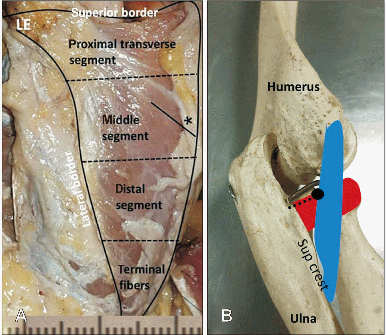
Fig. 2
Plain x-ray (A) anteroposterior and (B) lateral views of the elbow showing posteromedial radial head fracture.
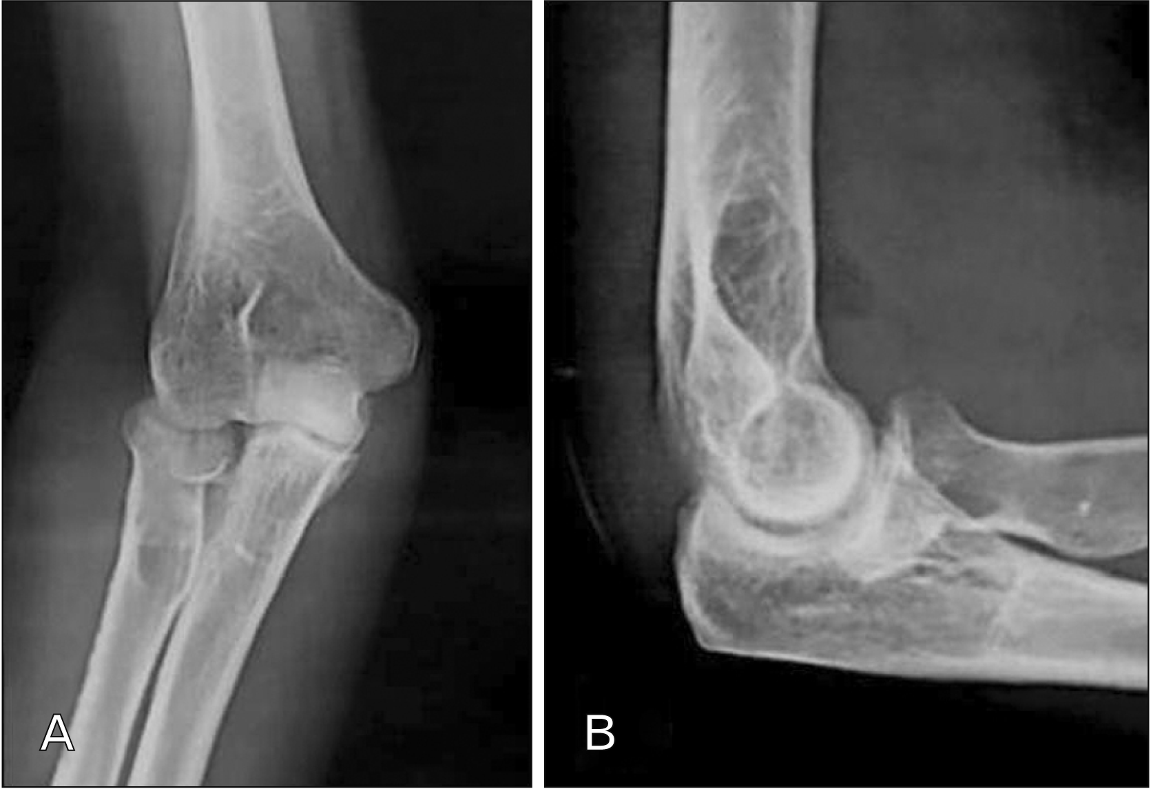
Fig. 3
Computed tomography scan (A)sagittal, (B) coronal and (C) axial cuts respectively showing posteromedial radial head fracture with the axial cut indicating the engagement of the fracture on the posterior edge of the radial notch.
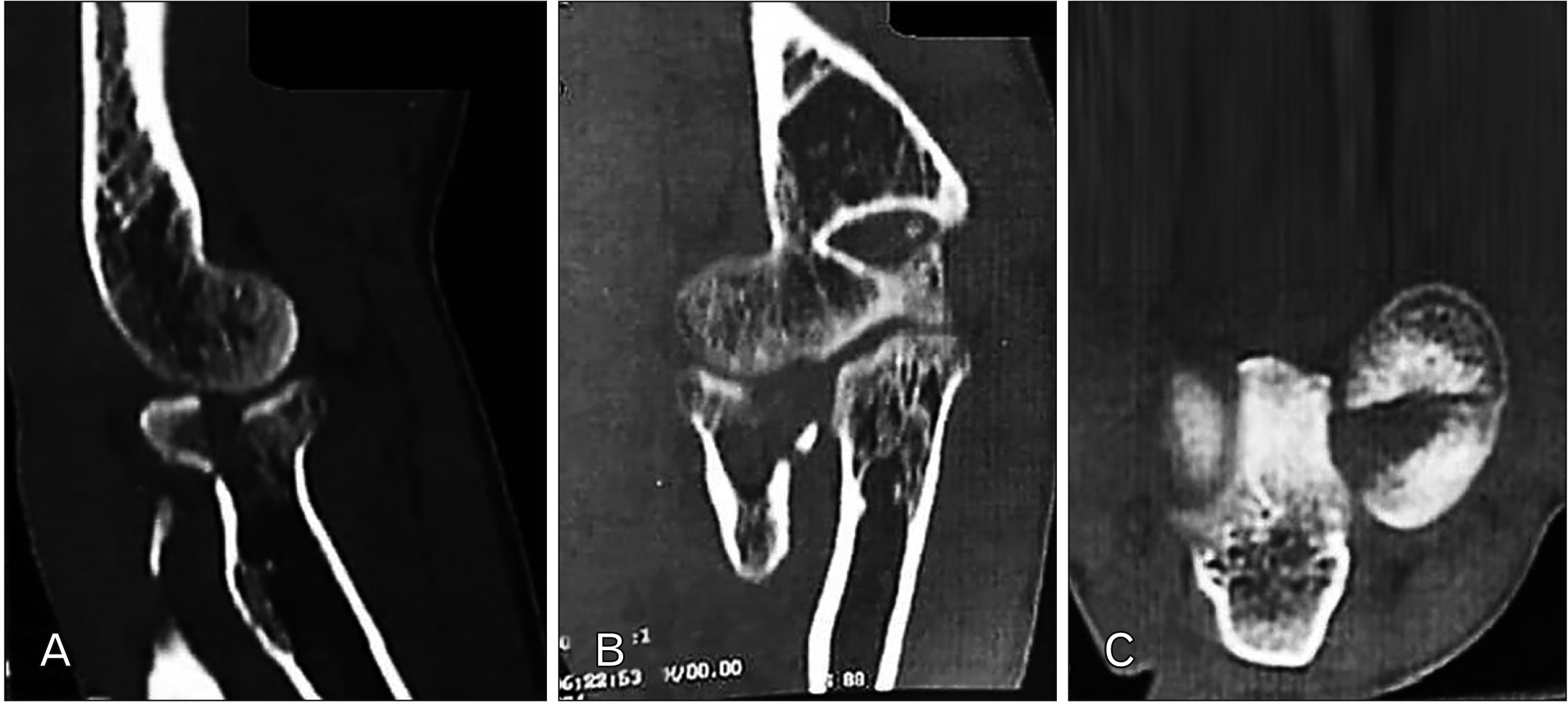
Fig. 4
Posterolateral view of the left proximal forearm in pronation. (A) The anconeus is a right angle triangular muscle attaching proximally to the LE of the humerus with common extensor group. The anconeus muscle is observed to have a sharp, tendinous expansion. (B) The overlying anconeus muscle has been incisied obliquely along the middle segment and extended till the distal segment. (C) The anconeus muscle retracted laterally, the LUCL is identified the window of the anconeus muscle approach exposes the posterior lateral articular capsule of the elbow joint. Cap, capitulum of the humerus; LE, lateral epicondyle; LUCL, lateral ulnar collateral ligament.
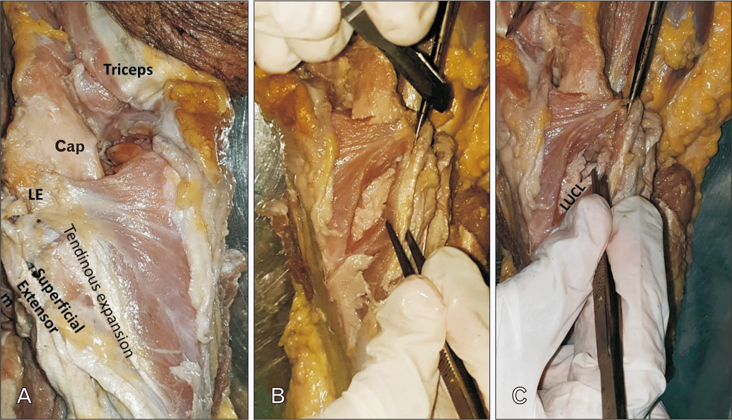
Fig. 5
Posterolateral view of elbow joint in pronation. (A) The articulated skeleton showing the LUCL (blue coloured) measurements in relation to anconeus (green coloured) segments. Dotted rectangle is direct way of posterolateral elbow approach (B) lateral view of elbow joint showing direct way of posterolateral elbow approach (metallic probe) passing through the middle segment of anconeus muscle. The superficial extensor muscle was reflected. LE, lateral epicondyle; LUCL, lateral ulnar collateral ligament.


 PDF
PDF Citation
 Print
Print


 XML Download