

 PDF
PDF Citation
 Print
Print


Macrodactyly of the toe is a rare congenital anomaly characterized by the overgrowth of a digit or multiple digits in the foot. Barsky1) and Kotwal and Farooque2) described that all components of the digits, including the phalanges, tendons, nerves, vessels, subcutaneous fat, fingernails, and skin, become enlarged, while the metatarsals remain unaffected. Owing to its low incidence, few reports or research articles exist on macrodactyly, and the etiology is largely unknown. Two distinguishing types of toe macrodactyly have been observed; the static type, where digits grow commensurately with the foot, and the progressive type, where digits grow faster than the rest of the limb. Additionally, Flatt3) described four types of macrodactyly; type 1 macrodactyly with nerve lipofibromatosis, type 2 macrodactyly with neurofibromatosis, type 3 macrodactyly associated with hyperostosis, and type 4 macrodactyly associated with hemihypertrophy.
Although social withdrawal seems to occur less frequently in those with macrodactyly of the toe than in those with macrodactyly of the fingers, functional discomfort, such as difficulty in wearing shoes or running, is an important factor. This condition is more extreme in the toe, leading to additional discomfort due to exacerbated pain. Macrodactyly is one of the most difficult anomalies to treat. Although there are various surgical techniques for toe macrodactyly, including amputation, osseous structure reduction, soft tissue debulking, and epiphysiodesis, the best treatment strategy remains unclear.4-7) Therefore, we present a case of second toe macrodactyly in a 6-year-old patient, who was treated with a two-stage operation over 7 years using a combination of surgical techniques, such as ostectomy, fusion, and soft tissue debulking.
CASE REPORT
This study was approved by the Institutional Review Board in Inje University Busan Paik Hospital (IRB No, 2022-031).
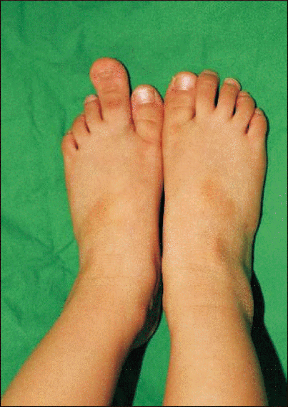
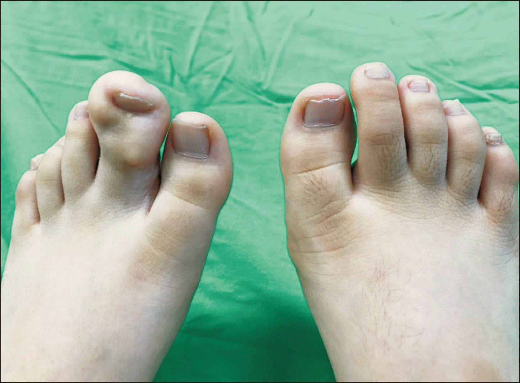
A 6-year-old girl visited our clinic as an outpatient complaining of left second toe enlargement that was identified at birth. She had difficulty wearing shoes, and her parents were concerned about the appearance of her foot. Compared with the second toe on the contralateral side, the soft tissues were thickened on both the plantar and dorsal sides (Fig. 1).
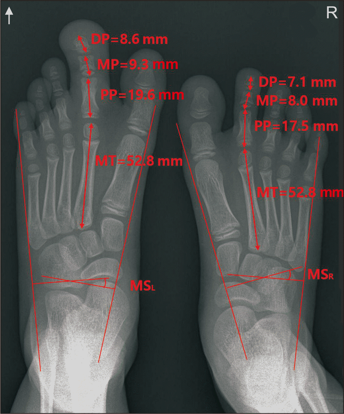
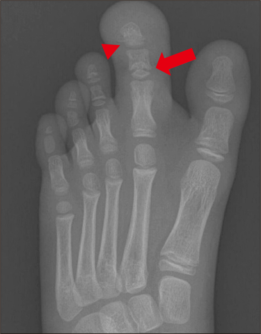
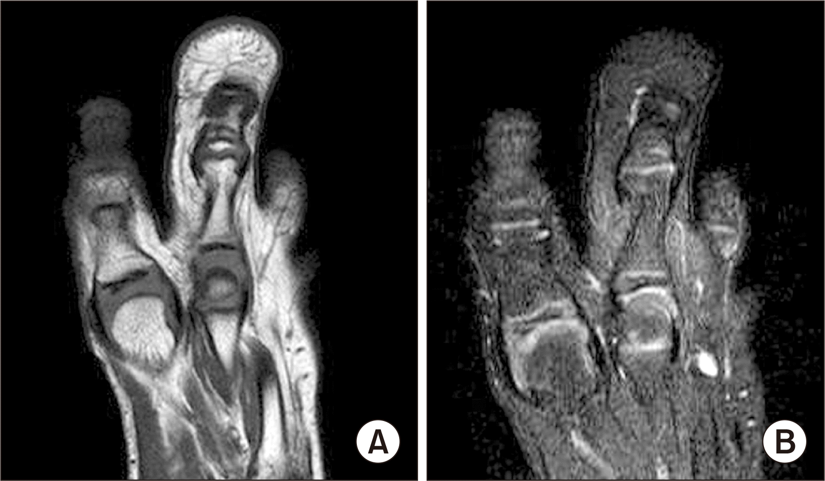
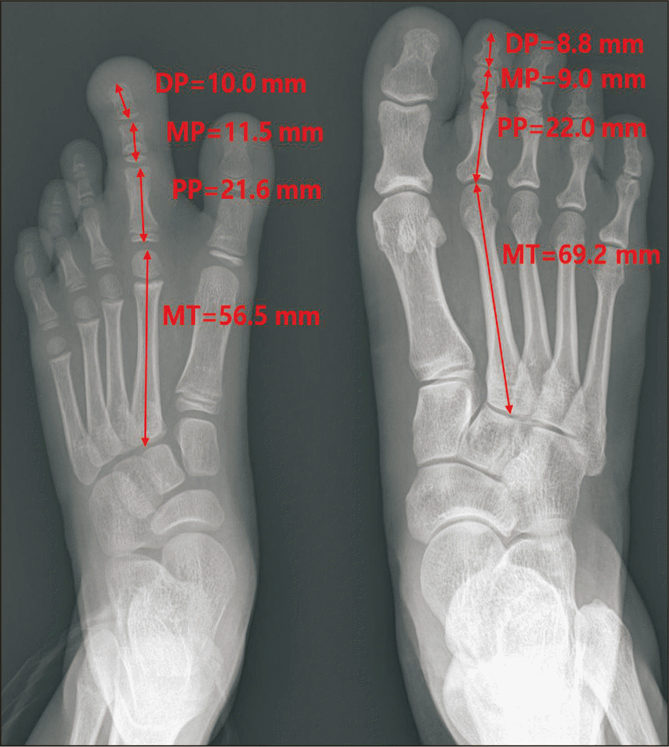
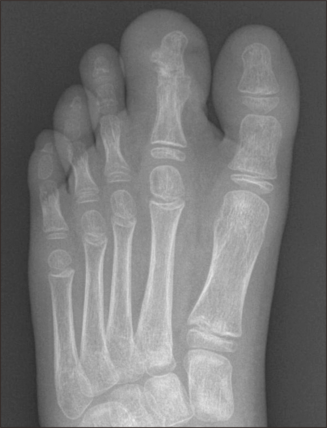
Standing anteroposterior (AP) radiographs of both feet revealed that the overall length of the left second toe phalanges was greater than that of the right side and that both metatarsal bones were of the same length (Fig. 2). The metatarsal spread angle of the left foot was 16.9°, whereas that of the right foot was 17.5°. While the proximal physeal plate of the proximal phalanx and the distal epiphyseal plate of the metatarsal bone were intact, the proximal physeal plate of the distal and middle phalanx seemed to be tethered in the left second ray (Fig. 3). There were no pathologic lesions, such as osteochondroma, or thickening of the ipsilateral lower extremity (Fig. 2, 3). Computed tomography angiography revealed no pathologic lesions, such as an arteriovenous malformation. Magnetic resonance imaging showed enlargement of the subcutaneous fat around the enlarged digits; however, there was no definite nerve enlargement (Fig. 4).
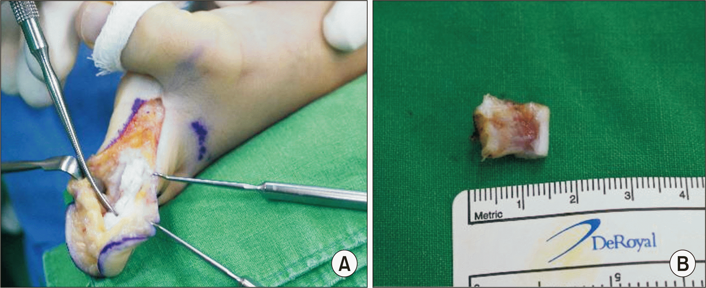
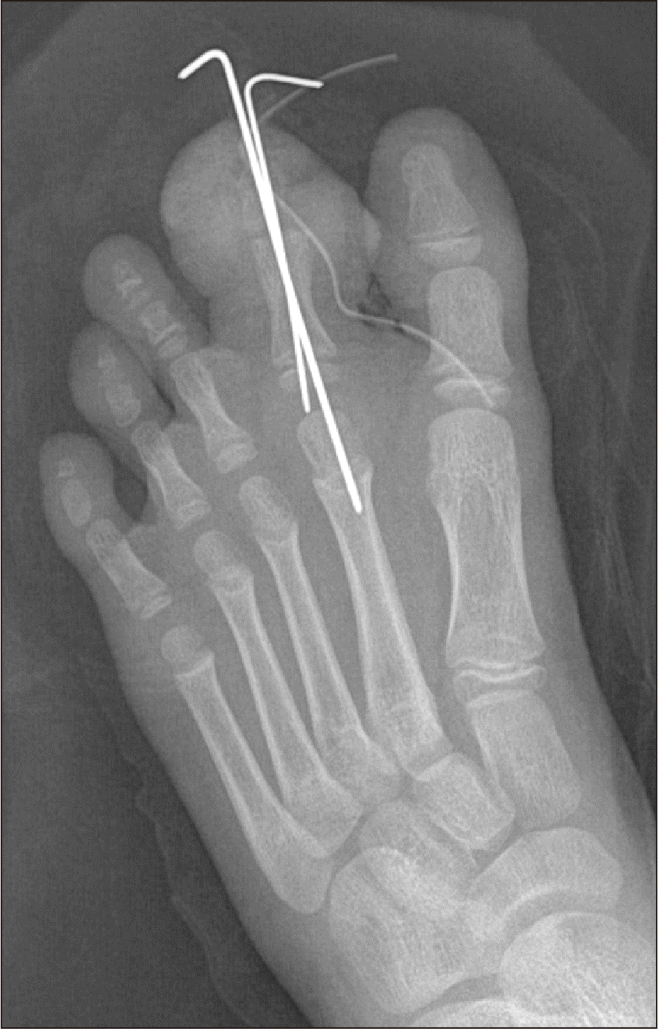
The patient’s parents desired surgical treatment; however, amputation was not desired as they were afraid that the patient might be bullied in the future. We focused on the possible tethered proximal physeal plate of the distal and middle phalanx. Surgical treatment, including ostectomy, interdigital fusion, and soft tissue volume reduction, was performed when the total length of the patient’s three phalanges was longer than that of her mother’s (Fig. 5). Subsequently, a V-shaped incision at the tip of the second toe and a large incision from the medial side were created so that subcutaneous fat tissue removal and osteotomy could be performed concurrently (Fig. 6). Next, thickened adipose tissue was observed and removed after securing the nerves and blood vessels. Ostectomy was subsequently performed at the middle phalanx and proximal epiphysis of the distal phalanx (Fig. 6). The remaining distal phalanx and proximal phalanx were finally fused using a K-wire (Fig. 7).
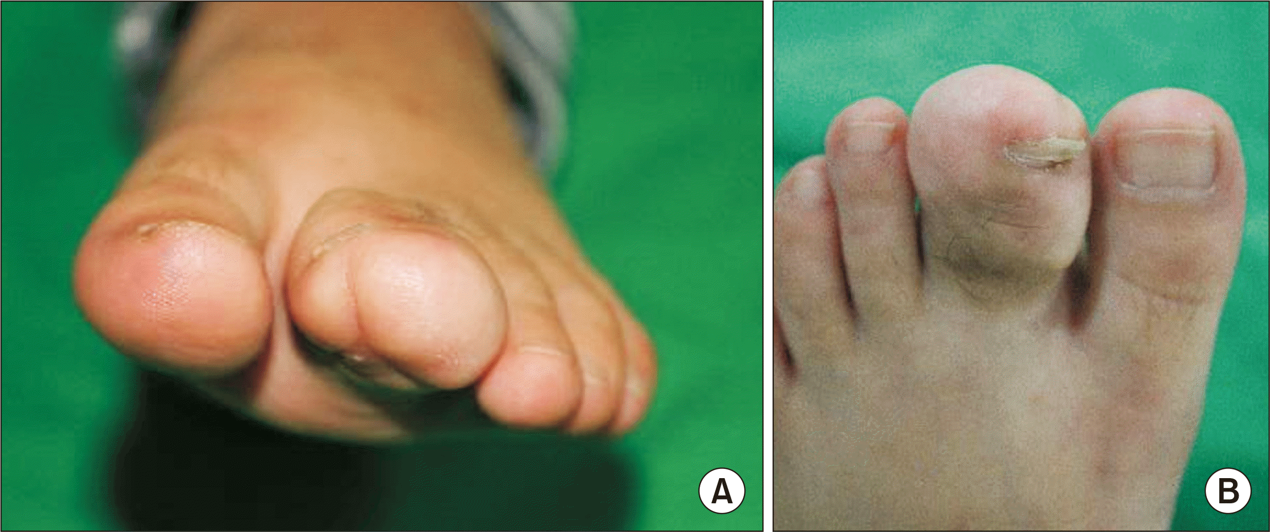
The outpatient follow-up conducted 3 weeks post operation revealed that the macroscopic length had significantly decreased than that observed before surgery. Additionally, the width had slightly decreased (Fig. 8). A plain radiograph obtained 5 weeks postoperatively showed bone union; therefore, the K-wire was removed (Fig. 9). Both the patient and her parents were satisfied with the cosmetic appearance and her ability to perform daily tasks. The patient was under close observation, and her parents were aware that additional surgery might be performed when necessary, depending on the progression of the affected toe.
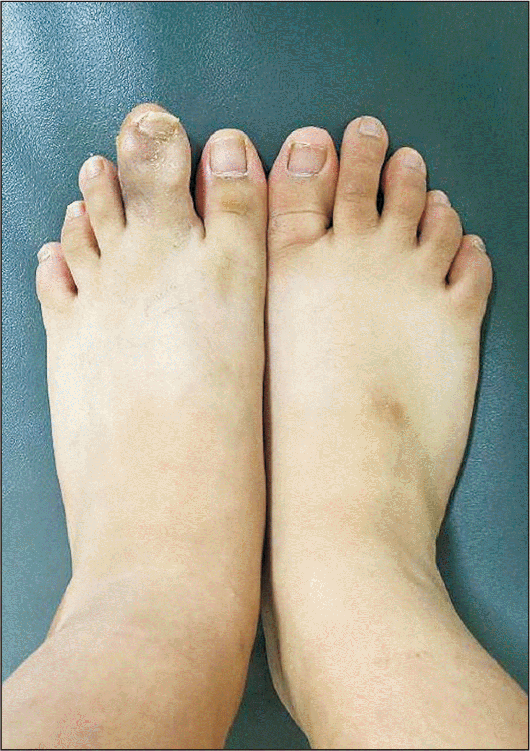
The patient and her parents were satisfied until the 7th year follow-up, during which the patient complained of difficulty wearing shoes. Soft-tissue thickening was observed in the outer part of the second toe (Fig. 10). Standing AP foot radiograph revealed a bony union state in the previously fused phalanges. There was no bony growth when compared with that on the unaffected side (Fig. 11). A second debulking operation was performed to remove the adipose tissue in the outer part of the second toe, and a large incision was created on the outside to preserve the nerves and blood vessels, following which the adipose tissue was removed. During the outpatient follow-up conducted 10 weeks post surgery (Fig. 12), both the patient and her parents were satisfied with the results, particularly its appearance.
DISCUSSION
Considering the negative impact of macrodactyly on function and cosmetic appearance, the treatment goal is to improve these two attributes, which may prevent the social withdrawal of patients. Various operative procedures, including amputation, osseous structure reduction, soft tissue debulking, and epiphysiodesis, have been introduced; however, the recurrence rate is high and patient dissatisfaction is common.4-7)
Phalangectomy or ray amputation may significantly reduce the volume of enlarged bony structures. Chang et al.8) and Kim et al.9) have argued that ray amputation is more effective than phalangectomy, especially for lesser toe macrodactyly, forefoot enlargement, and patients with a high metatarsal spread angle. However, despite a significant reduction in bony structures, the recurrence rate remains high, and subsequent soft tissue debulking is commonly performed. Additionally, cosmetic appearance must be considered while treating because the absence of one or more digits may not be acceptable for growing children or their parents.
Furthermore, osseous structure reduction also targets decreasing the bony structure volume. Tsuge7) introduced a reduction procedure comprising osteotomy of the proximal portion of the distal phalanx and the distal portion of the middle phalanx, and subsequent soft tissue debulking. Ray reduction is another reduction procedure where osteotomy is performed to shorten the metatarsal bone.10) Significant reduction in the bony structure volume and preservation of the digits and nails are the strengths of the osseous structure reduction procedure. However, the recurrence rate remains high, and Tsuge’s reduction procedure may be technically difficult.
In the current case, ostectomy of the middle phalanx was initially performed, and to the best of our knowledge, this is the first report of this procedure for macrodactyly. Amputation was not performed as the metatarsal spread angle was low, and both the patient and her parents had cosmetic concerns. As the sum of the lengths of the three phalanges was significantly higher than that of the normal side toe, soft tissue debulking alone seemed insufficient; therefore, osseous structure reduction was performed. Additionally, when determining the reduction level, we focused on the tethered proximal physeal line of the left second toe middle phalanx. Although the length between the left and right second middle phalanx was smaller than the length between the left and right proximal phalanges, we believed that the procedure involving the tethered physis might produce a better outcome, and ostectomy of the middle phalanx, including the tethered physis, was performed.
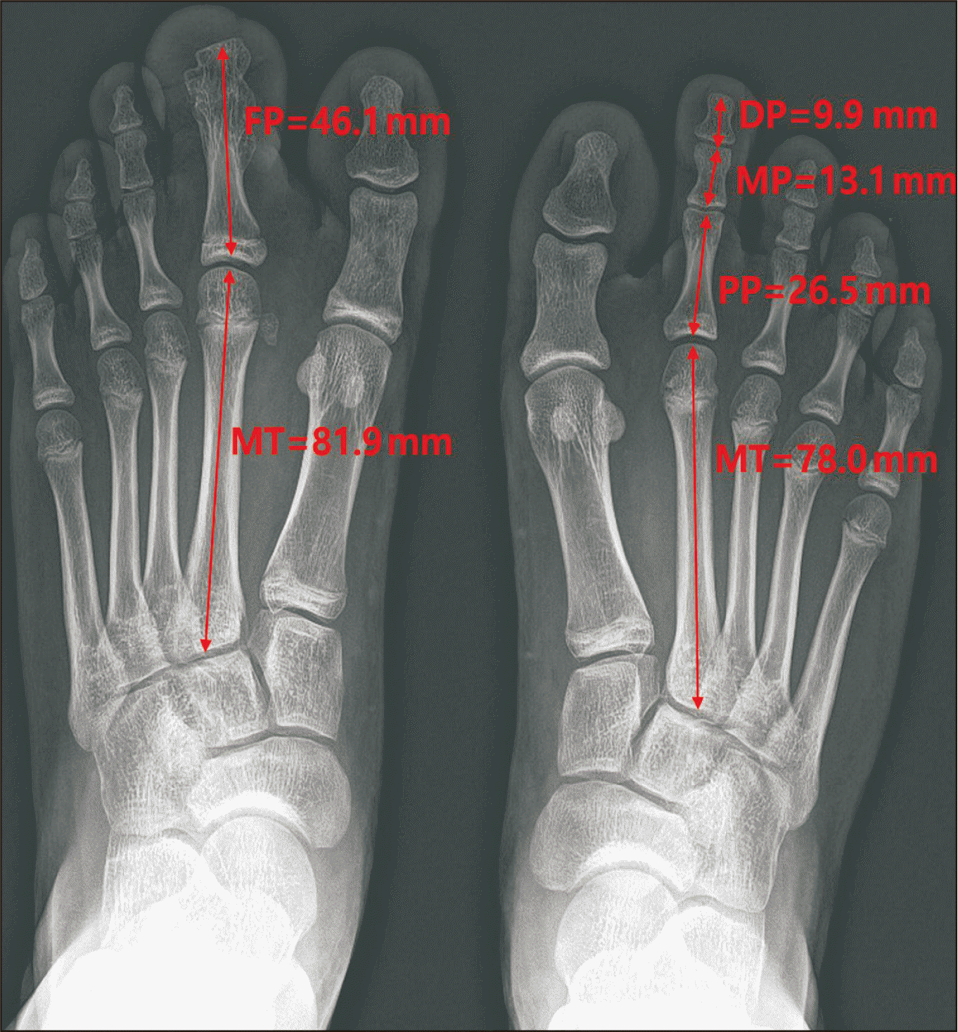
We suggest that ostectomy, including tethered physis, is another useful osseous structure reduction procedure for macrodactyly. The total length of the left foot of the three phalanges was 43.1 mm before the first surgery. Seven years later, the length of the fused phalanges was 46.1 mm, while that of the contralateral side was 49 mm. The patient’s second visit was due to soft tissue enlargement, not bony structure enlargement, and the size of the bony structure seemed to be successfully controlled. In addition to the preservation of the digit and nail, the patient and her parents were also satisfied with the cosmetic appearance.
This study had some limitations. First, this is a case report. The efficacy of performing ostectomy at the tethered physis cannot be generalized based on this case alone. Second, the patient’s age at her last follow-up was 13 years. Since growth usually does not end at this age, an outcome analysis should be performed in the future. However, all the physeal lines of the patient’s foot seemed to be nearly closed on plain radiographs. Therefore, we believe that the timing of assessing the surgical outcome does not matter in this study.
Despite these limitations, this case report has several strengths. First, to the best of our knowledge, this study is the first to introduce the concept of deciding the surgery level based on the tethered physis. As the exact pathophysiology of macrodactyly remains unknown, this may provide inspiration and direction for discovering macrodactyly pathogenesis and treatment. Second, our study included serial follow-up and measurements of the length of the enlarged ray.
In conclusion, ostectomy, including a tethered physis, is another useful osseous structure reduction procedure for macrodactyly. Further studies conducting this procedure on other digits and demonstrating the efficacy of this procedure in a larger study cohort are required to confirm our results.
 XML Download