

 PDF
PDF Citation
 Print
Print


INTRODUCTION
The natural course of Crohn’s disease (CD) is characterized by clinical remission alternating with recurrence.1 Persistent subclinical inflammation during clinical remission can lead to complications such as strictures, fistulas, and abscesses.2 The gluteal and presacral abscess are rare complications of CD, and the abscess can be made by fistula from nearby organs.3 Fistula is a common complication of CD because of the full thickness inflammation of the intestinal wall. Fistula affects up to 50% of CD patients within 20 years of initial diagnosis, and half of these are perianal fistulas.4 If the fistulas are not complete between the bowel and nearby organs, an intraabdominal abscesses may develop.5 Approximately 10-30% of patients with CD will develop an abdominal or pelvic abscess spontaneously over their lifetime.6 The most common site of fistula/abscess is perianal area, while gluteal and presacral regions are uncommon. There are few case reports of gluteal abscess combined with presacral abscess caused by CD,3,7-11 and the treatment has not been established.
Herein, we report a-21-year-old male with presentation of a gluteal abscess combined with presacral abscess due to fistulizing CD and its management. In addition, we will review other case reports regarding gluteal and presacral abscess caused by CD.
CASE REPORT
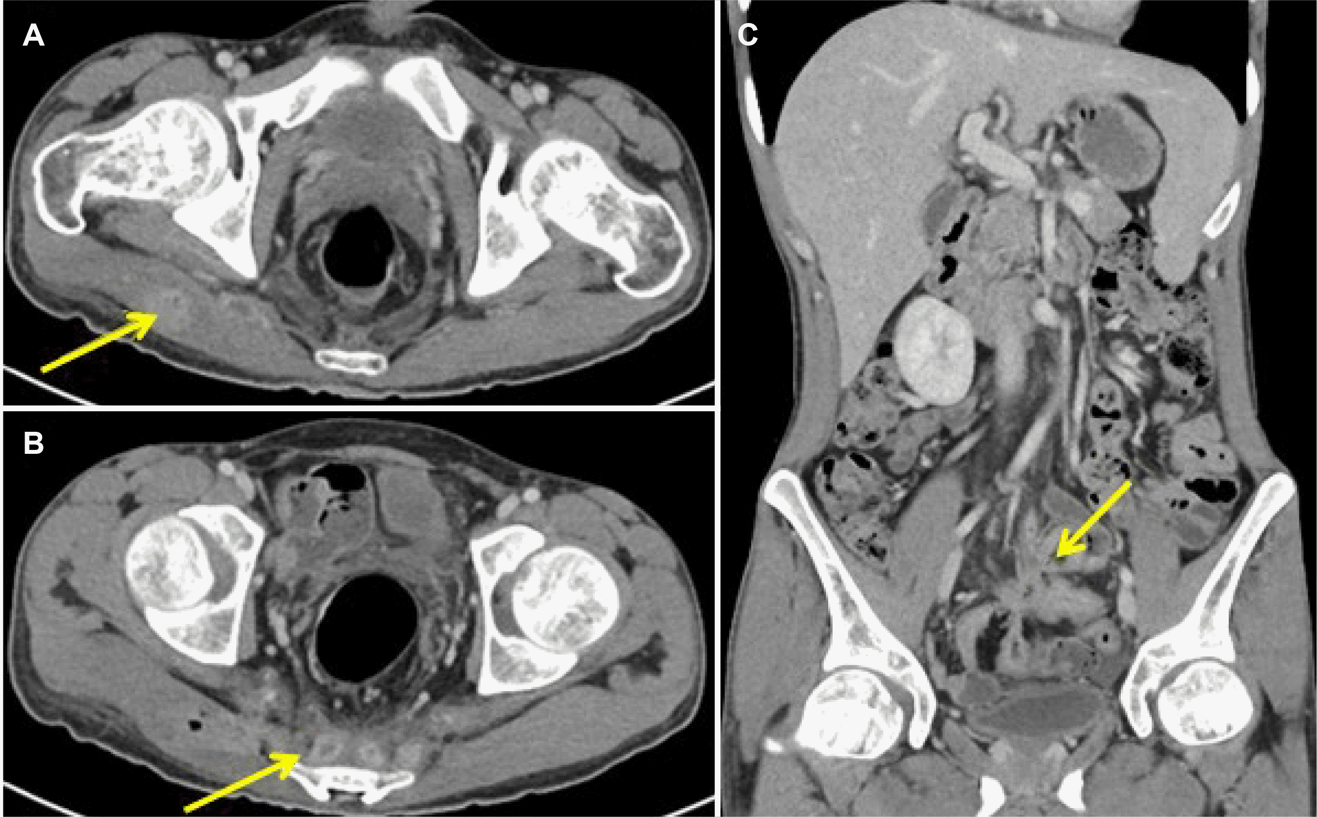
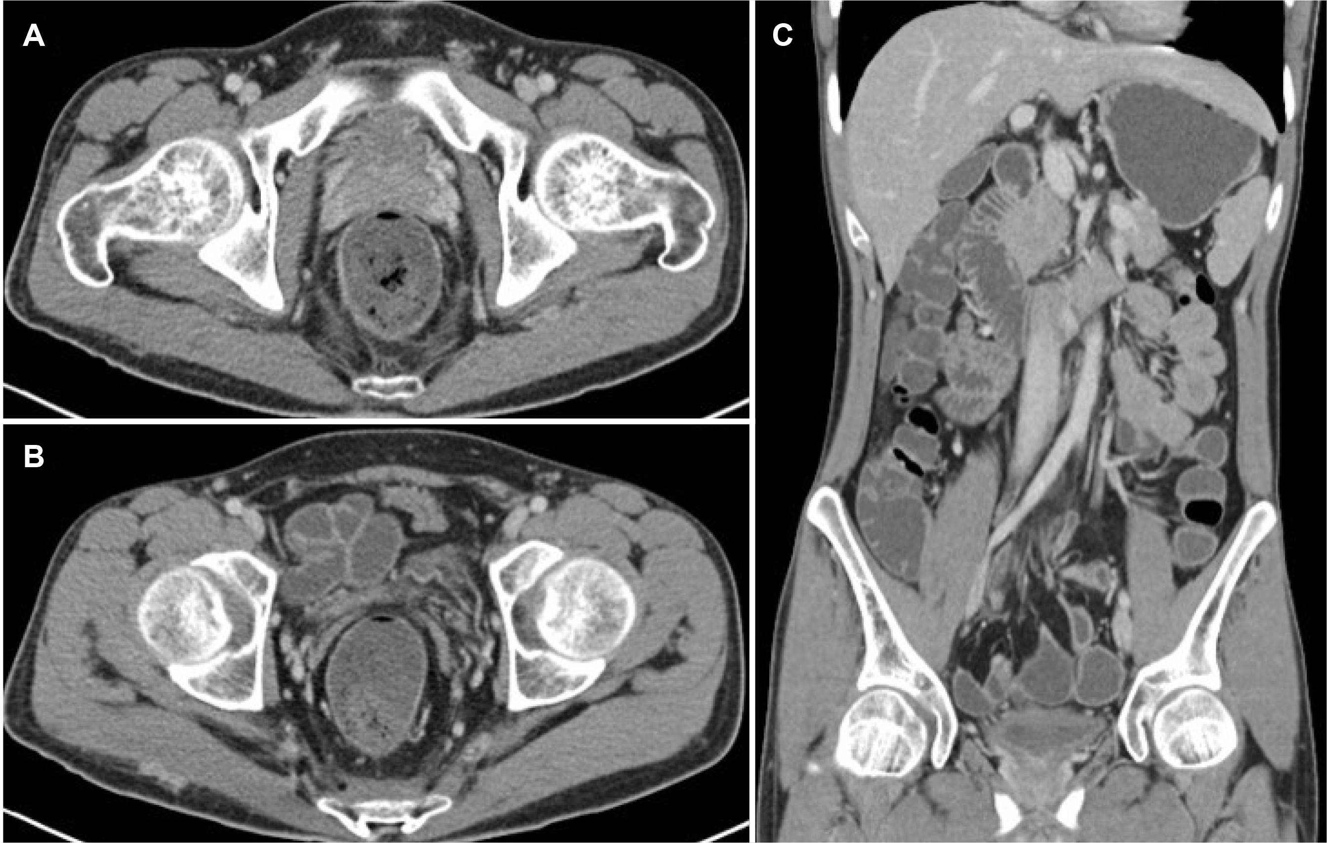
A 21-year-old man presented with CD in the small intestine diagnosed 10 months previously, complicated by anemia and osteopenia. He had been previously treated with mesalazine and steroids for 3 months. However, he did not return for follow-up care. Six months later, he was admitted to emergency department complaining of general weakness and right buttock and lower back pain for a duration of 3 months. No abdominal pain, fever, or weight loss was observed. Due to his lack of follow-up, he was not on any other long-term medication. On physical examination, his right buttock was swollen with focal tenderness. His laboratory test results were as following: hemoglobin of 10.6 g/dL, white blood cell (WBC) count of 11,500/mm3, C-reactive protein (CRP) level of 13.0 mg/dL, erythrocyte sedimentation rate (ESR) level of 53 mm/hr, and albumin of 2.9 g/dL. Abdominopelvic CT showed multiple segmental enhancing wall thickening of small bowel loops and indicated newly developed multiple abscess pockets in right gluteal region, including piriformis muscle and presacral space (Fig. 1). In addition, fistula tracts between small bowel loops and presacral space were observed. Patient’s Crohn’s Disease Activity Index (CDAI) was moderate activity (273.12 on the CDAI).
A recommendation was made for admission, but the patient refused and was started on antibiotic treatment through outpatient visit. During outpatient care begun since March 2021, he was treated with cefixime (200 mg/day) and metronidazole (1,500 mg/day) for 2 weeks and maintained on mesalazine (4,000 mg/day). The patient reported improvement in buttock and lower back pain in response to medication, but when the antibiotic drugs were stopped, the pain recurred. As the pain gradually worsened, he was admitted. Antibiotic treatment was started with Piperacillin/Tazobactam (4.5 g t.i.d/day) on the first day of hospitalization.
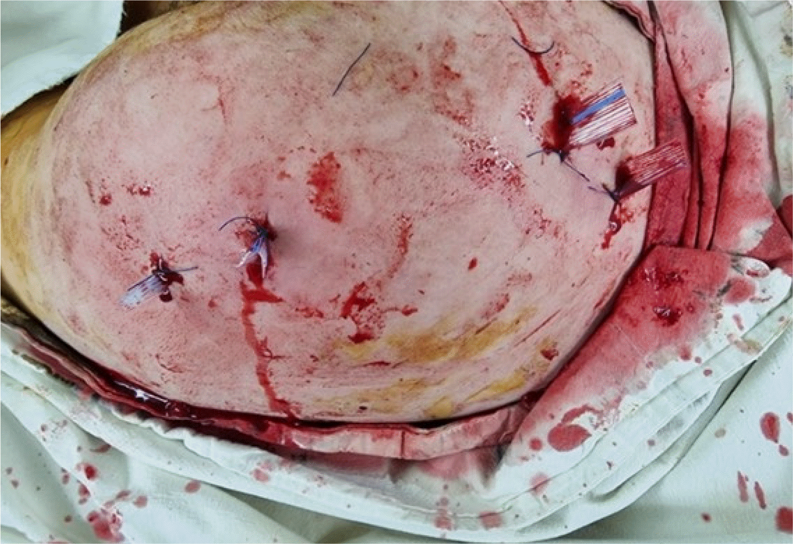
On the 3rd day, the patient had elevated temperature up to 38.3℃. Blood culture was performed to differentiate infection, and acetaminophen was prescribed to control temperature. On abdominopelvic CT, the abscess pocket extent remained unchanged. Despite the antibiotic treatment, there was no improvement in abscess extent and the patient complained of diarrhea and fever. Therefore, we determined that antibiotic treatment was unsuccessful, so surgical incision and drainage were done for abscesses on the 5th day. After initial drainage of purulence, massive irrigation was performed and 4 Penrose drains were inserted at surgical site (Fig. 2). Wound culture performed during surgery was negative. He did not complain of diarrhea or abdominal pain after surgery, and his pain and elevated temperature were resolved.
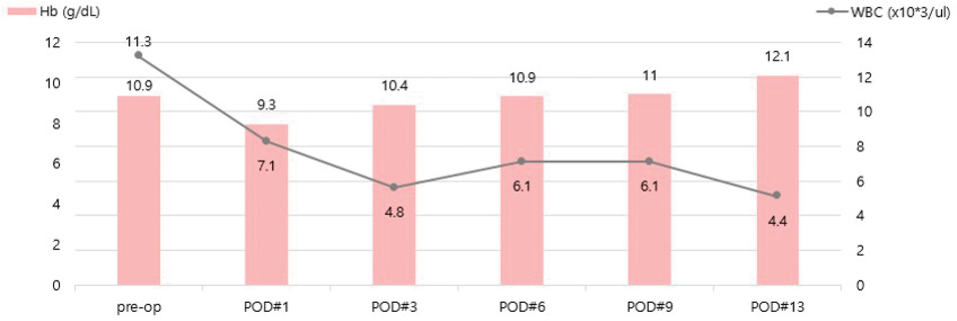
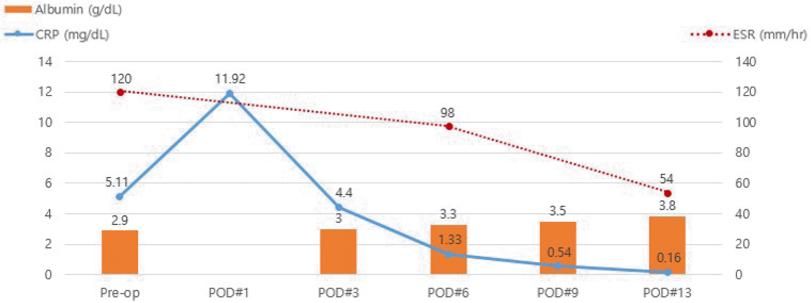
Following surgery, infliximab induction therapy was considered for the remaining complex fistulas in pelvic cavity. The first induction infliximab (230 mg, 5 mg/kg) was administered on the 12th day and the CDAI was moderate (220.72). About 2 weeks after the surgery, on the 18th day of hospitalization, the laboratory test results improved to near normal: hemoglobin of 12.1 g/dL, WBC of 4,400/mm3, CRP level of 0.16 mg/dL, ESR level of 54 mm/hr and albumin of 3.8 g/dL (Fig. 3, 4).
Postoperatively, buttock and lower back abscess resolved. The amount of secretion at the surgical site was also decreased, and 2 Penrose drains were removed. The patient was discharged with controlled symptoms following initial infliximab injection, and week 2 and 6 injections were done through the outpatient care.
After three doses in the induction regimen (on week 0, 2, and 6), abdominopelvic CT after 3 months showed that the extent of multiple abscess pockets in right gluteal muscle and presacral space was decreased and remaining multiple fistula tracts and multifocal enhancing wall thickening of small bowel loops were decreased (Fig. 5). The patient didn’t experience abdominal pain and diarrhea, and gained weight. The patient’s CDAI was remission (-0.12 on the CDAI). Following induction regimen, the patient continues to receive maintenance regimen at the outpatient care every 8 weeks, and shows no special complications for more than one-year follow-up period after surgery.
DISCUSSION
This case report has two significant clinical findings in that gluteal abscess combined with presacral abscess caused by CD is rare and infliximab induces closure of remained fistula, so that the patient recovered well without any surgical bowel resection. Although abscesses and fistulas are common complications of CD, the gluteal and presacral area are rare sites for abscess formation.3 The fistula to the retroperitoneal space uncommonly leads to the formation of presacral abscess. Furthermore, if the presacral abscess extends to bone directly or abscess erodes into the bone, osteomyelitis of pelvic bones is possible, which is rare as a complication of the presacral abscess.11 The presacral abscess continues to extend and passage through the greater sciatic foramen can result in a gluteal or posterior upper thigh abscess.8 Sangwan et al.12 reported three gluteal abscess cases among 1,735 patients with CD seen between 1980 and 1990. Denton et al.9 reported a case of abscess of the hamstring and adductor muscles resulting from the extension of pus in the pelvis complicated with CD. There was one case report of pure piriformis muscle abscess without involving gluteus muscles, due to a fistula from ileal loop to the piriformis muscle.13 In a study of 552 patients with CD, Brenner et al.14 reported 23 patients with purulent musculoskeletal complications caused by CD. These included four patients with gluteal muscle abscess/fistula, and four patients with sacral osteomyelitis, one of whom abscess extending into sacrosciatic foramen. There are several case reports of sacral osteomyelitis due to presacral abscess in patients with CD.3,11,13
There have been a few reports of gluteal abscess with presacral abscess complicating CD. Other cases were compared to this case, which showed gluteal abscess with or without presacral abscess associated with CD (Table 1).3,7-11 There were several common features among the cases, such as the initial clinical symptoms, treatment, and clinical courses. With the exception of one report showing only pure gluteal abscess, all cases were accompanied by gluteal abscess combined with presacral abscess. Common clinical symptoms of patients included buttock pain, lower back pain, difficulty in walking, elevated temperature, and weight loss. Three other cases used metronidazole or ampicillin/sulbactam, while the remaining patients did not receive antibiotic treatment and only had surgical intervention. Most of the patients underwent surgical incision and drainage of abscess and affected bowel resection. One study reported a direct endoscopic transanal drainage for presacral abscess as a safe and effective alternative drainage approach instead of surgery.3 No patients showed any sequelae and all fully recovered following intervention.
All patients in the papers we reviewed had or planned to have an affected bowel resection, but the patient in this case study recovered well only with abscess drainage performed at the appropriate time. Since CD frequently recurs and involves the entire intestine, it is important to reduce intestinal resection as much as possible. From the patient of this case study, if diagnosis and treatment are done quickly before abscesses lead to greater complications, we think timely surgical incision and drainage of abscess could be effective treatment options without intestinal resection.
Another probable influence on the patient’s recovery without intestinal resection is the use of biological agents. The treatment of fistula and abscess usually requires a combination of medical and surgical approach. Recently, biological agents are used for remaining fistulas instead of surgical bowel resection and it seems to be effective. Infliximab (Remicade®; Janssen Korea Ltd., Seoul, Korea), tumor necrosis factor-α (TNF-α) antagonists, has the strongest evidence in fistulizing CD treatment in terms of efficacy.15,16 Data from the literature show inspiring results after intravenous infusion for closure of fistulas, induction, and maintenance of remission in patients with moderate to severe CD unresponsive to other treatments.17 In this case, infliximab was administered for remaining complex fistulas in pelvic cavity after surgery. After the infliximab induction treatment, the patient improved and symptoms were resolved. He did not complain of symptoms such as diarrhea or abdominal pain, and gained weight. The complex fistulas remaining in the pelvic cavity also decreased.
In conclusion, the present case is a description of a newly developed gluteal and presacral abscess due to CD with multiple fistulas. This is a rare case of abscess in gluteal and presacral regions complicated by CD, according to the literature. There is no established treatment for gluteal abscess including presacral, so surgical incision and drainage of pus were performed followed by the administration of TNF antagonists for optimal treatment of multiple abscesses with fistulizing CD. This case is clinically significant in that he recovered well only with surgical incision and drainage followed by biological therapy without any intestinal resection. Neither procedure-related complications nor recurrence were observed in this patient.
 XML Download