

 PDF
PDF Citation
 Print
Print


I. Introduction
In 1894, nevoid basal cell carcinoma syndrome (NBCCS), also known as Gorlin-Goltz syndrome or basal cell nevus syndrome, was first introduced by Jarisch1. NBCCS is an autosomal dominant disorder caused by a PTCH1 gene mutation located on the 9q22.3-q31 chromosome1. This syndrome is characterized by basal cell nevus, odontogenic keratocysts (OKCs), cerebral calcification, and palmar and/or plantar pits. In addition, any of the following features may occur: oro-maxillofacial congenital malformations including cleft lip or palate, frontal and parietal protrusion, or hypertelorism; skeletal abnormalities; ovarian fibroma; or medulloblastoma2. The prevalence rate of NBCCS is 1:60,000, varies by age, and lacks sex predilection3,4. Basal cell carcinoma is found in 50%-97% of NBCCS patients, with some variation depending on race.
OKCs are the most common characteristic finding of NBCCS, occurring 65%-100% of NBCCS patients5. Clinically, they are invasive, grow rapidly, and tend to recur after surgical treatment. Basal epithelial cells exhibit increased cleavage and can potentially cause recurrence of OKC along with daughter hair follicles on the cyst wall. Spread to the mandible is more common than to the maxilla and occurs mainly in the posterior area of the mandible. The recurrence rate is 30% to 60%, and may vary depending on the surgical method used6.
The management of OKC can be generally classified into conservative or invasive treatment. Conservative treatment includes simple enucleation with or without curettage, or marsupialization and decompression followed by enucleation. Invasive treatment, which involves resection or complete removal of the lesion with peripheral ostectomy, is preferred in OKC with NBCCS, because of the clinically fast-growing or multi-locular intra-osseous lesions that can recur after marsupialization and curettage7. Whatever method is chosen, the goals of management are to successfully eliminate the potential for recurrence and minimizing the morbidity.
In this case report, we present our 11-year clinical follow-up experience with multiple OKCs in NBCCS managed by two-stage treatment, marsupialization and later surgical excision with peripheral ostectomy and marginal mandibulectomy.
II. Case Report
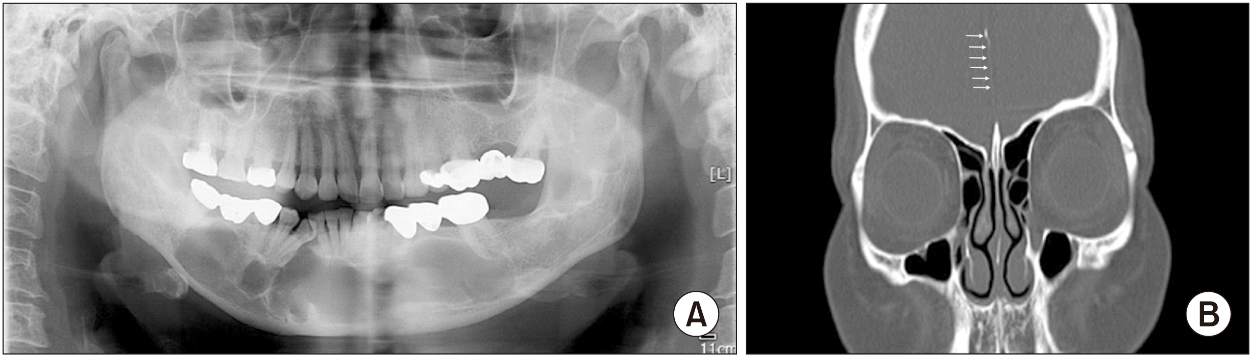
A 52-year-old female was referred to the Department of Oral and Maxillofacial Surgery at Ulsan University Hospital from a local dental clinic in 2009 due to multiple mandibular uni- and multi-cystic lesions. The patient had hypertelorism and a history of multiple surgeries for nevoid basal cell carcinomas on the face 2 years prior. Radiographs revealed multiple cystic lesions involving the mandibular symphysis, the right and left body of mandible, and left ramus regions.(Fig. 1. A) Falx cerebral calcification was also observed on computed tomographic (CT) scan.(Fig. 1. B)
Under a tentative diagnosis of OKC, the treatment plan included marsupialization followed by surgical excision to preserve both inferior alveolar nerves. Multiple openings were created and biopsy specimens were obtained from the opening sites. All specimens were sent for histopathological examination. For multi-cystic lesions, a single cystic lesion was created by perforating the septa, or multiple opening windows were created with the utmost care to preserve the inferior dental nerves. The examination findings of the biopsy specimens were consistent with the features of OKC, and a definitive diagnosis of NBCCS was reached according to the diagnostic criteria.
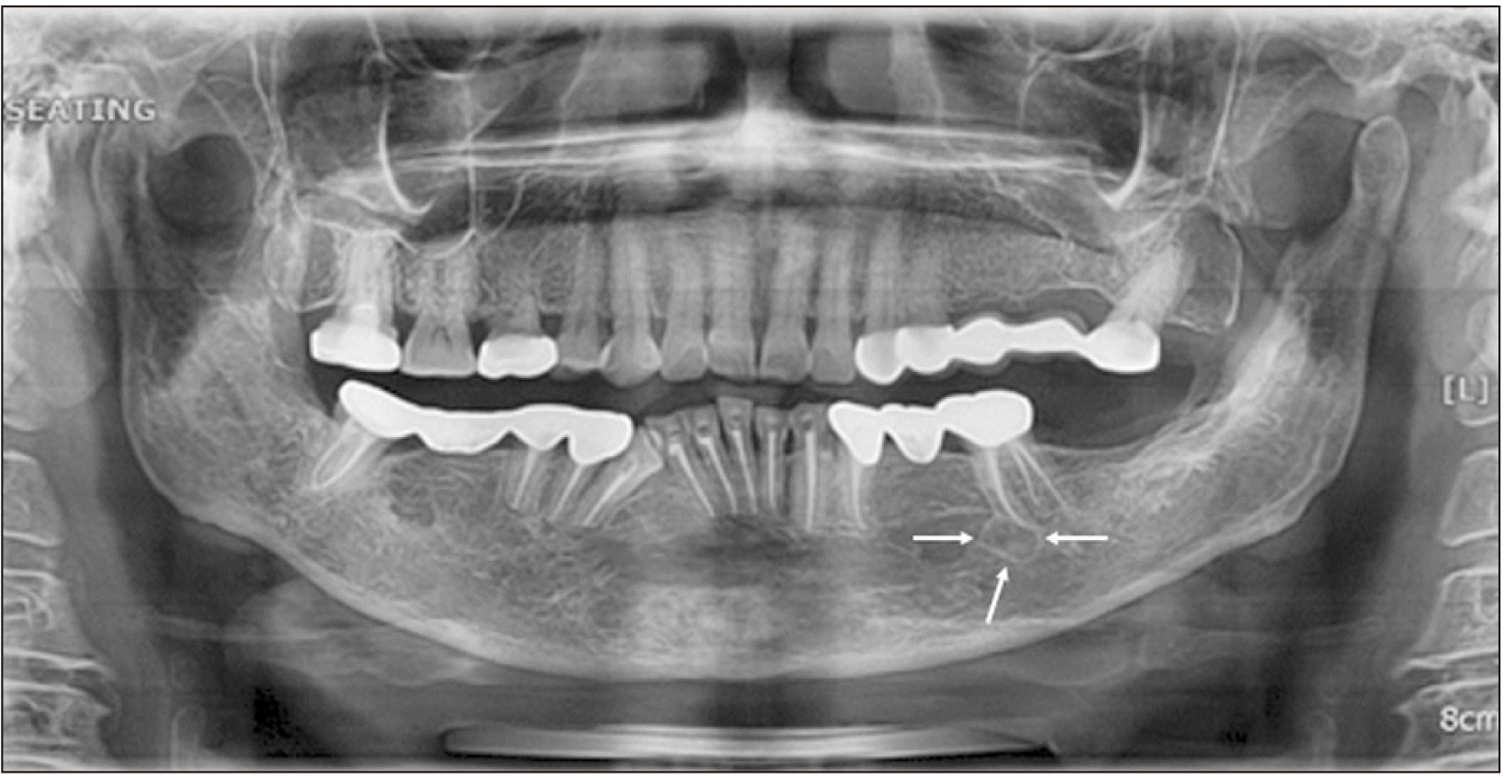
Clinical and radiographic examinations were carried out at 1-month intervals until the second surgery. Approximately one year later, cystic cavities were stable and the likelihood of damage to the inferior alveolar nerves was unlikely. The definitive second-stage surgery, which included surgical excision with peripheral ostectomy and marginal mandibulectomy on the left ramal region, was performed to eliminate residual pathological tissues.(Fig. 2)
After the definitive surgery, clinical and radiographic examinations were recommended every 6 months for at least 3 years, and every year thereafter because of the high recurrence rate of OKC in the NBCCS and the increased risk of developing new lesions in the future.
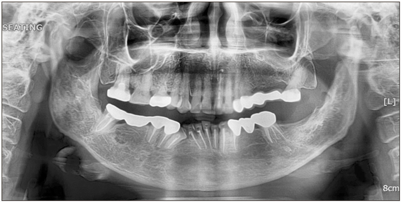
At the 8-year follow-up visit after surgical excision, a new lesion was observed in the mandibular left body region and excised under local anesthesia.(Fig. 3) Recurrence was presumed due to the high recurrence rate. Close follow-up, every 6 months during the first 2 years after treatment and once a year after that, was maintained thereafter and there was no sign of recurrence up until the last visit.(Fig. 4)
III. Discussion
The diagnostic criteria for NBCCS were introduced by Evans et al.8 in 1993 and then modified by Kimonis et al.9 in 1997. The diagnostic criteria for NBCCS established by Evans et al.8 were based on clinical and radiological findings, and a diagnosis is made when either two out of the five major criteria are satisfied or when one major criterion and two minor criteria are satisfied. Major diagnostic criteria include the following features: (1) Basal cell carcinoma; (2) OKC; (3) family history; and (4) plantar or palmar pits. Minor criteria are as follows: (1) Skeletal abnormality; (2) ovarian fibroma; (3) medulloblastoma; (4) macrocephaly; and (5) lymphomesenteric cyst. The patient in the present case had a history of surgery for basal cell carcinoma, hypertelorism, and multiple OKCs. CT examination revealed falx cerebri calcification. She was diagnosed with NBCCS, based on the identification of two major diagnostic criteria and one minor diagnostic criterion.
The major feature of OKC is its high recurrence rate, ranging from 0% to 62%10. The recurrence rate of OKC in patients with NBCCS was higher than that of OKC regardless of NBCCS. Manfredi et al.11 stated that the recurrence rate of OKC associated with NBCCS can reach up to 60%. When comparing sporadic OKC and syndromic OKC, syndromic OKC showed higher recurrence rates after both cyst enucleation and marsupialization. The recurrence rate of sporadic cysts was 17.6% after cyst enucleation and 22.2% after combination enucleation and marsupialization, while the recurrence rates of syndromic cysts were 25% and 68.8%, respectively7.
Cyst enucleation is the primary treatment for OKC; however, depending on the size of the OKC or location, enucleation may be performed after decompression. An aggressive surgical approach includes initial removal of all lesions; however, treatment with marsupialization and later enucleation with peripheral ostectomy allows for a less invasive approach, avoiding extensive damage. The major advantage of the two-stage procedure is preservation of the anatomical structures, bone, and teeth that are in close proximity to OKCs, particularly for multiple OKCs. However, Myoung et al.10 reported that patients with syndromic OKCs who underwent peripheral ostectomy with a 2- to 3-mm safety margin showed a recurrence rate of 47.6%.
Regarding the follow-up period, Chapelle et al.12 concluded that annual follow-up during the 5 years after removal of OKC was desirable because most recurrences appeared within this time period. However, Stoelinga13 recommended every 2 years for a period of at least 25 years. Nevertheless, the 25-year long-term follow-up period proposed by Stoelinga13 cannot completely avoid recurrence because recurrence can occur over a long period of time. Thus, the exact recurrence period cannot be specified. The recurrence rate after removal of OKCs may vary depending on the research setting14. Therefore, we should expect the possibility of OKC recurrence at any time and perform regular follow-up, especially in syndromic OKCs.
In this case, considering the wide area of involvement in the mandible by multiple OKCs, marsupialization was performed first to preserve both the inferior alveolar nerves and the involved teeth. Subsequently, additional procedures were performed, including surgical excision with peripheral ostectomy and marginal mandibulectomy for complete removal of the residual lesions, and the patient was followed annually. Although the lesion recurred after 8 years, simple excision was successfully performed under local anesthesia because of early detection. Considering the high recurrence rate of OKCs associated with NBCCS13, the safety margin of more than 2-3 mm that is typically recommended in routine peripheral ostectomy procedures should be maintained. Lastly, regular long-term follow-up is essential because of the high associated recurrence rate.

 XML Download